. 2004 Mar 19;32(5):1783-91.
doi: 10.1093/nar/gkh341. Print 2004.

Silencer elements as possible inhibitors of pseudoexon splicing

Affiliations

Silencer elements as possible inhibitors of pseudoexon splicing

Manuela Sironi et al. Nucleic Acids Res. .

Abstract

Human pre-mRNAs contain a definite number of exons and several pseudoexons which are located within intronic regions. We applied a computational approach to address the question of how pseudoexons are neglected in favor of exons and to possibly identify sequence elements preventing pseudoexon splicing. A search for possible splicing silencers was carried out on a pseudoexon selection that resembled exons in terms of splice site strength and exon splicing enhancer (ESE) representation; three motifs were retrieved through hexamer composition comparisons. One of these functions as a powerful silencer in transfection-based splicing assays and matches a previously identified silencer sequence with hnRNP H binding ability. The other two motifs are novel and failed to induce skipping of a constitutive exon, indicating that they might act as weak repressors or in synergy with other unidentified elements. All three motifs are enriched in pseudoexons compared with intronic regions and display higher frequencies in intronless gene-coding sequences compared with exons. We consider that a subpopulation of pseudoexons might rely on negative regulators for splicing repression; this hypothesis, if experimentally verified, might improve our understanding of exonic splicing regulatory sequences and provide the identification of a novel mutation target for human genetic diseases.


Figures
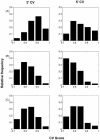
Figure 1
Figure 1
Consensus value frequency distributions. The 3′ (left panels) and 5′ (right panels) splice site CV distributions were calculated for Rex (A), Pex (B) and Pex1 (C).
Figure 2
Figure 2
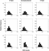
ESE frequency distribution. ESE frequencies were calculated for exon or pseudoexon bodies (central panels) and for the respective 3′- (left) and 5′- (right) flanking sequences (200 bp each side). (A) Rex; (B) Pex; (C) Pex1.
Figure 3
Figure 3
Consensus matrices for the three retrieved motifs. Matrices were defined as described in Materials and Methods; best scoring motifs are reported below each matrix.
Figure 4
Figure 4
Relative motif frequency in the initial set (A) and in the test set (B). The relative frequency of each motif was calculated in Pex1 and Pex1T (black), in non-Pex1/non-Pex1T pseudoexons (striped), whole intron sequences (dark gray), Rex and RexT (light gray) and intronless gene-coding sequences (white). Overall absolute frequencies for the three motifs in real exons (Rex + RexT) and in elite pseudoexons (Pex1 + Pex1T) were as follows: motif 1, 13 and 19; motif 2, 243 and 315; motif 3, 55 and 64.
Figure 5
Figure 5
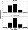
Comparison of ESE frequencies between motif-containing and motif-lacking real exons (Rex + RexT). ESE frequencies were calculated and expressed as histograms for motif-lacking (upper panel) and motif- containing (lower panel) real exons.
Figure 6
Figure 6
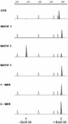
Transfection-based splicing assays and RT–PCR. Motifs 1, 2 and 3 were inserted in NF1 exon 24 and splicing efficiency was evaluated after transient transfection and RT–PCR; amplified products (shaded peaks) were run on a Genetic Analyzer. White peaks represent molecular weight markers. CTR, control vector (no insert); motif 1, 2 and 3, vectors carrying each of these motifs within exon 24, 7mer and 6mer, control vectors containing either an unrelated heptamer or hexamer (control of size variation on splicing efficiency).

References

    1. Maniatis T. and Tasic,B. (2002) Alternative pre-mRNA splicing and proteome expansion in metazoans. Nature, 418, 236–243. - PubMed
    1. Senapathy P., Shapiro,M.B. and Harris,N.L. (1990) Splice junctions, branch point sites and exons: sequence statistics, identification and applications to genome project. Methods Enzymol., 183, 252–278. - PubMed
    1. Manley J.L. and Tacke,R. (1996) SR proteins and splicing control. Genes Dev., 10, 1569–1579. - PubMed
    1. Mayeda A., Screaton,G.R., Chandler,S.D., Fu,X.D. and Krainer,A.R. (1999) Substrate specificities of SR proteins in constitutive splicing are determined by their RNA recognition motifs and composite pre-mRNA exonic elements. Mol. Cell. Biol., 19, 1853–1863. - PMC - PubMed
    1. Schaal T.D. and Maniatis,T. (1999) Selection and characterization of pre-mRNA splicing enhancers: identification of novel SR protein-specific enhancer sequences. Mol. Cell. Biol., 19, 1705–1719. - PMC - PubMed