. 2004 Mar 19;32(5):1818-23.
doi: 10.1093/nar/gkh349. Print 2004.

The major form of MeCP2 has a novel N-terminus generated by alternative splicing

Affiliations

The major form of MeCP2 has a novel N-terminus generated by alternative splicing

Skirmantas Kriaucionis et al. Nucleic Acids Res. .

Abstract

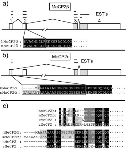
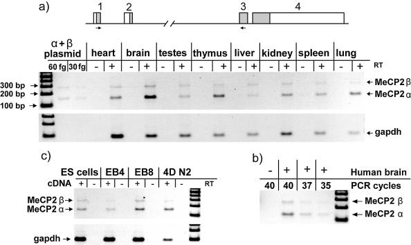
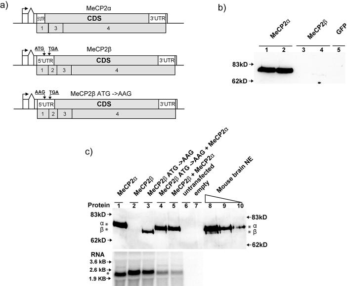
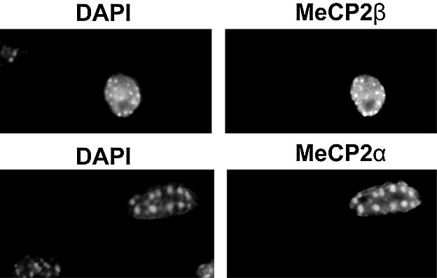
MeCP2 is a methyl-CpG binding protein that can repress transcription of nearby genes. In humans, mutations in the MECP2 gene are the major cause of Rett syndrome. By searching expressed sequence tag (EST) databases we have found a novel MeCP2 splice isoform (MeCP2alpha) which encodes a distinct N-terminus. We demonstrate that the MeCP2alpha mRNA splice variant is more abundant than the previously annotated MeCP2 mRNA (MeCP2beta) in mouse tissues and human brain. Furthermore, MeCP2beta mRNA has an upstream open reading frame that inhibits its translation. As a result of these differences, >90% of MeCP2 in mouse brain is MeCP2alpha. Both protein isoforms are nuclear and colocalize with densely methylated heterochromatic foci in mouse cells. The presence of a previously unknown MeCP2 isoform has implications for the genetic screening of Rett syndrome patients and for studies of the functional significance of MeCP2.


Figures

Figure 1
Figure 1
Alternative splicing of MeCP2 mRNA. (a) Previously described MeCP2β is encoded when all known exons are sequentially spliced. (b) The novel MeCP2α isoform arises when exon 1 is spliced onto exon 3, skipping exon 2. Shaded boxes are protein coding and open boxes are non-coding. ESTs suggesting the occurrence of each isoform in vivo are mapped as lines above the genomic DNA. (c) Alignment of mouse and human MeCP2β and MeCP2α N-termini with zebrafish and frog MeCP2. MeCP2α is more similar to zebrafish and frog orthologs than is MeCP2β.
Figure 2
Figure 2
Relative abundance of splice variant mRNAs in mouse tissue, human brain and differentiating ES cells. (a) A mouse tissue cDNA panel was analysed by semiquantitative PCR with primers that anneal to exons 1 and 3 (short arrows on map). The α isoform was more abundant in lung, thymus, brain and heart. Amplification of an equimolar mixture of α- and β-encoding plasmids indicated no preference for amplification of either PCR band. (b) Human brain cDNA showed dominance of the α isoform mRNA. (c) ES cells were differentiated to give embryoid bodies and neuronal cells in culture. EB4 and EB8 refer to embryoid bodies on day 4 and day 8 of differentiation. 4DN2 refers to day 4 after embryoid bodies were split and plated on serum-free neurobasal medium.
Figure 3
Figure 3
MeCP2β is inefficiently translated to give a protein that can be separated from MeCP2α by PAGE. (a) Diagrams of MeCP2α, MeCP2β and mutated MeCP2β constructs used for transfection of Mecp2-null tail fibroblasts. An SV-40 promoter and artificial intron is followed by the MeCP2 5′UTR, coding sequence, 3′UTR, MeCP2 polyadenylation signal and SV40 polyadenylation signal. MeCP2 exons are numbered below each map. The start codon of the wild-type upstream ORF in MeCP2β is labeled as ATG (middle diagram) and is mutated to AAG in the MeCP2β ATG-AAG construct (bottom diagram). (b) Weak expression of the MeCP2β construct in transfected cells. Two independent plasmid preparations were used for each type of transfection (MeCP2α, lanes 1 and 2; MeCP2β, lanes 3 and 4). The product of the MeCP2β plasmid was barely visible in lane 4 only. (c) Western blot analysis (upper panel) of transfected Mecp2-null mouse fibroblasts and native MeCP2 from mouse brain nuclear extracts. MeCP2 protein isoforms (asterisks) migrated at different sizes on this 8% PAGE gel (compare lanes 1, 3 and 4). The upstream ORF inhibited the translation of MeCP2β (lanes 2 and 3). MeCP2α is the predominant isoform in mouse brain nuclear extract (lanes 8, 9 and 10). The northern blot (lower panel) showed that different levels of translated MeCP2 protein are not due to differential transcription of the constructs, as similar amounts of MeCP2 mRNA were seen in lanes 1–3. In the absence of transfection, no endogenous MeCP2 RNA is detectable in these Mecp2-null cells.
Figure 4
Figure 4
Both MeCP2 isoforms colocalized with methyl-CpG-rich DAPI bright spots when Mecp2-null fibroblasts were transfected with wild-type MeCP2α and MeCP2β expression constructs.

References

    1. Hendrich B. and Bird,A. (1998) Identification and characterization of a family of mammalian methyl-CpG binding proteins. Mol. Cell. Biol., 18, 6538–6547. - PMC - PubMed
    1. Nan X., Meehan,R.R. and Bird,A. (1993) Dissection of the methyl-CpG binding domain from the chromosomal protein MeCP2. Nucleic Acids Res., 21, 4886–4892. - PMC - PubMed
    1. El Osta A. and Wolffe,A.P. (2001) Analysis of chromatin-immunopurified MeCP2-associated fragments. Biochem. Biophys. Res. Commun., 289, 733–737. - PubMed
    1. Nan X., Tate,P., Li,E. and Bird,A. (1996) DNA methylation specifies chromosomal localization of MeCP2. Mol. Cell Biol., 16, 414–421. - PMC - PubMed
    1. Lorincz M.C., Schubeler,D. and Groudine,M. (2001) Methylation-mediated proviral silencing is associated with MeCP2 recruitment and localized histone H3 deacetylation. Mol. Cell. Biol., 21, 7913–7922. - PMC - PubMed

Publication types