. 2004 Jul 15;381(Pt 2):537-46.
doi: 10.1042/BJ20031499.

Intracellular transport of human lysosomal alpha-mannosidase and alpha-mannosidosis-related mutants

Affiliations

Intracellular transport of human lysosomal alpha-mannosidase and alpha-mannosidosis-related mutants

Gaute Hansen et al. Biochem J. .

Abstract

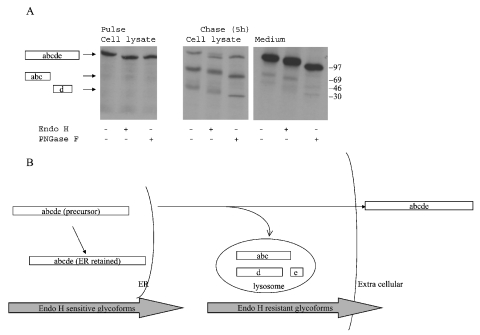
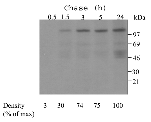
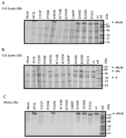
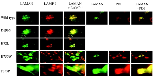
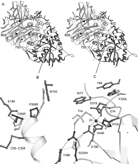
Human LAMAN (lysosomal a-mannosidase) was synthesized as a 120 kDa precursor in transfected COS cells [African-green-monkey kidney cells], which was partly secreted as a single-chain form and partly sorted to the lysosomes being subsequently cleaved into three peptides of 70, 40 and 15 kDa respectively. Both the secreted and the lysosomal forms contained endo H (endoglucosidase H)-resistant glycans, suggesting a common pathway through the trans-Golgi network. A fraction of LAMAN was retained intracellularly as a single-chain endo H-sensitive form, probably in the ER (endoplasmic reticulum). The inherited lack of LAMAN causes the autosomal recessive storage disease a-mannosidosis. To understand the biochemical consequences of the disease-causing mutations, 11 missense mutations and two in-frame deletions were introduced into human LAMAN cDNA by in vitro mutagenesis and the resulting proteins were expressed in COS cells. Some selected mutants were also expressed in Chinese-hamster ovary cells. T355P (Thr355Pro), P356R, W714R, R750W and L809P LAMANs as well as both deletion mutants were misfolded and arrested in the ER as inactive single-chain forms. Six of the mutants were transported to the lysosomes, either with less than 5% of normal specific activity (H72L, D196E/N and R220H LAMANs) or with more than 30% of normal specific activity (E402K LAMAN). F320L LAMAN resulted in much lower activity in Chinese-hamster ovary cells when compared with COS cells. Modelling into the three-dimensional structure revealed that the mutants with highly reduced specific activities contained substitutions of amino acids involved in the catalysis, either co-ordinating Zn2+ (His72 and Asp196), stabilizing the active-site nucleophile (Arg220) or positioning the active-site residue Asp319 (Phe320).


Figures

Figure 1
Figure 1. Intracellular transport of human LAMAN
(A) COS-7 cells were transfected with pcDNA constructs containing wild-type human LAMAN cDNA using LIPOFECTAMINE™ 2000. The cells were pulsed 48 h later with [35S]methionine/cysteine for 30 min and the cells were subsequently harvested or chased for 5 h. Human LAMAN was precipitated from the cell lysates or the media with a polyclonal antiserum [7]. The resulting immunoprecipitates were treated with endo H or PNGase F, separated by SDS/PAGE and analysed by autoradiography. The positions of the respective LAMAN peptide fragments are indicated on the left. (B) A model of the intracellular transport of human LAMAN in COS cells, based on the results shown in (A).
Figure 2
Figure 2. Secretion kinetics of human LAMAN
COS-7 cells were transfected with pcDNA constructs containing a human LAMAN cDNA construct as described in Figure 1. Subsequent to the pulse period of 30 min with [35S]methionine/cysteine, the cells were chased for the indicated times. Human LAMAN was immunoprecipitated from the medium and analysed by autoradiography. The bands were scanned and the intensity of the respective bands was correlated with that of the band representing the 24 h chase.
Figure 3
Figure 3. Intracellular transport of mutant human LAMANs
COS-7 cells were transfected with pcDNA constructs containing missense mutations obtained by site-directed mutagenesis (Table 1) and metabolically labelled as described in Figure 1. Subsequent to the pulse period of 30 min with [35S]methionine/cysteine, the cells were either harvested (A) or subjected to 5 h of chase (B, C). Human LAMAN was immunoprecipitated from the cell extracts (A, B) or the media (C) and analysed by autoradiography.
Figure 4
Figure 4. Intracellular distribution of human LAMAN in COS cells observed with immunofluorescence microscopy
Double immunofluorescence staining was performed using antibodies against LAMAN, LAMP-1 and PDI. Yellow indicates overlap of the LAMAN peptides (green) and LAMP-1 (red) or PDI (red).
Figure 5
Figure 5. Western-blot analyses of mutant human LAMANs
Cell lysate and medium from COS cells transfected with various mutant LAMAN constructs were subjected to SDS/PAGE and electroblotted on to a PVDF membrane. The membrane was incubated in diluted antiserum against native recombinant human LAMAN as described in the Materials and methods section.
Figure 6
Figure 6. Amino acid substitutions modelled in the bovine LAMAN structure
(A) Stereographic representation of the complete structure. The active-site domain is darker than the rest of the structure. The affected residues are indicated with black balls. Mutations outside the active-site domain are labelled, and mutations in the N-terminal domain are shown in detail in (B, C). (B) Mutations affecting the folding of LAMAN. The mutated wild-type residues are shown in dark grey and the modelled mutations in white. Hydrogen bonds are displayed with broken lines and metal co-ordination with solid lines. The T355P mutation disrupts a hydrogen bond to Glu149 and, probably, to the free cysteine residue Cys358, which in the mature enzyme participates in a disulphide bridge. Pro356 is in optimal position to initialize helix formation and is important for the rate of folding of LAMAN. (C) Mutations that inactivate the enzyme, but allow folding, are located close to the active site: although His220 hydrogen-bonds to the nucleophile Asp196, it cannot co-ordinate the substrate as Arg220 probably does. His72 is involved in the metal binding, whereas Phe320 follows the active-site residue Asp319 and makes a stacking interaction with Tyr84. Probably, the hydrophobic stacking also stabilizes Trp77, which is involved in substrate binding.

References

    1. Henrissat B., Bairoch A. Updating the sequence-based classification of glycosyl hydrolases. Biochem. J. 1996;316:695–696. - PMC - PubMed
    1. DeGasperi R., al Daher S., Daniel P. F., Winchester B. G., Jeanloz R. W., Warren C. D. The substrate specificity of bovine and feline lysosomal α-D-mannosidases in relation to α-mannosidosis. J. Biol. Chem. 1991;266:16556–16563. - PubMed
    1. Aronson N. N., Kuranda M. J. Lysosomal degradation of Asn-linked glycoproteins. FASEB J. 1989;3:2615–2622. - PubMed
    1. Van den Elsen J. M., Kuntz D. A., Rose D. R. Structure of Golgi α-mannosidase II: a target for inhibition of growth and metastasis of cancer cells. EMBO J. 2000;20:3008–3017. - PMC - PubMed
    1. Heikinheimo P., Helland R., Leiros H.-K., Karlsen S., Evjen G., Ravelli R., Schoen G., Ruigrok R., Tollersrud O. K., McSweeney S., et al. The structure of bovine lysosomal α-mannosidase suggests a novel mechanism for low pH activation. J. Mol. Biol. 2003;327:631–644. - PubMed

Publication types

MeSH terms