. 2004 Mar;25(3):370-6.

Diffusion tensor imaging as potential biomarker of white matter injury in diffuse axonal injury

Affiliations

Diffusion tensor imaging as potential biomarker of white matter injury in diffuse axonal injury

Thierry A G M Huisman et al. AJNR Am J Neuroradiol. 2004 Mar.

Abstract

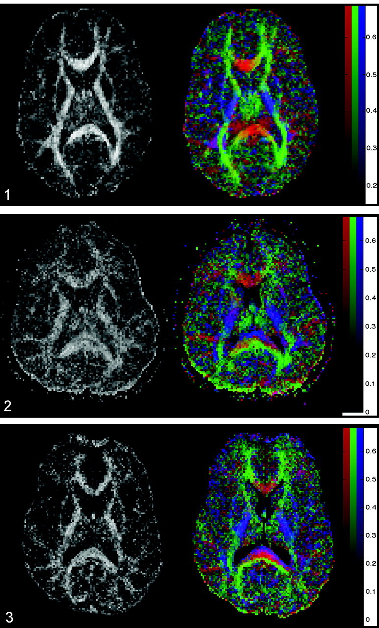
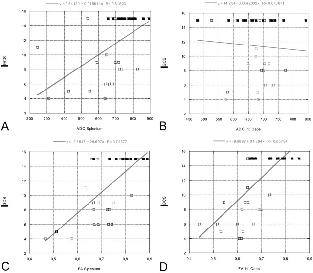
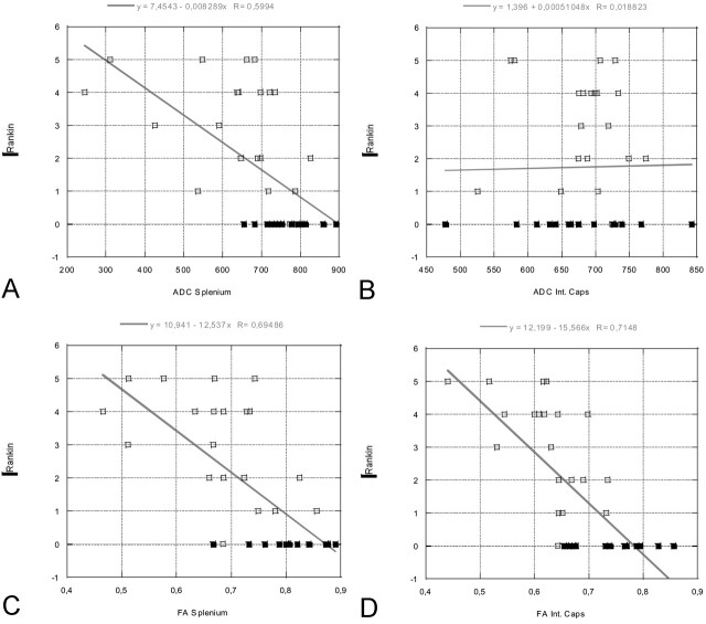
Background and purpose: Multiple biomarkers are used to quantify the severity of traumatic brain injury (TBI) and to predict outcome. Few are satisfactory. CT and conventional MR imaging underestimate injury and correlate poorly with outcome. New MR imaging techniques, including diffusion tensor imaging (DTI), can provide information about brain ultrastructure by quantifying isotropic and anisotropic water diffusion. Our objective was to determine if changes in anisotropic diffusion in TBI correlate with acute Glasgow coma scale (GCS) and/or Rankin scores at discharge.

Methods: Twenty patients (15 male, five Female; mean age, 31 years) were evaluated. Apparent diffusion coefficients (ADCs) and fractional anisotropy (FA) values were measured at multiple locations and correlated with clinical scores. Results were compared with those of 15 healthy control subjects.

Results: ADC values were significantly reduced within the splenium (Delta18%, P =.001). FA values were significantly reduced in the internal capsule (Delta14%; P <.001) and splenium (Delta16%; P =.002). FA values were significantly correlated with GCS (r = 0.65-0.74; P <.001) and Rankin (r = 0.68-0.71; P <.001) scores for the internal capsule and splenium. The correlation between FA and clinical markers was better than for the corresponding ADC values. No correlation was found between ADC of the internal capsule and GCS/Rankin scores.

Conclusion: DTI reveals changes in the white matter that are correlated with both acute GCS and Rankin scores at discharge. DTI may be a valuable biomarker for the severity of tissue injury and a predictor for outcome.


Figures

F<sc>ig</sc> 1.
Fig 1.
Axial FA map (left) and color coded map of mean diffusion direction (right) at the level of the basal ganglia, thalami and internal capsulae in a healthy control subject. Left, Gray-scale FA map displays a high degree of anisotropic diffusion (bright) within the internal capsule and the splenium of the corpus callosum. The cortex and the central gray matter are dark because of their low degree of anisotropic diffusion. Right, Color-coded image displays a predominant left-right-left mean diffusion direction (red) within the center of the splenium of the corpus callosum, an anteroposterior direction (green) within the optic radiations, and a superior-inferior direction (blue) within the posterior internal capsule.
F<sc>ig</sc> 2.
Fig 2.
Images in a 24-year-old man with severe TBI. Acute GCS, 5. Rankin score at discharge, 3. Left, FA map shows a reduced FA index of the splenium of the corpus callosum (FA = 0.511 ± 0.036, mean control FA = 0.808 ± 0.060) and internal capsule (FA = 0.531 ± 0.036, mean control FA = 0.735 ± 0.066). Right, Color-coded map shows that, within the center of the splenium of the corpus callosum, the normally predominant red voxels are missing and replaced by a mixture of blue and green voxels (compare with Fig 1). This finding suggests that fiber tracts that connect both cerebral hemispheres are injured or disrupted within the center of the splenium.
F<sc>ig</sc> 3.
Fig 3.
Images in a 37-year-old man with severe TBI. His GCS score at the time of MR imaging was 3, and his Rankin score at discharge was 4. Left, FA map shows a reduced FA index of the corpus callosum (FA = 0.634 ± 0.036). Right, Color-coded map shows a layered blue, red, and green aspect of the splenium of the corpus callosum. This could indicate a partial, selective injury of the most anterior and posterior left-right-left fiber tracts.
F<sc>ig</sc> 4.
Fig 4.
Linear regression plots of ADC and FA values of the splenium and internal capsule versus GCS at the time of acute MR imaging (in patients) or at time of comparison MR imaging (control subjects, all with GCS scores of 15). A statistically significant correlation is seen between the FA values of the splenium/internal capsule and GCS, as well as between the ADC values within the splenium and GCS. GCS scores vary between 3 and 15, where 3 represents the worst score, and 15, the best score. Open rectangles indicate patients; solid rectangles, control subjects. A, ADC splenium versus GCS. B, ADC internal capsule versus GCS. C, FA splenium versus GCS. D, FA internal capsule versus GCS.
F<sc>ig</sc> 5.
Fig 5.
Linear regression plots of ADC and FA values of the splenium and internal capsule versus Rankin score at the time of discharge (in patients) or at time of comparison MR imaging (control subjects, all with Rankin scores of 0). A statistically significant correlation is seen between the FA values of the splenium/internal capsule and Rankin score, as well as between the ADC values of the splenium and Rankin score. Rankin scores vary between 0 and 5, where 0 represents the best score and 5, the worst score. Open rectangles indicate patients; solid rectangles, control subjects. A, ADC splenium versus Rankin. B, ADC internal capsule versus Rankin. C, FA splenium versus Rankin. D, FA internal capsule versus Rankin.

Similar articles

Cited by

References

    1. Gean AD. White matter shearing injury and brainstem. injury In: Imaging of Head Trauma. New York: Raven,1994. :207–248
    1. Murray JG, Gean AD, Evans SJ. Imaging of acute head injury. Semin Ultrasound CT MR. 1996;17:185–205 - PubMed
    1. Gentry LR. Head trauma. In: Atlas SW, ed. Magnetic Resonance Imaging of the Brain and Spine. New York: Raven,1996;611–647
    1. Strich SJ. Shearing of nerve fibres as a cause of brain damage due to head injury: a pathological study of twenty cases. Lancet 1961;2:443–448
    1. Adams JH, Graham DI, Murray LS, Scott G. Diffuse axonal injury due to nonmissile head injury in humans: an analysis of 45 cases. Ann Neurol 1982;12:557–563 - PubMed

Publication types

MeSH terms

LinkOut - more resources