. 2004 Mar;25(3):431-40.

Magic angle effects in MR neurography

Affiliations

Magic angle effects in MR neurography

Karyn E Chappell et al. AJNR Am J Neuroradiol. 2004 Mar.

Abstract

Background and purpose: Magic angle effects are well recognized in MR imaging of tendons and ligaments, but have received virtually no attention in MR neurography. We investigated the hypothesis that signal intensity from peripheral nerves is increased when the nerve's orientation to the constant magnetic induction field (B(0)) approaches 55 degrees (the magic angle).

Methods: Ten volunteers were examined with their peripheral nerves at different orientations to B(0) to detect any changes in signal intensity and provide data to estimate T2. Two patients with rheumatoid arthritis also had their median nerves examined at 0 degrees and 55 degrees.

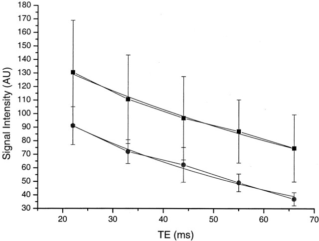
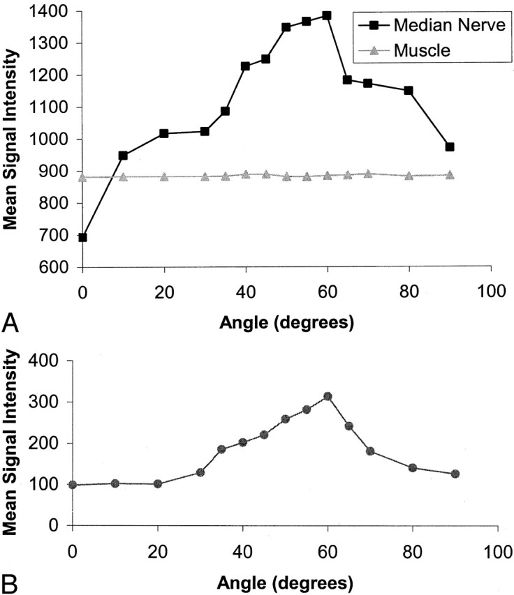
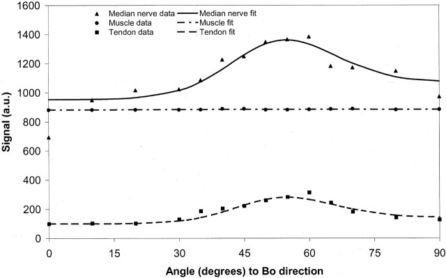
Results: When examined with a short TI inversion-recovery sequence with different TEs, the median nerve showed a 46-175% increase in signal intensity between 0 degrees and 55 degrees and an increase in mean T2 from 47.2 to 65.8 msec. When examined in 5 degrees to 10 degrees increments from 0 degrees to 90 degrees, the median nerve signal intensity changed in a manner consistent with the magic angle effect. No significant change was observed in skeletal muscle. Ulnar and sciatic nerves also showed changes in signal intensity depending on their orientation to B(0). Components of the brachial plexus were orientated at about 55 degrees to B(0) and showed a higher signal intensity than that of nerves in the upper arm that were nearly parallel to B(0). A reduction in the change in signal intensity in the median nerve with orientation was observed in the two patients with rheumatoid arthritis.

Conclusion: Signal intensity of peripheral nerves changes with orientation to B(0). This is probably the result of the magic angle effect from the highly ordered, linearly orientated collagen within them. Differences in signal intensity with orientation may simulate disease and be a source of diagnostic confusion.


Figures

F<sc>ig</sc> 1.
Fig 1.
A and B, Transverse STIR (2500/30/160 TR/TEeffective/TI) MR images of the median nerve at 0° (A) and 55° (B). The median nerve (arrow) has a higher signal intensity in B. There is no apparent change in signal intensity in the flexor tendons (see also Fig 3).
F<sc>ig</sc> 2.
Fig 2.
Plot of signal intensity against TE at 55° (▪) and 0° (•) on STIR images (1500/22, 33, 44, 55, 66/107 TR/TE/TI) with a monoexponential fit, in an adult volunteer. The signal intensity is higher at 55°. The mean T2 was also longer at 55° than at 0° (65.8 msec versus 47.2 msec).
F<sc>ig</sc> 3.
Fig 3.
Plots of signal intensity against orientation for the median nerve and hypothenar muscle and for an adjacent flexor tendon. Same subject as in Fig 1. A, The median nerve shows a 98% increase in signal intensity, which peaks at about 60° and decreases as the angulation is increased to 90°. Muscle shows no significant change in signal intensity. B, The adjacent flexor tendon, examined with the same sequence and plotted on the same scale, follows the same pattern but has a lower initial signal intensity and shows less change.
F<sc>ig</sc> 4.
Fig 4.
A and B, STIR images of the brachial plexus (A) and nerves entering upper arm (B). The components of the brachial plexus (arrows in A) have a higher signal intensity than that of skeletal muscle, whereas nerves in the upper arm (arrows in B), emerging from the brachial plexus and nearly parallel to B0, are isointense or slightly hyperintense to muscle.
F<sc>ig</sc> 5.
Fig 5.
Sagittal MR images of the ulnar nerve with the elbow flexed to 125°. A, This most medial section shows that the nerve is isointense to muscle in the upper arm, but at the level of the condyle (arrow) signal intensity increases as the nerve rotates toward 55°. B, Middle section shows the nerve (arrow) perpendicular to B0 where it is isointense to muscle. C, Lateral section shows the nerve (arrows) at 125° to B0 where it is hyperintense to muscle.
F<sc>ig</sc> 6.
Fig 6.
Sagittal MR image of the median nerve with the wrist flexed to 55°. The nerve is parallel to B0 in the upper forearm and is isointense with muscle, but where the nerve is flexed toward 55° it shows increased signal intensity (arrow). This may simulate disease.
F<sc>ig</sc> 7.
Fig 7.
A and B, Transverse MR images of the sciatic nerve orientated at 0° to B0 (A) and 55° to B0 (B). The signal intensity in the nerve (arrow) in B is higher than that in A.
F<sc>ig</sc> 8.
Fig 8.
Electron micrograph of a human peripheral nerve (stain was aqueous uranyl nitrate followed by Reynolds lead citrate; original magnification, X 14,000). The numerous small dots are collagen fibers seen in cross section.
F<sc>ig</sc> 9.
Fig 9.
Diagram of brachial plexus. A black line at 55° to the body axis and B0 is superimposed. The components of the plexus are generally orientated parallel to this line, which is at the magic angle. (Reprinted from reference 33 with permission of Icon Learnings Systems.)
F<sc>ig</sc> 10.
Fig 10.
Fit of median nerve, muscle, and flexor tendon data to the model described in the Appendix. There is a close fit for median nerve and flexor tendon, assuming a significant fraction of tissue with dipolar interactions. The muscle data fit without need for significant dipolar interactions.

Comment in

References

    1. Maravilla KR, Bowen BC. Imaging of the peripheral nervous system: evaluation of peripheral neuropathy and plexopathy. ANJR Am J Neuroradiol 1998;19:1011–1023 - PMC - PubMed
    1. Aagaard B, Maravilla K, Kliot M. Magnetic resonance neurography: magnetic resonance imaging of the peripheral nerves. Neuroimaging Clin N Am 2001;11:131–146 - PubMed
    1. Blair DN, Rapoport S, Sortman HD, Blair OC. Normal brachial plexus MR imaging. Radiology 1987;165:763–767 - PubMed
    1. Rapoport S, Blair DN, McCarthy SM, Desser TS, Hammes LW, Sortman HD. Brachial plexus: correlation of MR imaging with CT and pathologic findings. Radiology 1987;167:161–165 - PubMed
    1. Posniak HV, Olson MC, Dudiak CM, Wisniewska R, O’Malley C. MR imaging of the brachial plexus. AJR Am J Roentgenol 1993;161:373–379 - PubMed

MeSH terms