In vivo proton MR spectroscopy of primary and nodal nasopharyngeal carcinoma
- PMID: 15037477
- PMCID: PMC8158563
In vivo proton MR spectroscopy of primary and nodal nasopharyngeal carcinoma
Abstract
Background and purpose: The aim of this study was to determine the feasibility of performing in vivo proton ((1)H) MR spectroscopy of nasopharyngeal carcinoma (NPC) and to document the (1)H spectrum of this cancer.
Methods: Twenty-seven patients with NPC lesions >1 cm(3) underwent localized (1)H MR spectroscopy performed at 1.5 T. Water-suppressed spectra from both primary tumors (nine cases) and metastatic nodes (18 cases) were obtained at TE 136 and 272. Spectra were analyzed in the time domain by using a nonlinear least squares fitting algorithm with incorporation of previous knowledge. Choline (Cho)/creatine (Cr) ratios for primary NPC and metastatic nodes were calculated and compared. Spectra from normal neck muscle of five volunteers were acquired as control data.
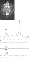
Results: (1)H MR spectroscopy was successfully obtained in seven (78%) of nine primary tumors and 16 (89%) of 18 metastatic nodes. Intense lipid signals in the range of 0.89 to 2.02 ppm were observed in 95% of spectra at TE 136 and 91% of spectra at TE 272. At TE 136, Cho/Cr for metastatic nodes (5.3 +/- 1.6) was significantly higher than the ratio for primary (2.6 +/- 0.5) NPC lesions (P =.02). Cho/Cr ratios for NPC lesions were higher than those for normal neck muscles, for which values ranged from 0 to 0.97 and 0 to 1.1 at TE 136 and 272, respectively.
Conclusion: (1)H MR spectroscopy is a feasible technique for the evaluation of NPC tumors >1 cm(3). Cho/Cr ratios for the lesions were high compared with those for normal neck muscle.
Figures


Similar articles
-
Proton MR spectroscopy of squamous cell carcinoma of the extracranial head and neck: in vitro and in vivo studies.AJNR Am J Neuroradiol. 1997 Jun-Jul;18(6):1057-72. AJNR Am J Neuroradiol. 1997. PMID: 9194433 Free PMC article.
-
In vivo 1H MR spectroscopy of thyroid carcinoma.Eur J Radiol. 2005 Apr;54(1):112-7. doi: 10.1016/j.ejrad.2004.05.003. Eur J Radiol. 2005. PMID: 15797300
-
The choline/creatine ratio in five benign neoplasms: comparison with squamous cell carcinoma by use of in vitro MR spectroscopy.AJNR Am J Neuroradiol. 2000 Nov-Dec;21(10):1930-5. AJNR Am J Neuroradiol. 2000. PMID: 11110549 Free PMC article.
-
Diagnostic Value of Magnetic Resonance Spectroscopy in Radiation Encephalopathy Induced by Radiotherapy for Patients with Nasopharyngeal Carcinoma: A Meta-Analysis.Biomed Res Int. 2016;2016:5126074. doi: 10.1155/2016/5126074. Epub 2016 Feb 3. Biomed Res Int. 2016. PMID: 26953103 Free PMC article. Review.
-
Management of the neck in nasopharyngeal carcinoma (NPC).Otolaryngol Clin North Am. 1998 Oct;31(5):785-802. doi: 10.1016/s0030-6665(05)70087-6. Otolaryngol Clin North Am. 1998. PMID: 9735107 Review.
Cited by
-
Primary B cell lymphoma of the sphenoid sinus: CT and MRI characteristics with correlation to perfusion and spectroscopic imaging features.Eur Arch Otorhinolaryngol. 2007 Oct;264(10):1207-13. doi: 10.1007/s00405-007-0322-0. Epub 2007 May 4. Eur Arch Otorhinolaryngol. 2007. PMID: 17479272
-
Quantitative proton MR spectroscopy as a biomarker of tumor necrosis in the rabbit VX2 liver tumor.J Vasc Interv Radiol. 2011 Aug;22(8):1175-80. doi: 10.1016/j.jvir.2011.03.016. Epub 2011 May 28. J Vasc Interv Radiol. 2011. PMID: 21620723 Free PMC article.
-
In vivo proton MR spectroscopy of primary tumours, nodal and recurrent disease of the extracranial head and neck.Eur Radiol. 2007 Jan;17(1):251-7. doi: 10.1007/s00330-006-0294-2. Epub 2006 May 16. Eur Radiol. 2007. PMID: 16703309
-
Functional MRI for Treatment Evaluation in Patients with Head and Neck Squamous Cell Carcinoma: A Review of the Literature from a Radiologist Perspective.Curr Radiol Rep. 2018;6(1):2. doi: 10.1007/s40134-018-0262-z. Epub 2018 Jan 22. Curr Radiol Rep. 2018. PMID: 29416951 Free PMC article. Review.
-
The role of magnetic resonance imaging in oncology.Clin Transl Oncol. 2010 Sep;12(9):606-13. doi: 10.1007/s12094-010-0565-x. Clin Transl Oncol. 2010. PMID: 20851801
References
-
- Smith IC, Stewart LC. Magnetic resonance spectroscopy in medicine: clinical impact. Prog Nucl Magn Reson Spectroscopy 2002;40:1–34
-
- Huang W, Roche P, Shindo M, Madoff D, Geronimo C, Button T. Evaluation of head and neck tumor response to therapy using in vivo 1H MR spectroscopy: correlation with pathology. Proc Int Soc Magn Reson Med 2000;8:552
MeSH terms
Substances
LinkOut - more resources
Full Text Sources
Medical
