. 2004 Mar 1;18(5):572-83.
doi: 10.1101/gad.1171704.

Ephrin-B1 forward and reverse signaling are required during mouse development

Affiliations

Ephrin-B1 forward and reverse signaling are required during mouse development

Alice Davy et al. Genes Dev. .

Abstract

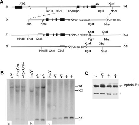
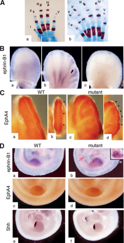
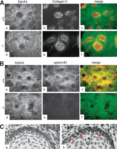
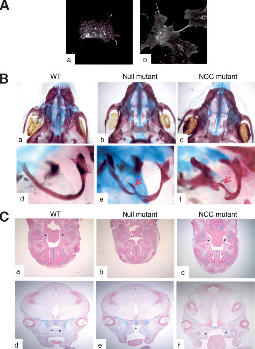
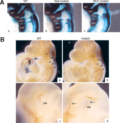
Eph receptors and ephrin ligands are key players in many developmental processes including embryo patterning, angiogenesis, and axon guidance. Eph/ephrin interactions lead to the generation of a bidirectional signal, in which both the Eph receptors and the ephrins activate downstream signaling cascades simultaneously. To understand the role of ephrin-B1 and the importance of ephrin-B1-induced reverse signaling during embryonic development, we have generated mouse lines carrying mutations in the efnb1 gene. Complete ablation of ephrin-B1 resulted in perinatal lethality associated with a range of phenotypes, including defects in neural crest cell (NCC)-derived tissues, incomplete body wall closure, and abnormal skeletal patterning. Conditional deletion of ephrin-B1 demonstrated that ephrin-B1 acts autonomously in NCCs, and controls their migration. Last, a mutation in the PDZ binding domain indicated that ephrin-B1-induced reverse signaling is required in NCCs. Our results demonstrate that ephrin-B1 acts both as a ligand and as a receptor in a tissue-specific manner during embryogenesis.


Figures

Figure 1.
Figure 1.
Targeting of the efnb1 locus. (A) Wild-type efnb1 locus (panel a) was targeted by homologous recombination of a targeting vector (panel b) carrying exons 2–5 of efnb1 gene flanked by loxP sites (triangles) and a positive selection cassette flanked by FRT sites (diamonds). The conditional allele (lox; panel c) is recombined in the presence of the Cre recombinase, producing a null allele (del; panel d). (B) Tail DNA from P10 pups was analyzed by Southern blot analysis in order to confirm proper targeting and recombination in the presence of the Cre recombinase (XbaI digest with a probe indicated in gray in panels c and d). Restriction digests of DNA from first-generation pups (panel a) as well as second-generation pups (panels b,c) are presented. (C) Western blot analysis showing loss of ephrin-B1 at the protein level in homozygous null E10.5 embryos (–/–), and decreased levels of ephrin-B1 in heterozygous embryos (+/–), as compared with wild-type (Y/+) littermates. Nonspecific bands are indicated (asterisks).
Figure 2.
Figure 2.
Altered expression of ephrin-B1 in heterozygous females correlates with polydactyly. (A) Skeletal preparations from an ephrin-B1 null male (panel a) and a heterozygous female (panel b) were stained with Alcian blue and Alizarin red. Polydactyly, affecting digit II, is observed in the heterozygous females but not the null male. (B) In situ hybridization of E11.5 limb buds shows an altered ephrin-B1 expression pattern in heterozygous limb buds (panel b) as compared with wild-type limb bud (panel a). (Panel c) No expression is detected in a null limb bud. (C) EphA4 expression pattern, detected by immunohistochemistry, is perturbed in heterozygous limb buds. Wild-type limb buds present a homogenous EphA4 expression (panel a) and three distinct areas of condensation can be seen (asterisks), corresponding to the prospective digits II, III, and IV (panel b). In ephrin-B1 heterozygous limb buds, EphA4 is distributed in multiple stripes (panel c) and multiple areas of condensation are observed (asterisks, panel d). Panels a, b, c, and d are different views of the same limb bud. (D) In situ hybridization of E10.5 wild-type (panels a,c,e) and heterozygous mutant (panels b,d,f) embryos. Expression of ephrin-B1 is patchy in the heterozygous mutants (panel b), with ephrin-B1-positive cells (red arrows) sorting out from ephrin-B1-negative territories. (Panel d) Insert is a different heterozygous limb bud. (Panel f) EphA4 distribution is unchanged in the mutant embryos. No ectopic Shh expression is detected in the mutant limb buds.
Figure 3.
Figure 3.
Loss of ephrin-B1 does not affect the chondrogenic capacity of limb bud cells in vitro. (A) High-density micromass cultures from limb bud cells isolated from wild-type (panels a–c) or ephrin-B1 null (panels d–f) E11.5 embryos were analyzed by immunofluorescence using the EphA4 antibody (green) and a collagen II-specific antibody (red), as indicated. Collagen II-positive cells (red arrows) can be detected outside the chondrogenic micromasses in the culture isolated from mutant embryos. (B) High-density micromass cultures from limb bud cells isolated from wild-type (panels a–c) or ephrin-B1 null (panels d–f) E11.5 embryos were analyzed by immunofluorescence using the EphA4 antibody (green) and a monoclonal antibody specific for ephrin-B1 (25H11; red). Both EphA4 and ephrin-B1 are expressed in the cells surrounding the chondrogenic micromasses, including the perichondrium. (C) H&E staining of sections from wild-type (panel a) or ephrin-B1 heterozygous (panel b) E15.5 embryos. Space between cells can clearly be seen in mutant (arrows) but not in wild-type perichondrium. (Panel c) Chondrocytes.
Figure 4.
Figure 4.
Ephrin-B1 acts autonomously in neural crest cells. (A) Primary cells isolated from branchial arches of E10.5 wild-type embryos were passaged once and analyzed by immunofluorescence using the ephrin-B1-specific antibody (25H11; panel a) or the ephrin-B1-Fc recombinant protein to detect EphB receptors (panel b). (B) Alcian blue/Alizarin red-stained skeleton preparations of wild-type (panels a,d), ephrin-B1null (panels b,e), or ephrin-B1NCC (panels c,f). Palatal shelves (asterisks) are fused in the wild-type animal (panel a), but remain separated in ephrin-B1null (panel b) and in ephrin-B1NCC (panel c). Tympanic rings are defective bilaterally in ephrin-B1null (panel e) and in ephrin-B1NCC (panel f). (C) Histological sections of wild-type (panels a,d), ephrin-B1null (panels b,e), or ephrin-B1NCC (panels c,f), stained with Alcian blue and Nuclear Fast red. (Panels a–c) At E13.5, palatal shelves (asterisks) can be observed on transverse sections on each side of the tongue of wild-type (panel a) and NCC-deleted ephrin-B1 mutant (panel c). (Panel b) Embryo-wide deletion of ephrin-B1 precludes formation of palatal shelves. (Panels d,e) At E15.5, palatal shelves have elevated and fused (asterisks) in wild-type embryos (panel d). (Panel e) In ephrin-B1null embryos, the roof of the mouth has elevated but no palatal shelves are present. (Panel f) At E14.5, palatal shelves (asterisks) are formed in ephrin-B1NCC mutants, but they failed to elevate.
Figure 5.
Figure 5.
Loss of ephrin-B1 induces NCC migration defects. (A) Whole-mount X-gal staining of E9.5 wild-type (panel a), ephrin-B1null (panel b), or ephrin-B1NCC (panel c). Mutant NCC exhibit a wandering behavior in ephrin-B1null (panel b) and ephrin-B1NCC embryos (panel c), as compared with wild-type (panel a). Mutant cells invade territories that are normally devoid of crest cells (red arrows). (B) Whole-mount immunohistochemistry staining of E10.5 wild-type (panels a,c) and ephrin-B1null (panels b,d) embryos using a neurofilament antibody. Cranial ganglia V-X appear normal in the mutant embryos, but branching (asterisk) and fasciculation (arrow) defects can be observed. (ba) Branchial arches; (o) otic vesicle; (OM) occulomotor nerve.
Figure 6.
Figure 6.
Generation of ephrin-B1ΔPDZ mutant. (A) Wild-type ephrin cDNA (WT), or mutant cDNA (ΔPDZ and P-tyr) were transfected into 293T cells together with a flag-tagged PDZ domain. The EphB2-Fc recombinant protein was used to pull down ephrin and the amount of PDZ domain coimmunoprecipitated was evaluated with an anti-flag antibody. The right panel shows a quantification of the amount of PDZ domain associating with wild-type and mutant ephrin. The asterisk indicates a nonspecific band. (B, panel a) Wild-type efnb1 locus. (Panel b) Homologous recombination of the vector for the ΔPDZ mutation introduces a HindIII site. (C, panel a) Southern blot analysis of targeted ΔPDZ ES cells. Genomic DNA was digested with HindIII and the probe used is indicated in gray in panel b of part B. Genomic DNA from different ES clones was used as a template for PCR (panel b), using either mutation-specific primers (top) or primers specific to exon 5 (bottom). (D) ES cells treated with retinoic acid (RA) were analyzed by Western blot using the ephrin antibody (C18). Ephrin-B1 could be detected in wild-type ES cells (WT), ES cells targeted with the conditional allele of ephrin-B1 (lox), and ES cells targeted with ΔPDZ mutation (ΔPDZ). To ascertain that the signal was specific for ephrin-B1, we also tested ES cells deficient for ephrin-B1 (null). These cells were obtained by transiently expressing the Cre recombinase in ephrin-B1lox ES cells and screening for recombined clones. The asterisks indicate nonspecific bands. (E) Embryos obtained by injection of ES cells carrying the ephrin-B1ΔPDZ mutation into ROSA26 blastocysts were stained with X-gal to evaluate the degree of contribution of the mutant ES cells. Two different tissues (spinal chord, top and limb bud, bottom) from four different E14.5 embryos are presented (ch1–ch4). The embryos with the most contribution of the mutant ES cells (ch3 and ch4) presented a cleft palate (+), whereas the embryos with very little contribution of the mutant ES cells (ch1 and ch2) did not have a cleft palate (–). (F) Histological examination of E14.5 embryos obtained by injection of ES cells carrying the ephrin-B1ΔPDZ mutation into wild-type blastocysts. (Panels b,d) Palatal shelves (asterisks) were formed but failed to elevate and fuse in the chimeric embryo. (Panels a,c) Littermate without a cleft palate. Sections were stained with Alcian blue and Nuclear Fast red.

References

    1. Adams R.H. 2002. Vascular patterning by Eph receptor tyrosine kinases and ephrins. Semin. Cell Dev. Biol. 13: 55–60. - PubMed
    1. Adams R.H., Diella, F., Hennig, S., Helmbacher, F., Deutsch, U., and Klein, R. 2001. The cytoplasmic domain of the ligand ephrinB2 is required for vascular morphogenesis but not cranial neural crest migration. Cell 104: 57–69. - PubMed
    1. Bouillet P., Oulad-Abdelghani, M., Vacaire, S., Garnier, J.M., Schuhbaur, B., Dolle, P., and Chambon, P. 1995. Efficient cloning of cDNAs of retinoic acid-responsive genes in P19 embryonal carcinoma cells and characterization of a novel mouse gene, Stra1 (mouse LERK-2/Eplg2). Dev. Biol. 170: 420–433. - PubMed
    1. Bourgeois P., Bolcato-Bellemin, A.-L., Danse, J.-M., Bloch-Zupan, A., Yoshiba, K., Stoetzel, C., and Perrin-Schmitt, F. 1998. The variable expressivity and incomplete penetrance of the twist-null heterozygous mouse phenotype resemble those of human Saethre-Chotzen syndrome. Hum. Mol. Genet. 7: 945–957. - PubMed
    1. Bruckner K., Pasquale, E.B., and Klein, R. 1997. Tyrosine phosphorylation of transmembrane ligands for Eph receptors. Science 275: 1640–1643. - PubMed

Publication types

MeSH terms