. 2004 Mar 29;164(7):1009-20.
doi: 10.1083/jcb.200310061. Epub 2004 Mar 22.

YSK1 is activated by the Golgi matrix protein GM130 and plays a role in cell migration through its substrate 14-3-3zeta

Affiliations

YSK1 is activated by the Golgi matrix protein GM130 and plays a role in cell migration through its substrate 14-3-3zeta

Christian Preisinger et al. J Cell Biol. .

Abstract

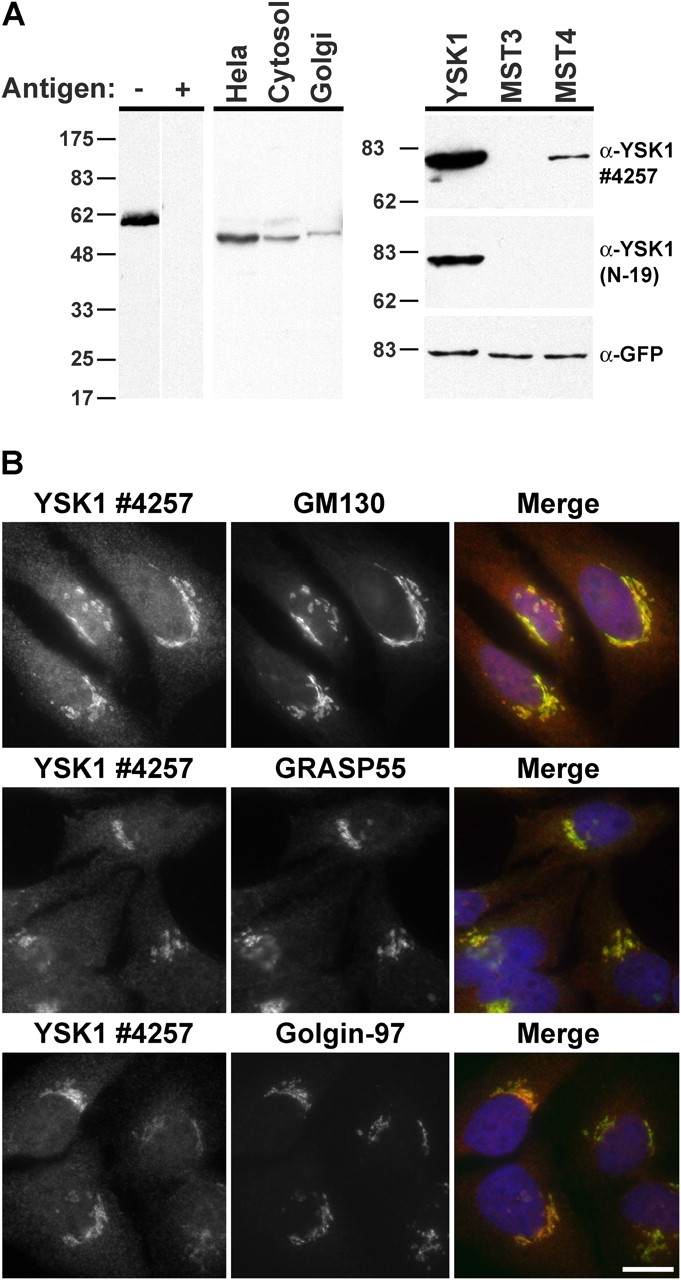
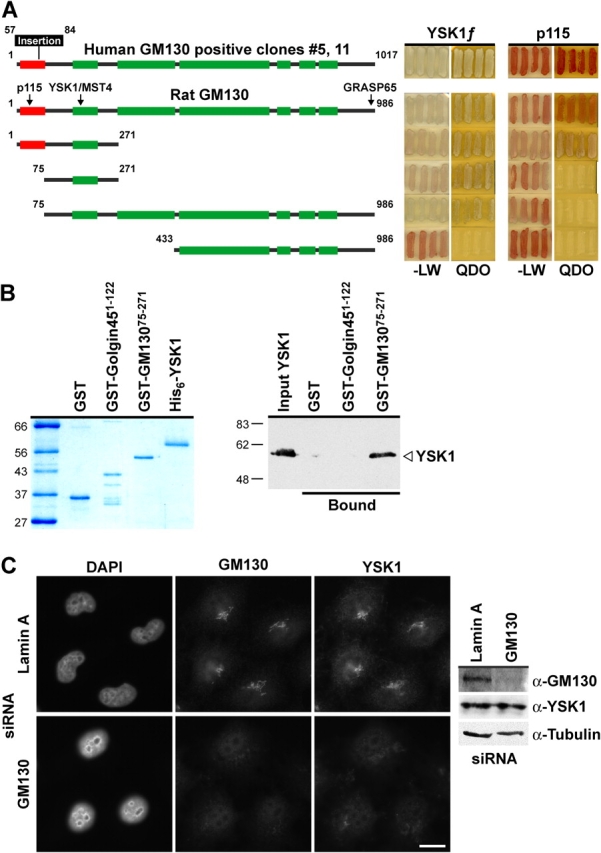
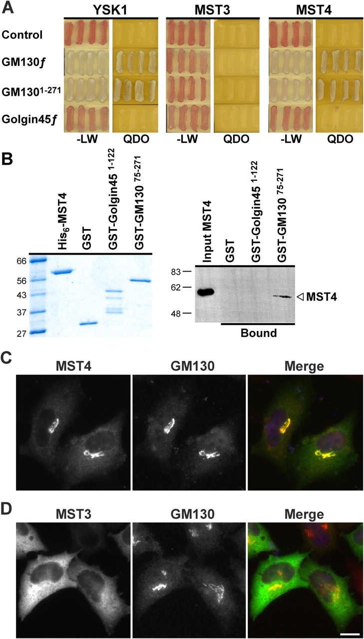
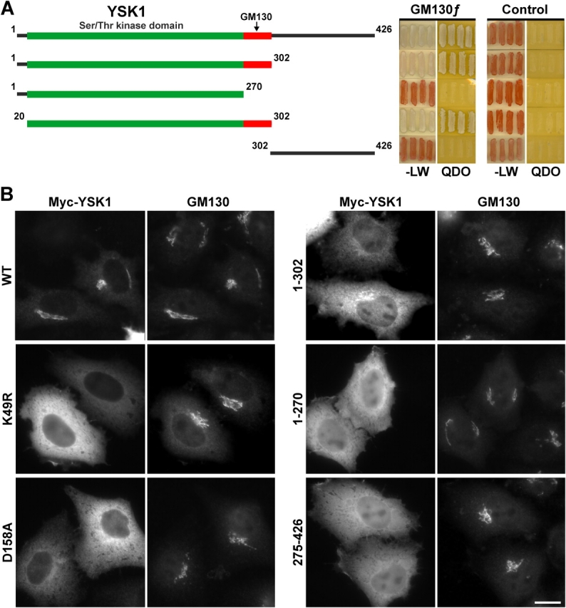
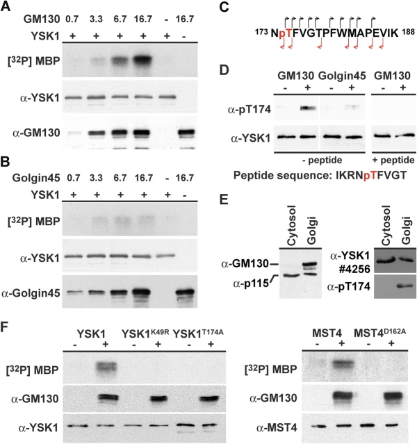
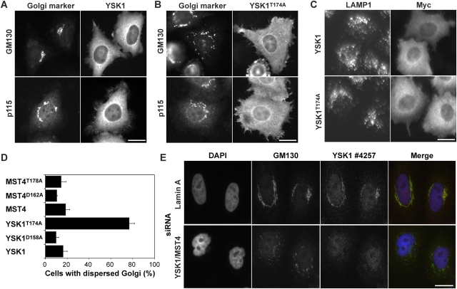
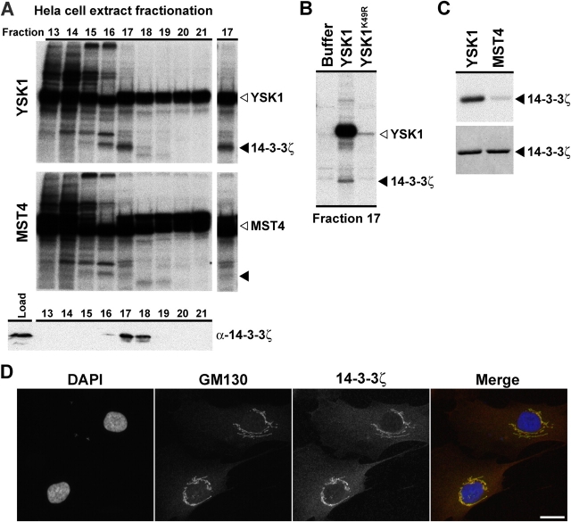
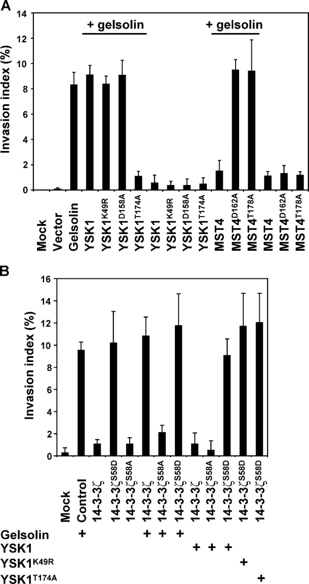
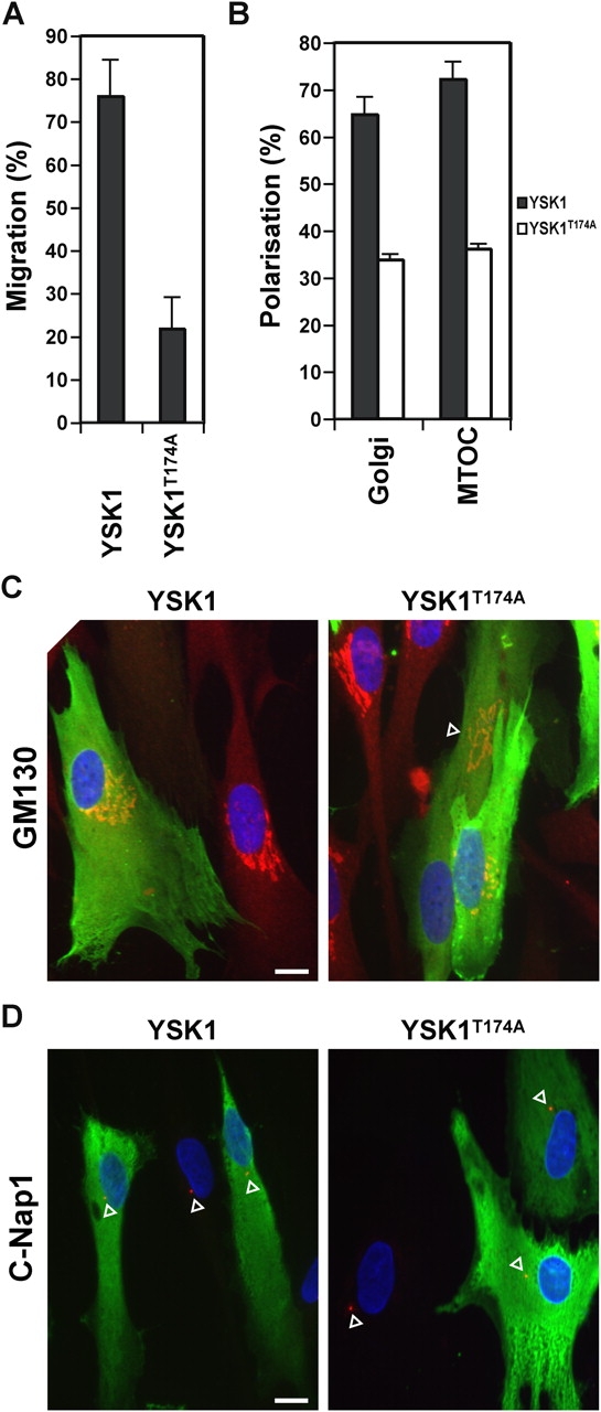
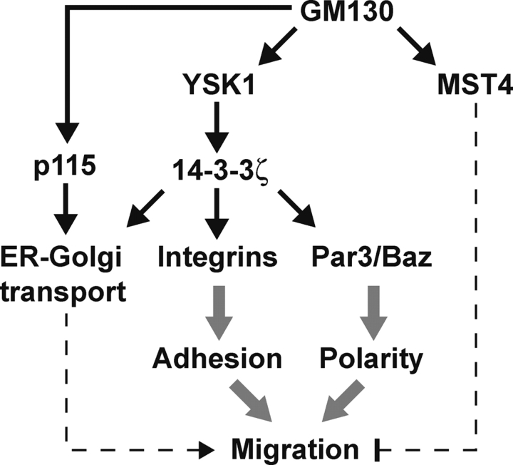
The Golgi apparatus has long been suggested to be important for directing secretion to specific sites on the plasma membrane in response to extracellular signaling events. However, the mechanisms by which signaling events are coordinated with Golgi apparatus function remain poorly understood. Here, we identify a scaffolding function for the Golgi matrix protein GM130 that sheds light on how such signaling events may be regulated. We show that the mammalian Ste20 kinases YSK1 and MST4 target to the Golgi apparatus via the Golgi matrix protein GM130. In addition, GM130 binding activates these kinases by promoting autophosphorylation of a conserved threonine within the T-loop. Interference with YSK1 function perturbs perinuclear Golgi organization, cell migration, and invasion into type I collagen. A biochemical screen identifies 14-3-3zeta as a specific substrate for YSK1 that localizes to the Golgi apparatus, and potentially links YSK1 signaling at the Golgi apparatus with protein transport events, cell adhesion, and polarity complexes important for cell migration.


Figures

Figure 1.
Figure 1.
YSK1 localizes to the Golgi apparatus. (A) Samples containing 25 μg of protein from insect cells expressing His6-tagged human YSK1 (up shifted due to the presence of the tag), HeLa cells, rat liver cytosol, or 10 μg of rat liver Golgi membranes were analyzed by Western blotting with 1 μg/ml of affinity-purified rabbit antibody 4257 to human YSK1. For antibody competition, 10 μg/ml of the antigen was preincubated with the antibody for 1 h before use. Samples containing 20 μg of protein from HeLa cells transfected with constructs for GFP-tagged YSK1, MST3, and MST4 for 18 h were blotted with 1 μg/ml of affinity-purified rabbit antibody 4257 to YSK1, a peptide antibody raised against the unique first 19 aa of YSK1 (N19), or a sheep antibody to GFP. (B) HeLa cells were costained with rabbit antibodies to YSK1 and sheep antibodies to GM130, GRASP55, or golgin97. Bar, 10 μm.
Figure 2.
Figure 2.
Identification of the Golgi protein GM130 as a binding partner for YSK1. (A) Bait constructs comprising the full-length human GM130 splice variant rescued from the library screen, full-length rat GM130 or the indicated deletion constructs of rat GM130 were tested against full-length human YSK1 for the ability to grow on selective medium (QDO), compared with nonselective medium (−LW) in the yeast two-hybrid system. Lighter colony color on QDO is an indicator of a strong signal. Boxed regions indicate the p115 binding site (red) and predicted coiled-coil domains (green). (B) His-tagged YSK1 was incubated with GST-tagged fragments of rat GM130, golgin45, or GST and recovered on glutathione-agarose. Recovered complexes were analyzed by Western blotting. (C) HeLa cells depleted for lamin A or GM130 for 67 h using siRNA were stained and Western blotted for GM130, YSK1, and α-tubulin. Bar, 10 μm.
Figure 3.
Figure 3.
The YSK1-related kinase MST4 is Golgi apparatus localized. (A) Full-length YSK1, MST3, and MST4 were tested against empty vector (control), full-length GM130, GM1301-271, and golgin45 for the ability to grow on selective medium (QDO), compared with nonselective medium (−LW) in the yeast two-hybrid system. Lighter colony color on QDO is an indicator of a strong signal. (B) His-tagged MST4 was incubated with GST-tagged fragments of rat GM130, golgin45, or GST and recovered on glutathione-agarose. Recovered complexes were analyzed by Western blotting. (C and D) HeLa cells transfected with myc-tagged MST3 and MST4 constructs for 18 h were costained with the 9E10 mAb to the myc epitope (green) and a sheep antibody to GM130 (red). Bar, 10 μm.
Figure 4.
Figure 4.
YSK1 shows activity-dependent targeting to the Golgi apparatus. (A) Bait constructs comprising full-length YSK1, or the indicated deletion constructs were tested against full-length rat GM130 or an empty bait plasmid (control) for the ability to grow on selective medium (QDO), compared with nonselective medium (−LW) in the yeast two-hybrid system. Lighter colony color on QDO is an indicator of a strong interaction. Boxed regions indicate the canonical serine/threonine kinase domain (green), and the extension to this necessary for binding to GM130 (red). (B) HeLa cells transfected with myc-tagged YSK1 constructs for 18 h were costained with the 9E10 mAb to the myc epitope and a sheep antibody to GM130. Bar, 10 μm.
Figure 5.
Figure 5.
Binding to the NH2-terminal domain of GM130 triggers autophosphorylation and activation of YSK1. (A) Purified YSK1, 0.8 pmoles, was incubated in the absence or presence of 0.7, 3.3, 6.7, and 16.7 pmoles of His-tagged GM13075-271 or (B) His-tagged golgin451-122 at 37°C for 30 min. After this preincubation, samples were analyzed for kinase activity toward the model substrate MBP using γ-[32P]ATP, and by Western blotting for YSK1 and GM130 or golgin45 to control for gel loading. (C) Fragmentation of the phosphorylated NTFVGTPFWMAPEVIK peptide (Fig. S4) by tandem mass spectrometry gives daughter ions derived from the NH2 and COOH termini of the peptide indicating that threonine 174 (pT), shown in red, is the phosphorylated residue. (D) Activated and mock-activated YSK1 was Western blotted with affinity-purified antibody N19 to YSK1 and the pT174 antibody to the phosphorylated T-loop sequence IKRNpTFVGT, in the presence and absence of 5 μg of blocking peptide. (E) 25 μg of rat liver cytosol and 10 μg of Golgi membranes were Western blotted with antibodies to GM130, p115, YSK1 4256, and pT174 to phosphorylated YSK1. (F) Purified YSK1, YSK1K49R, YSK1T174A, MST4, and MST4D162A, 0.8 pmoles, were incubated in the absence or presence of 16.7 pmoles of His-tagged GM13075-271 at 37°C for 30 min. After this preincubation, samples were analyzed for kinase activity toward the model substrate MBP using γ-[32P]ATP, and by Western blotting for YSK1 MST4 and GM130 to control for gel loading.
Figure 6.
Figure 6.
YSK1/MST4 are required for Golgi apparatus localization in the perinuclear region. (A) HeLa cells were transfected with expression constructs for the wild-type myc-tagged (A) YSK1 or (B) YSK1T174A for 44 h and then stained with mouse antibodies to the myc epitope and sheep antibodies to either GM130 or p115. (C) HeLa cells were transfected with expression constructs for the wild-type myc-tagged YSK1 or YSK1T174A for 44 h and then stained with rabbit antibodies to the myc epitope and mouse antibodies to LAMP1. (D) Golgi apparatus dispersal was scored after 48 h in HeLa cells transfected with YSK1 and MST constructs indicated in the legend. Mean values are plotted (n = 3) with at least 300 cells counted per experiment for each condition. (E) HeLa cells were transfected with siRNA duplexes for YSK1 and MST4 for 112 h then stained with antibodies to GM130 (green) and YSK1/MST4 (red), and with DAPI for DNA (blue). Exposure times of 1 s were used for all images. Bars, 10 μm.
Figure 7.
Figure 7.
A biochemical screen for YSK1 targets identifies 14-3-3ζ. (A) A modified KESTREL approach was used to identify substrates for YSK1 and MST4 as described in the Materials and methods. Phosphorylations with YSK1 and MST4 of fractions 13–21 from a fractionation of HeLa S3 cell extract by gel filtration on Superose-6 together with a Western blot for 14-3-3ζ are shown. Fraction 17 is shown as a cut out to the right, with the position of 14-3-3ζ indicated by a closed arrowhead and the respective kinases indicated by open arrowheads. (B) Superose-6 column fraction 17 was treated with buffer, YSK1, or kinase-dead YSK1K49R using the KESTREL method, and analyzed by SDS-PAGE and autoradiography. Closed and open arrowheads indicate phosphorylation of 14-3-3ζ and YSK1 autophosphorylation, respectively. (C) Kinase assays were performed for 60 min at 37°C in KESTREL assay buffer with 2 μg of recombinant His-tagged 14-3-3ζ, and 500 ng of preactivated YSK1 or MST4. An autoradiograph of 14-3-3ζ phosphorylations by YSK1 and MST4 is shown in the top panel, and the corresponding region of a Coomassie blue stained gel in the bottom panel. Closed arrowheads indicate 14-3-3ζ. (D) HS68 cells were costained with a sheep antibody to GM130 and the mouse mAb 22-II-D8 to 14-3-3ζ. Bar, 10 μm.
Figure 8.
Figure 8.
Cell migration and invasion is modulated by YSK1 and MST4. (A) Cotransfection of YSK1T174A but not other forms of YSK1 abrogates gelsolin-induced invasion of collagen type I by HEK293T cells. HEK293T cells were left untransfected, or cotransfected with plasmids encoding gelsolin and constructs for wild-type YSK1 and MST4, or point mutants of these kinases, as indicated in the figure (n = 3). (B) Effects of 14-3-3ζ phosphorylation mutants on invasion into collagen type I. HEK293T cells were left untransfected, or cotransfected with plasmids encoding wild-type and point mutant forms of 14-3-3ζ, gelsolin, YSK1, YSK1K49R, and YSK1T174A, as indicated in the figure (n = 3). Collagen invasion assays were performed and quantitated as described in the Materials and methods. The invasive index is the percentage of cells invading the collagen gel over the total number of cells.
Figure 9.
Figure 9.
Golgi apparatus and centrosome polarization is inhibited by YSK1T174A. (A and B) Cells were microinjected with plasmids encoding myc-tagged YSK1 and YSK1T174A, fixed after 16 h, and stained with antibodies to GM130 or c-Nap1, and 9E10 monoclonal to the myc epitope. Bar graphs show the percentage of YSK1 or YSK1T174A expressing cells migrating at the wound edge or into the wound (n = 12), and the percentage of cells with Golgi apparatus and centrosomes (MTOC) polarized toward the wound edge (n = 4). (C and D) Images of cells expressing YSK1 or YSK1T174A (green) and stained for GM130 or c-Nap1 (red) are shown. The position of the wound corresponds to the bottom of the figure. Arrowheads indicate the position of the Golgi apparatus and centrosome. Bars, 10 μm.
Figure 10.
Figure 10.
MST kinases: linking Golgi apparatus function with cell migration? Together with the vesicle tethering factor p115, GM130 is part of a landmark complex on the Golgi apparatus important for protein transport and Golgi structure. Binding to GM130 activates YSK1 and MST4. Activated YSK1 phosphorylates 14-3-3ζ and potentially other downstream targets needed for normal cell migration, whereas MST4 acts via an uncharacterized pathway. Known targets of 14-3-3ζ important for regulating cell migration and polarity are depicted.

Similar articles

Cited by

References

    1. Barr, F.A., and B. Short. 2003. Golgins in the structure and dynamics of the Golgi apparatus. Curr. Opin. Cell Biol. 15:405–413. - PubMed
    1. Barr, F.A., M. Puype, J. Vandekerckhove, and G. Warren. 1997. GRASP65, a protein involved in the stacking of Golgi cisternae. Cell. 91:253–262. - PubMed
    1. Barr, F.A., N. Nakamura, and G. Warren. 1998. Mapping the interaction between GRASP65 and GM130, components of a protein complex involved in the stacking of Golgi cisternae. EMBO J. 17:3258–3268. - PMC - PubMed
    1. Benton, R., I.M. Palacios, and D. St Johnston. 2002. Drosophila 14-3-3/PAR-5 is an essential mediator of PAR-1 function in axis formation. Dev. Cell. 3:659–671. - PubMed
    1. Bialkowska, K., Y. Zaffran, S.C. Meyer, and J.E. Fox. 2003. 14-3-3 zeta mediates integrin-induced activation of Cdc42 and Rac. Platelet glycoprotein Ib-IX regulates integrin-induced signaling by sequestering 14-3-3 zeta. J. Biol. Chem. 278:33342–33350. - PubMed

Publication types

MeSH terms