. 2004 Mar 22;32(5):1836-47.
doi: 10.1093/nar/gkh344. Print 2004.

The CtBP2 co-repressor is regulated by NADH-dependent dimerization and possesses a novel N-terminal repression domain

Affiliations

The CtBP2 co-repressor is regulated by NADH-dependent dimerization and possesses a novel N-terminal repression domain

Sharon S C Thio et al. Nucleic Acids Res. .

Abstract

The C-terminal binding protein 2 (CtBP2) is a 48 kDa phosphoprotein reported to function as a co- repressor for a growing list of transcriptional repressors. It was recently demonstrated that CtBP is a dimeric NAD+-regulated d-isomer-specific 2-hydroxy acid dehydrogenase. However, the specific substrate(s) of CtBP enzymatic activity and the relationship of this activity to its co-repression function remain unknown. The ability of a human CtBP to bind and serve as a co-repressor of E1A has been shown to be regulated by nuclear NADH levels. Here we extend the functional characterization of CtBP by demonstrating that amino acid substitutions at Gly189 in the conserved NAD+-binding fold both abrogate the ability of CtBP2 to homodimerize and are associated with a dramatic loss of co-repressor activity. Consistent with the known enzymatic activity of CtBP2, mutations at Arg272 in the substrate-binding domain and at His321 in the catalytic domain result in significant loss of CtBP2 transcriptional co-repressor activity. High resolution serial C-terminal deletion analysis of CtBP2 also revealed a novel N-terminal repression domain that is distinct from its dehydrogenase domain. Our results suggest a model in which CtBP2 co-repressor function is regulated, at least in part, through the effect of NADH on CtBP2 homodimerization.


Figures

Figure 1
Figure 1
Sequence alignment of the putative NAD+/NADH-binding domain (A), the substrate-binding domain (B) and the regulatory domain (C) of mCtBP2 and three representative members of the d-isomer-specific 2-hydroxy acid dehydrogenase (2HAD) family of enzymes. A schematic representation of mCtBP2 highlighting 2HAD conserved regions (patterned boxes and denoted amino acid positions) is shown above. Detailed consensus sequences are shown below each figure. Completely conserved (five out of five) residues are denoted by an asterisk. Strict but not completely conserved (four out of five) residues are denoted by a hash sign. Amino acids that play significant roles in enzyme activity are in bold.
Figure 2
Figure 2
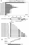
C-terminal deletion analysis of mCtBP2 reveals an additional N-terminal repression domain. Patterned boxes denote the functional 2HAD domains in mCtBP2. Bracketed numbers following the name of each construct indicate amino acid residues present in each construct. (A) C33A cells were transiently transfected with 100 ng each of Gal4-tagged full-length mCtBP2 and deletion mutants. These constructs were tested for their ability to repress a heterologous SV40 enhancer reporter plasmid pG5-GL3(SV) (3 µg). (B) Western blot analysis of whole-cell lysates of C33A cells transfected with 100 ng of wild-type and deletion mutants of Gal4/mCtBP2. Membranes were probed with both anti-Gal4DBD and anti-cdk2 mouse monoclonal antibodies. The top panel shows the expression levels of Gal4/mCtBP2 C-terminal deletion mutants (FL and numbers 1–12 represent full-length Gal4/mCtBP2 and mutants 1–12, respectively). Determination of the protein level of the constitutively expressed cyclin-dependent kinase 2 (cdk2) is included as a control for equal protein loading.
Figure 3
Figure 3
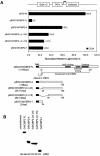
Identification of a novel N-terminal transcriptional repression domain. Patterned boxes denote the functional 2HAD domains in mCtBP2. Bracketed numbers following the name of each construct indicate amino acid residues present in each construct. (A) C33A cells were transiently transfected with 100 ng each of Gal4-tagged full-length mCtBP2 and mCtBP2 deletion constructs. These constructs were tested for their ability to repress a heterologous basal reporter plasmid pG5-GL3 (3 µg). (B) Western blot analysis of whole-cell lysates of C33A cells transfected with 100 ng of full-length and deletion constructs of Gal4/mCtBP2. Membranes were probed with both anti-Gal4DBD and anti-cdk2 mouse monoclonal antibodies.
Figure 4
Figure 4
Gal4-tagged mCtBP2 mutants display loss of transcriptional repression activity. (A) Partial protease digestion of wild-type and mutant mCtBP2. [35S]Methionine-labeled in vitro translated mutant proteins with papain. (B) Western blot analysis of whole-cell lysates of C33A cells transfected with 100 ng of wild-type and mutant Gal4-tagged mCtBP2. Membranes were probed with both anti-Gal4DBD and anti-cdk2 mouse monoclonal antibodies. (C) C33A cells were transiently transfected with 100 ng each of Gal4-tagged mCtBP2, Gal4-tagged mCtBP2 single and double mutants, together with 3 µg of the SV40 enhancer reporter plasmid pG5-GL3(SV).
Figure 5
Figure 5
Mutants of conserved amino acid G189 in mCtBP2 show a reduced ability to dimerize with wild-type mCtBP2. (A) Gal4-tagged and untagged mCtBP2 IVT products were synthesized. A 1 µl aliquot of each IVT product was loaded per lane. (B) [35S]Methionine-labeled Gal4/mCtBP2, but not mCtBP2, is specifically immunoprecipitated by a mouse monoclonal antibody against Gal4DBD. (C) A schematic diagram illustrating the co-immunoprecipitation and dimerization assays. The shaded sphere represents the protein A bead that binds to the anti-Gal4DBD monoclonal antibody. The antibody recognizes the Gal4DBD tag on the unlabeled, cold Gal4/mCtBP2, which in turn dimerizes to non-tagged [35S]methionine-labeled wild-type or mutant mCtBP2 (upper complex). Homodimerization of Gal4/CtBP2 through protein–protein interactions mediated by either CtBP2 (middle complex) or the Gal4DBD (lower complex), which is known to homodimerize (e.g. CtBP2/Gal4–Gal4/CtBP2), cannot be detected by autoradiography since Gal4DBD proteins are not [35S]methionine labeled. (D) Co-immunoprecipitation of [35S]methionine-labeled mCtBP2 by Gal4/CtBP2 is specific. ‘+’ indicates IVT products present in each binding reaction. (E) Co-immunoprecipitation of [35S]methionine-labeled mutant G189A, G189R and G189S mCtBP2 IVT products with unlabeled Gal4/mCtBP2 using a mouse monoclonal antibody against Gal4DBD. A 5 µl aliquot of labeled and unlabeled IVT products was included in each binding reaction.
Figure 6
Figure 6
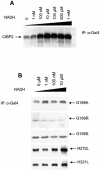
NADH regulates homodimerization of mCtBP2 in vitro. (A) NADH enhances homodimerization of wild-type mCtBP2 in a dose-dependent manner. (B) Increasing concentrations of NADH do not affect dimerization of G189 mutants of mCtBP2 with wild-type mCtBP2. Dimerized R272L and H321L mutants are still responsive to increasing doses of NADH. Co-immunoprecipitations of [35S]methionine-labeled G189A, G189R, G189S, R272L and H321L proteins with cold Gal4/mCtBP2 were performed using a mouse monoclonal antibody against Gal4DBD in the presence of increasing concentrations of NADH. In both (A) and (B), 5 µl of labeled and unlabeled IVT products were included in each binding reaction.
Figure 7
Figure 7
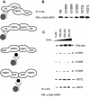
NADH regulates homodimerization of mCtBP2 in vivo. (A) A schematic diagram illustrating the in vivo co-immunoprecipitation and dimerization assays is shown. The shaded sphere represents the protein A agarose bead that binds to the anti-HA monoclonal antibody. This antibody recognizes the HA tag on HA/mCtBP2, which in turn dimerizes to Gal4/mCtBP2 wild-type or mutant proteins. Homodimerization of HA/CtBP2 through protein interactions mediated by CtBP2 (second complex) cannot be detected by the Gal4DBD monoclonal antibody used for the western blot. Similarly, homodimerization of Gal4/CtBP2 through protein–protein interactions mediated by either CtBP2 (third complex) or the Gal4DBD (fourth complex), which is known to homodimerize (e.g. CtBP2/Gal4–Gal4/CtBP2), cannot be detected since Gal4DBD proteins cannot be immunoprecipitated by the anti-HA antibody. (B) Co-immunoprecipitation of mutant G189A, G189R, G189S, R272L and H321L mCtBP2 proteins using a mouse monoclonal antibody against HA. (C) Co-immunoprecipitations of G189A, G189R, G189S, R272L and H312L proteins with HA/mCtBP2 were performed using a mouse monoclonal antibody against HA in the presence of increasing concentrations of CoCl2.

Similar articles

Cited by

References

    1. Turner J. and Crossley,M. (2001) The CtBP family: enigmatic and enzymatic transcriptional co-repressors. Bioessays, 23, 683–690. - PubMed
    1. Chinnadurai G. (2002) CtBP, an unconventional transcriptional corepressor in development and oncogenesis. Mol. Cell, 9, 213–224. - PubMed
    1. Boyd J.M., Subramanian,T., Scheaper,U., La Regina,M., Bayley,S. and Chinnadurai,G. (1993) A region in the C-terminus of adenovirus 2/5 E1A protein is required for association with a cellular phosphoprotein and important for the negative modulation of T24-ras mediated transformation, tumorigenesis and metastasis. EMBO J., 12, 469–478. - PMC - PubMed
    1. Scheaper U., Boyd,J.M., Verma,S., Uhlmann,E., Subramanian,T. and Chinnadurai,G. (1995) Molecular cloning and characterization of a cellular phosphoprotein that interacts with a conserved C-terminal domain of adenovirus E1A involved in negative modulation of oncogenic transformation. Proc. Natl Acad. Sci. USA, 92, 10467–10471. - PMC - PubMed
    1. Subramanian T., La Regina,M. and Chinnadurai,G. (1989) Enhanced ras oncogene mediated cell transformation and tumorigenesis by adenovirus 2 mutants lacking the C-terminal region of E1A protein. Oncogene, 4, 415–420. - PubMed

Publication types

MeSH terms

Associated data