. 2004 Apr 6;101(14):4758-63.
doi: 10.1073/pnas.0401123101. Epub 2004 Mar 22.

Structural basis of androgen receptor binding to selective androgen response elements

Affiliations

Structural basis of androgen receptor binding to selective androgen response elements

Paul L Shaffer et al. Proc Natl Acad Sci U S A. .

Abstract

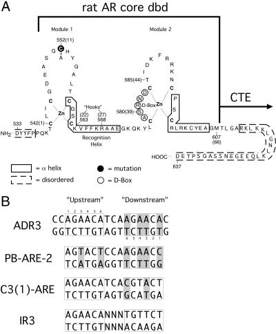
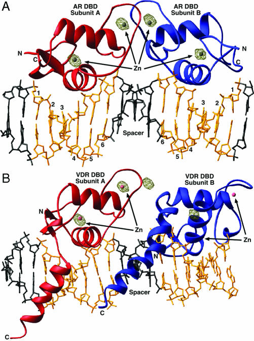
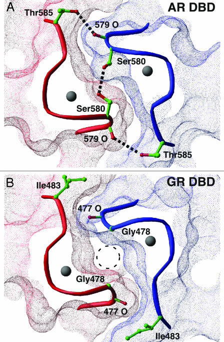
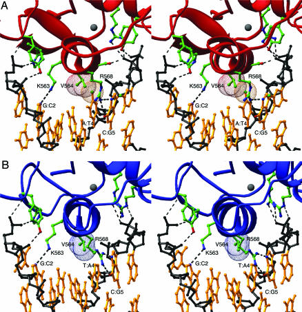
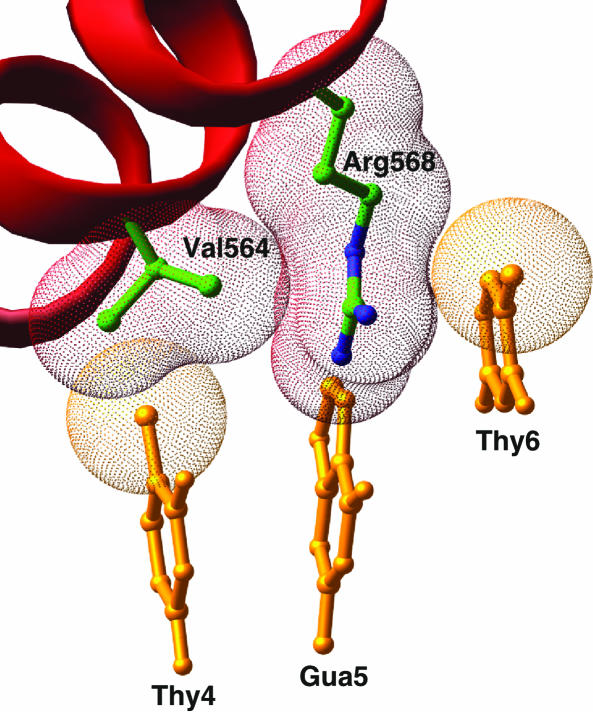
Steroid receptors bind as dimers to a degenerate set of response elements containing inverted repeats of a hexameric half-site separated by 3 bp of spacer (IR3). Naturally occurring selective androgen response elements have recently been identified that resemble direct repeats of the hexameric half-site (ADR3). The 3D crystal structure of the androgen receptor (AR) DNA-binding domain bound to a selective ADR3 reveals an unexpected head-to-head arrangement of the two protomers rather than the expected head-to-tail arrangement seen in nuclear receptors bound to response elements of similar geometry. Compared with the glucocorticoid receptor, the DNA-binding domain dimer interface of the AR has additional interactions that stabilize the AR dimer and increase the affinity for nonconsensus response elements. This increased interfacial stability compared with the other steroid receptors may account for the selective binding of AR to ADR3 response elements.


Figures

Fig. 1.
Fig. 1.
Protein and DNA constructs. (A) The rat AR DBD. Sequence numbers in parentheses refer to the common receptor DBD-numbering scheme. Residues in dashed boxes are disordered in both protomers of the homodimeric complex. (B) The DNA used in cocrystallization, labeled ADR3, two naturally occurring AR response elements, PB-ARE-2 and C3 (1)-ARE, and a canonical IR3 steroid response element. Differences from the IR3 sequence are shaded gray.
Fig. 2.
Fig. 2.
Crystal packing of the AR DBD–ADR3 complex. Red and blue ribbons are the upstream and downstream subunits, respectively, with the DNA backbone shown in gold. The view is parallel to the c axis of the crystal, and the unit cell is shown.
Fig. 3.
Fig. 3.
Overall architecture of the AR DBD–ADR3 and VDR DBD–DR3 complexes. (A) The AR DBD–ADR3 complex. The two protomers are in red and blue, the hexameric half-site DNA is gold, and the spacer and flanking base pairs are black. In brown is a 20-σ contour of the experimental anomalous Fourier difference map. (B) The VDR DBD–DR3 complex. VDR DBD protomer A is shown in the same orientation as the AR DBD subunit A in A. The zincs of subunit B fail to occupy the peaks in the anomalous difference Fourier map in this dimeric arrangement, indicating the AR DBD does not form a head-to-tail dimer.
Fig. 4.
Fig. 4.
(A) The AR DBD dimer interface. The molecular surfaces of the AR subunits are shown in red and blue. Dashed black lines are hydrogen bonds. (B) A similar view of the GR DBD dimer interface. The “glycine hole” is noted by the dashed circle.
Fig. 5.
Fig. 5.
Stereoview of the AR DBD–DNA interfaces. (A) The upstream, cognate, protein–DNA interface. (B) The downstream, noncognate interface. The protein is shown in the same orientation as in A.
Fig. 6.
Fig. 6.
The arginine “sandwich.” Val-564 and Arg-568 of the AR DBD subunit along with bases T4, G5, and T6 of the antisense strand of the upstream, cognate half-site are shown. The C5 methyl group of T6 forms van der Waals interactions with one face of Arg-568, whereas the other side packs against Val-564.

References

    1. McPhaul, M. J. (1999) J. Steroid Biochem. Mol. Biol. 69, 315–322. - PubMed
    1. Gottlieb, B., Lehvaslaiho, H., Beitel, L. K., Lumbroso, R., Pinsky, L. & Trifiro, M. (1998) Nucleic Acids Res. 26, 234–238. - PMC - PubMed
    1. Mangelsdorf, D. J., Thummel, C., Beato, M., Herrlich, P., Schutz, G., Umesono, K., Blumberg, B., Kastner, P., Mark, M., Chambon, P., et al. (1995) Cell 83, 835–839. - PMC - PubMed
    1. Luisi, B. F., Xu, W. X., Otwinowski, Z., Freedman, L. P., Yamamoto, K. R. & Sigler, P. B. (1991) Nature 352, 497–505. - PubMed
    1. Khorasanizadeh, S. & Rastinejad, F. (2001) Trends Biochem. Sci. 26, 384–390. - PubMed

Publication types

Associated data