. 2004 Apr 6;101(14):5018-23.
doi: 10.1073/pnas.0308052101. Epub 2004 Mar 22.

Both chemotaxis and net motility greatly influence the infectivity of Vibrio cholerae

Affiliations

Both chemotaxis and net motility greatly influence the infectivity of Vibrio cholerae

Susan M Butler et al. Proc Natl Acad Sci U S A. .

Abstract

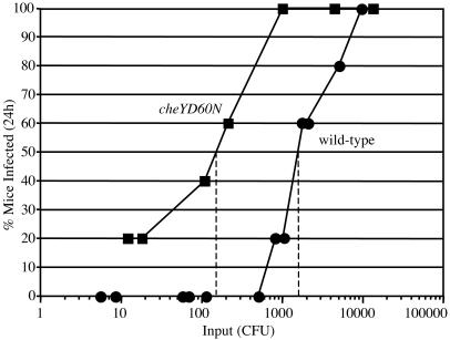
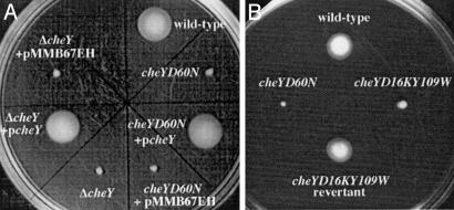
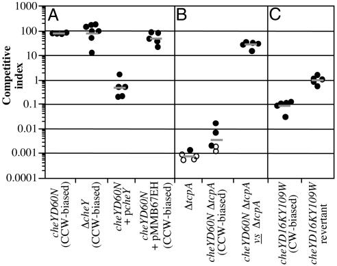
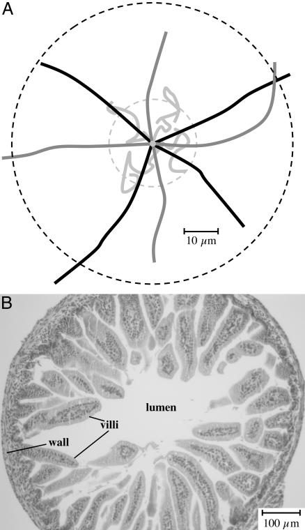
The role of chemotaxis in the virulence of gastrointestinal pathogens is ill defined. Counterintuitively, nonchemotactic mutants of the polarly flagellated pathogen Vibrio cholerae greatly out-compete the wild-type strain during infection of the small intestine. We show that the out-competition phenotype is dependent on the direction of flagellar rotation and independent of Toxin Co-regulated Pilus function. Specifically, the out-competition associated with the loss of chemotaxis required the presence of counterclockwise-biased flagellar rotation and smooth straight runs by the bacteria. In contrast, a nonchemotactic strain with clockwise-biased flagellar rotation was confined to small-scale net movement and was attenuated for infection. The significance of the out-competition phenotype was examined and was shown to correlate with a true increase in infectivity. Counterclockwise-biased mutants are aberrantly distributed throughout the infant mouse small intestine and we find that the expression of virulence factors occurs normally in all segments. Thus, alteration of the chemotactic properties of V. cholerae allows it to exploit additional niches in the host intestine.


Figures

Fig. 3.
Fig. 3.
Determination of the ID50 for wild-type and cheYD60N strains. Groups of five mice were infected with varying doses (x axis) of the wild type (filled circles) or the cheYD60N mutant (filled squares). Each data point represents the percentage of the five mice that were infected (y axis) after a 24-h period. The limit of detection of the mouse output was 10 cfu per small intestine; however, amongst mouse outputs with detectable numbers of V. cholerae, 1,000 cfu was the lowest output observed. The ID50 value for each strain was determined graphically.
Fig. 1.
Fig. 1.
Analysis of chemotactic ability by using swarm plates. Genetic backgrounds are indicated next to each swarm location.
Fig. 2.
Fig. 2.
Competition assays in infant mice between wild-type and mutant V. cholerae strains. Mutant strain backgrounds for each experiment are indicated on the x axis. Each data point represents the competitive index (CI) from one mouse. The CI is given as the ratio of mutant to wild type after infection divided by the ratio of mutant to wild type after overnight growth in LB. Horizontal bars indicate the geometric mean of the CIs for each experiment. (A) Competition between the cheYD60N, ΔcheY, cheYD60N (pcheY), and cheYD60N (pMMB67EH) strains (all LacZ-) and the wild-type strain (LacZ+). (B) Competition between either the cheYD16KY109W mutant or cheYD16KY109W reversion strain (both LacZ-) and wild type (LacZ+). (C) Competition between either the ΔtcpA mutant or the cheYD60NΔtcpA double mutant and wild type, and between the cheYD60NΔtcpA mutant (LacZ-) and the ΔtcpA mutant (LacZ+). Open symbols indicate that the mutant was below the limit of detection.
Fig. 4.
Fig. 4.
(A) Analysis of net movement by individual V. cholerae bacteria. Video images of motile V. cholerae were recorded under dark-field microscopy. The net linear distances traveled by individual bacteria over time were calculated, and the means and standard deviations are presented in the text. The mean net linear distance traveled over a 1-s interval by the CW-biased strain is indicated by the inner hatched circle, and that for the wild-type and CCW-biased strains (which were indistinguishable) is indicated by the outer hatched circle. The paths traveled by four representative bacteria of the wild-type (black lines), CCW-biased (dark gray lines), and CW-biased (light gray lines) strains are shown. Note that we have superimposed the original position of each bacterium at the center of the figure and have rotated and distributed the paths to facilitate their visualization. (B) Light micrograph (×10 magnification) of a hematoxylin/eosin (H&E)-stained cross section through the small intestine of a 5-day-old mouse. Note that the mucus gel normally overlaying the villi is not present in this micrograph.

Similar articles

Cited by

References

    1. Herrington, D. A., Hall, R. H., Losonsky, G., Mekalanos, J. J., Taylor, R. K. & Levine, M. M. (1988) J. Exp. Med. 168, 1487-1492. - PMC - PubMed
    1. Taylor, R. K., Miller, V. L., Furlong, D. B. & Mekalanos, J. J. (1987) Proc. Natl. Acad. Sci. USA 84, 2833-2837. - PMC - PubMed
    1. Heidelberg, J. F., Eisen, J. A., Nelson, W. C., Clayton, R. A., Gwinn, M. L., Dodson, R. J., Haft, D. H., Hickey, E. K., Peterson, J. D., Umayam, L., et al. (2000) Nature 406, 477-483. - PMC - PubMed
    1. Gosink, K. K., Kobayashi, R., Kawagishi, I. & Hase, C. C. (2002) J. Bacteriol. 184, 1767-1771. - PMC - PubMed
    1. Eisenbach, M. (1990) Mol. Microbiol. 4, 161-167. - PubMed

Publication types

Substances

LinkOut - more resources