

Overexpression of pigment epithelium-derived factor decreases angiogenesis and inhibits the growth of human malignant melanoma cells in vivo
- PMID: 15039211
- PMCID: PMC1615357
- DOI: 10.1016/s0002-9440(10)63210-5
Overexpression of pigment epithelium-derived factor decreases angiogenesis and inhibits the growth of human malignant melanoma cells in vivo
Abstract
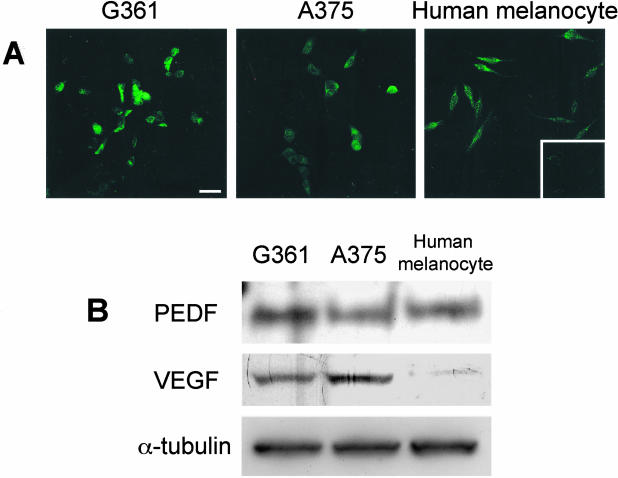
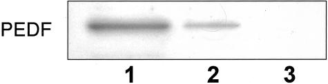
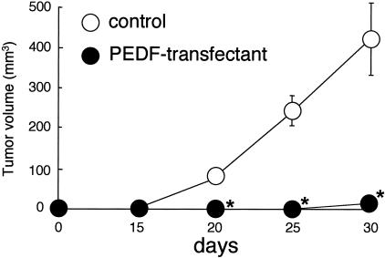
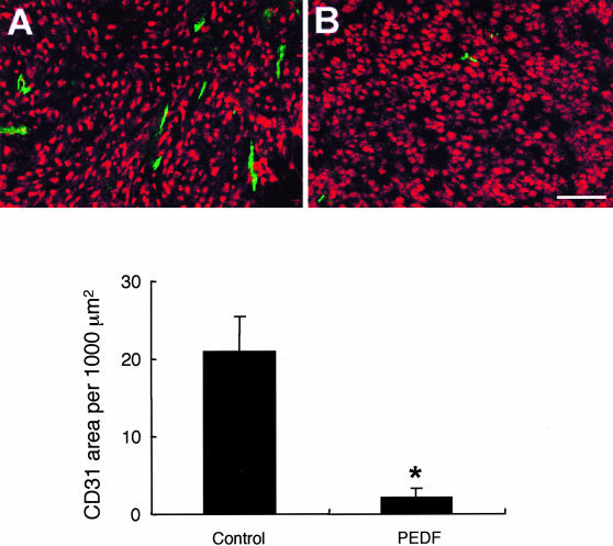
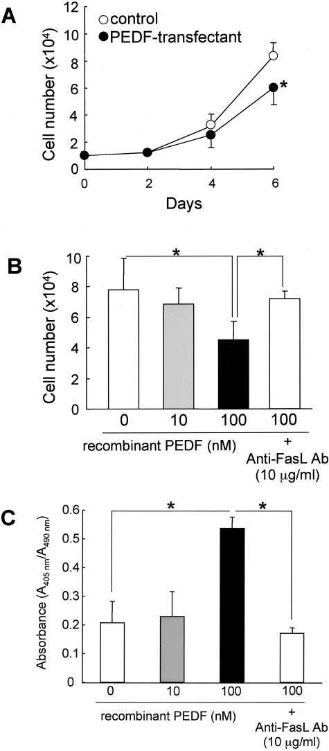
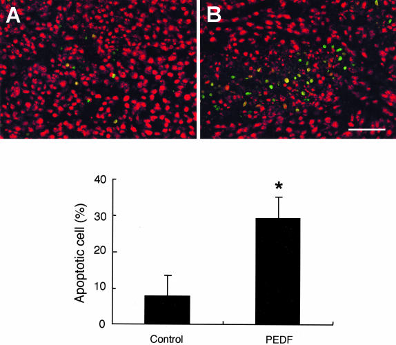
Pigment epithelium-derived factor (PEDF) has recently been shown to be the most potent inhibitor of angiogenesis in the mammalian eye, and is involved in the pathogenesis of angiogenic eye disease such as proliferative diabetic retinopathy. However, a functional role for PEDF in tumor growth and angiogenesis remains to be determined. In this study, we have investigated both the in vitro and in vivo growth characteristics of human malignant melanoma G361 cell lines, stably transfected to overexpress human PEDF. Expression levels of PEDF proteins in melanoma cell lines G361 and A375 were comparable with that of human cultured melanocytes, whereas vascular endothelial growth factor levels in two tumor cell lines were much stronger than that in normal melanocytes. Overexpression of PEDF was found to significantly inhibit tumor growth and vessel formation in G361 nude mice xenografts. Furthermore, in vitro proliferation rates of G361 cells were decreased in PEDF-transfected cells. PEDF proteins showed dose-dependent induced growth retardation and apoptotic cell death in nontransfected G361 cells, which were completely prevented by treatment with antibodies against the Fas ligand. Our present study highlights two beneficial effects of PEDF treatment on melanoma growth and expansion; one is the suppression of tumor angiogenesis, and the other is induction of Fas ligand-dependent apoptosis in tumor cells. PEDF therefore might be a promising novel therapeutic agent for treatment of patients with melanoma.
Figures
References
-
- Holmgren L, O’Reilly MS, Folkman J. Dormancy of micrometastases: balanced proliferation and apoptosis in the presence of angiogenesis suppression. Nat Med. 1995;1:149–153. - PubMed
-
- Carmeliet P, Jain RK. Angiogenesis in cancer and other diseases. Nature. 2000;407:249–257. - PubMed
-
- Scappaticci FA. Mechanisms and future directions for angiogenesis-based cancer therapies. J Clin Oncol. 2002;20:3906–3927. - PubMed
-
- Tombran-Tink J, Chader CG, Johnson LV. PEDF: a pigment epithelium-derived factor with potent neuronal differentiative activity. Exp Eye Res. 1991;53:411–414. - PubMed
-
- Dawson DW, Volpert OV, Gillis P, Crawford SE, Xu HJ, Benedict W, Bouck NP. Pigment epithelium-derived factor: a potent inhibitor of angiogenesis. Science. 1999;285:245–248. - PubMed
Publication types
MeSH terms
Substances
LinkOut - more resources
Full Text Sources
Other Literature Sources
Research Materials
Miscellaneous
