. 2004 Apr;164(4):1481-93.
doi: 10.1016/S0002-9440(10)63234-8.

Characteristic multiorgan pathology of cystic fibrosis in a long-living cystic fibrosis transmembrane regulator knockout murine model

Affiliations

Characteristic multiorgan pathology of cystic fibrosis in a long-living cystic fibrosis transmembrane regulator knockout murine model

Peter R Durie et al. Am J Pathol. 2004 Apr.

Abstract

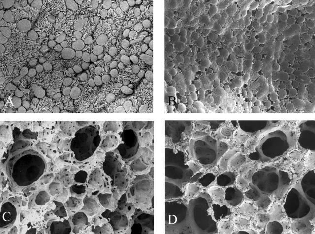
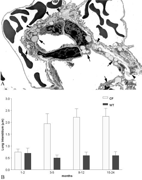
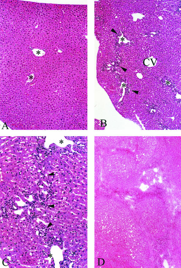
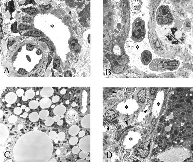
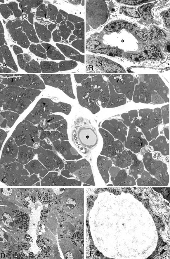
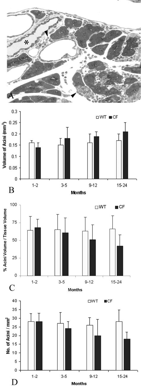
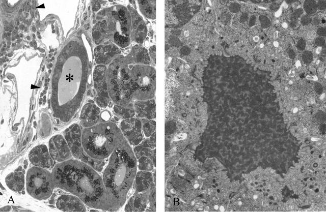
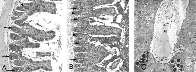
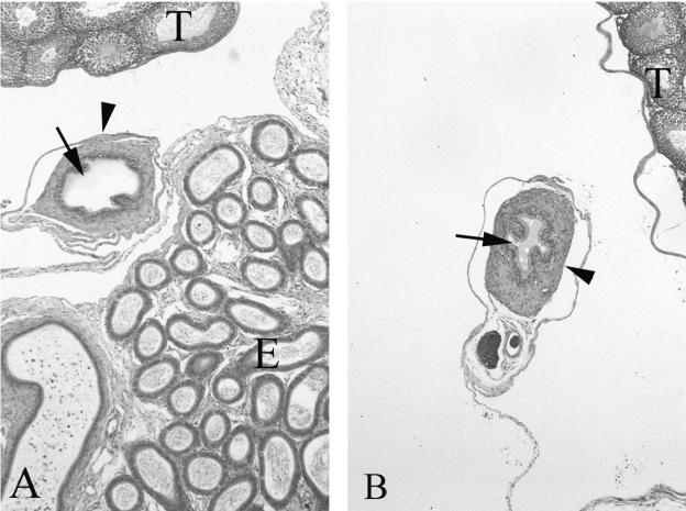
The lack of an appropriate animal model with multiorgan pathology characteristic of the human form of cystic fibrosis has hampered our understanding of the pathobiology of the disease. We evaluated multiple organs of congenic C57BL/6J cystic fibrosis transmembrane regulator (Cftr)(-/-) and Cftr(+/+) mice maintained from weaning on a liquid diet then sacrificed between 1 and 24 months of age. The lungs of the Cftr(-/-) animals showed patchy alveolar overdistention, interstitial thickening, and fibrosis, with progression up to 6 months of age. The proximal and distal airway surface was encased with mucus-like material but lacked overt evidence of chronic bacterial infections or inflammation. All Cftr(-/-) animals showed progressive liver disease, with hepatosteatosis, focal cholangitis, inspissated secretions, and bile duct proliferation; after 1 year of age there was progression to focal biliary cirrhosis. The intercalated, intralobular and interlobular ducts and acinar lumina of the exocrine pancreas, the parotid and submaxillary glands of the Cftr(-/-) animals were dilated and filled with inspissated material, as well as mild inflammation and acinar cell drop out. Quantitative measurements of the pancreas showed significant acinar atrophy and increased acinar volume in comparison with age-matched Cftr(+/+) littermates. The ileal lumen and crypts were filled with adherent fibrillar material. After 3 months of age the vas deferens of the Cftr(-/-) animals could not be identified. None of the aforementioned pathological changes were observed in the Cftr(+/+) littermates fed the same liquid diet. We show, for the first time, that long-lived C578L/6J Cftr(-/-) mice develop manifestations of cystic fibrosis-like disease in all pathologically affected organs in the human form of cystic fibrosis.


Figures

Figure 1
Figure 1
A: SEM of the surface of the respiratory epithelium from a terminal bronchiole in an 11-month-old wild-type animal. Note the numerous ciliated and nonciliated cells. B: Terminal bronchiole from a Cftr−/− littermate. Respiratory epithelium is encrusted in mucus-like material. C: Alveoli from the wild-type animal. D: Alveoli from the affected animal. Distal airways were caked with mucus-like material. Original magnifications: ×1000 (A, B); ×650 (C, D).
Figure 2
Figure 2
A: TEM of alveoli from a lung of a 5-month-old Cftr−/− animal. Note the numerous lipid-containing fibroblasts (arrows) and abundant collagen (arrowheads) in the interstitium. B: Summary histogram comparing the interstitial thickness between wild-type and Cftr−/− animals as well as between age groups. Note the marked increase in interstitial thickness in the 3- to 5-month-old and older groups in the Cftr−/− animals. Scale bar, 2 μm. Original magnification, ×12,500.
Figure 3
Figure 3
A: H&E from an 11-month-old wild-type animal maintained on the liquid diet showing no pathological abnormalities. Portal tracts are evident in the field of view (asterisks). B: Cftr−/− littermate shows portal tract inflammation, ductular cell proliferation, and some portal-to-portal bridging (arrowheads). Parenchymal and sinusoidal cells that surround the central vein (CV) appear normal. C: Higher power of B. Portal-to-portal bridging is evident. Arrowheads indicate ductular and inflammatory cells. D: Low power of liver from a 22-month-old Cftr−/− animal that has developed foci of advanced lobular cirrhosis. Original magnifications: ×250 (A, B); ×400 (C); ×200 (D).
Figure 4
Figure 4
A: TEM of a portal tract from a 22-month-old wild-type animal containing a bile duct. The lumen (asterisk) appears void of any material. B: TEM of a portal tract from a 12-month-old Cftr−/− animal that has developed severe cholangitis showing macrophages and polymorphs both within the lumen of one of the ducts (asterisks) and in the periportal spaces. One of the ducts is filled with inspissated material. C: TEM of several hepatocytes from a 3-month-old Cftr−/− animal filled with lipid droplets, which is consistent with steatosis. D: Periportal fibrosis and duct proliferation. Bundles of collagen fibrils (arrowheads) and several bile ducts are seen in the field (asterisks). Original magnifications: ×2500 (A, B, D); ×5000 (C).
Figure 5
Figure 5
A: Toluidine blue-stained epoxy section of pancreas from a wild-type 9-month-old animal. Note the uniform size of all of the acini and the near absence of any material in any of the ducts (arrows). B: TEM of an interlobular duct from the same animal in Figure 3A. Note the absence of any material in the lumen of the duct (asterisk). An islet (IS) is seen at the bottom right. C: Toluidine blue-stained section of pancreas from a Cftr−/− littermate of the animal seen in Figure 3, A and B. Note the inspissated material found in the interlobular duct (asterisk) and intralobular ducts (arrows). Many of the lumina of the acini appear dilated (arrowheads). D: TEM of dilated acinar lumen (asterisk) from pancreas of animal in Figure 3C. Note the fibrillar material in the lumen. E: TEM of interlobular duct in Figure 3C. Lumen (asterisk) is filled with inspissated material. Note the flattened appearance of the ductular cells. Original magnifications: ×450 (A); ×3500 (B); ×825 (C); ×5000 (D); ×3200 (E).
Figure 6
Figure 6
A: Toluidine blue-stained section of pancreas from a 12-month-old Cftr−/− animal showing periacinar and periductal (asterisk) inflammation (arrowheads) and acinar loss and atrophy. B: Histogram of acinar volume. Acinar volume increased with age in the Cftr−/− age groups. C: Histogram of percentage of acinar volume to tissue volume. Unlike the acinar volumethere was a progressive decrease. D: Histogram of acinar numerical density. Density also decreased, which is suggestive of acinar cell drop-out in the older Cftr−/− animals. Original magnification, ×400.
Figure 7
Figure 7
A: Toluidine blue-stained section from a 12-month-old Cftr−/− submaxillary gland. Note the periductal inflammation (arrowheads). The duct is filled with inspissated material (asterisk). B: TEM of material found in the lumen of a parotid duct from a 5-month-old Cftr−/− animal. In all of the affected animals many of the acinar and ductular lumina were filled with this material. Original magnifications: ×800 (A); ×10,000 (B).
Figure 8
Figure 8
A: Periodic acid-Schiff (PAS) staining of a cross-section of ileum from a 3-month-old wild-type animal. Note the intense staining of the goblet cells on the villi and in the crypts (arrows). Some of the crypts were filled with PAS-stained material (arrowheads). B: Cftr−/− littermate showing crypts filled and distended with PAS-positive material (arrows) and an increase of PAS-positive material within the lumen (asterisk). C: TEM of crypt filled with inspissated material (asterisk). Original magnifications: ×300 (A, B); ×2500 (C).
Figure 9
Figure 9
A: H&E-stained section of a median section through the testes (T), epididymis (E), and vas deferens from a 3-month-old wild-type mouse. Arrow indicates the lumen of the vas deferens and an arrowhead indicates the surrounding smooth muscle layer. B: Similar section from an age-matched affected animal. Although the vas deferens was not detected in any of the animals older than 3 months, two animals in the 3-month group had a visible vas deferens. In comparison with the wild-type littermates the vas deferens had a reduction in luminal diameter (arrows) and thickening of the smooth muscle layer. The lumen also contained accumulation of viscous material. Original magnifications, ×100.

References

    1. Rommens JM, Iannuzzi MC, Kerem B, Drumm ML, Melmer G, Dean M, Rozmahel R, Cole JL, Kennedy D, Hidaka N. Identification of the cystic fibrosis gene: chromosome walking and jumping. Science. 1989;245:1059–1065. - PubMed
    1. Anderson MP, Rich DP, Gregory RJ, Smith AE, Welsh MJ. Generation of cAMP-activated chloride currents by expression of CFTR. Science. 1991;251:679–682. - PubMed
    1. Oppenheimer EH, Esterly JR. Pathology of cystic fibrosis review of the literature and comparison with 146 autopsied cases. Perspect Pediatr Pathol. 1975;2:241–278. - PubMed
    1. Scriver CR, Fujiwara TM. Cystic fibrosis genotypes and views on screening are both heterogeneous and population related. Am J Hum Genet. 1992;51:943–950. - PMC - PubMed
    1. Imrie JR, Fagan DG, Sturgess JM. Structural and developmental abnormalities of the exocrine pancreas in cystic fibrosis. Am J Pathol. 1979;95:697–707. - PMC - PubMed

Publication types

MeSH terms

Substances