. 2004 Apr;72(4):1856-65.
doi: 10.1128/IAI.72.4.1856-1865.2004.

Mechanisms involved in apoptosis of human macrophages induced by lipopolysaccharide from Actinobacillus actinomycetemcomitans in the presence of cycloheximide

Affiliations

Mechanisms involved in apoptosis of human macrophages induced by lipopolysaccharide from Actinobacillus actinomycetemcomitans in the presence of cycloheximide

Takao Suzuki et al. Infect Immun. 2004 Apr.

Abstract

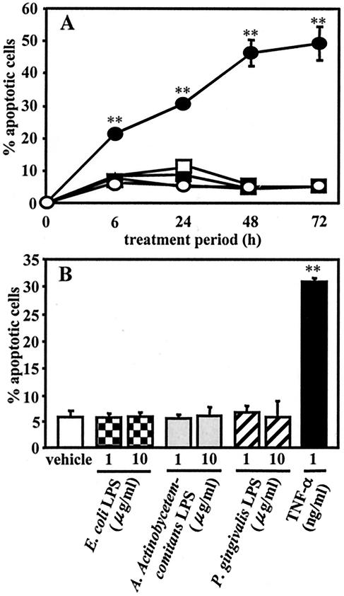
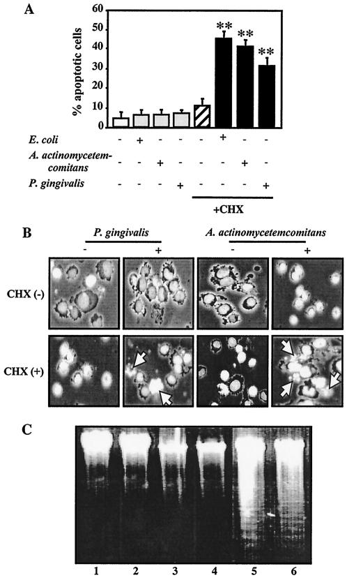
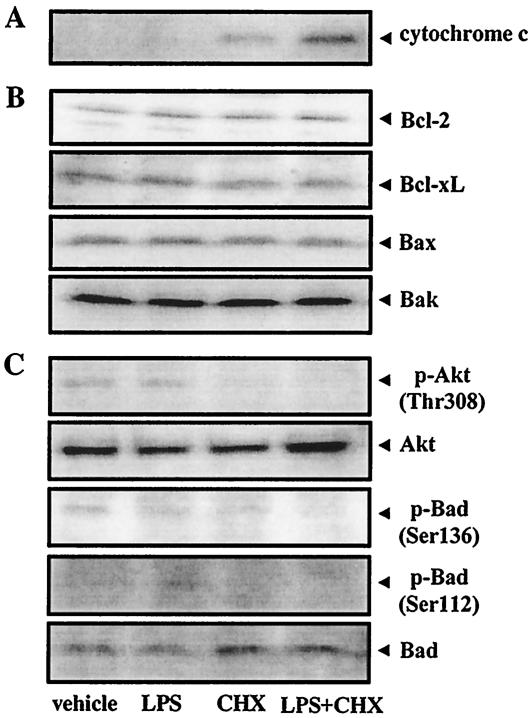
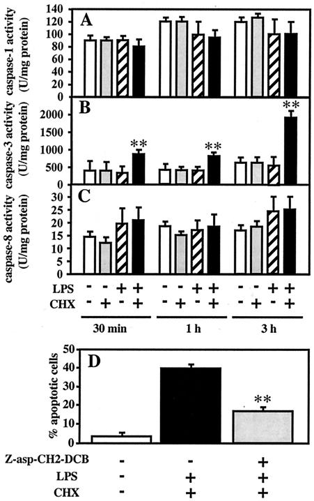
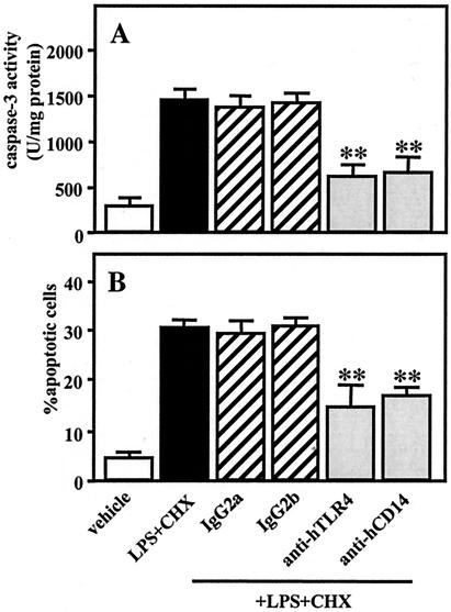
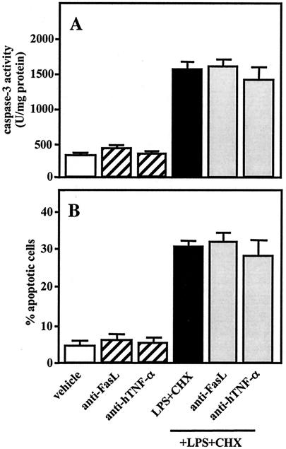
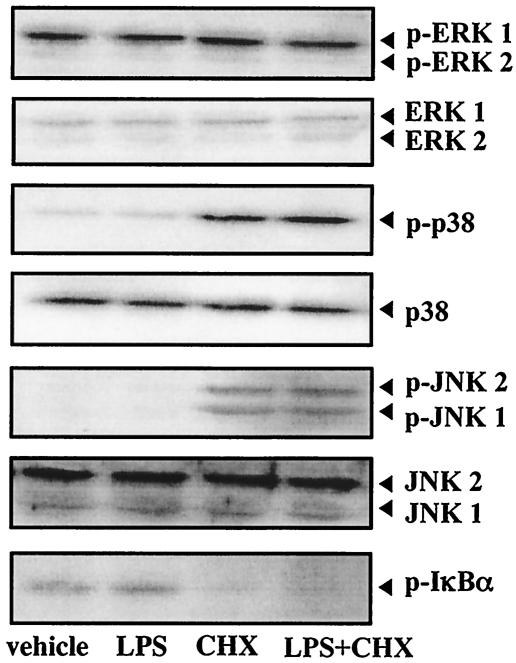
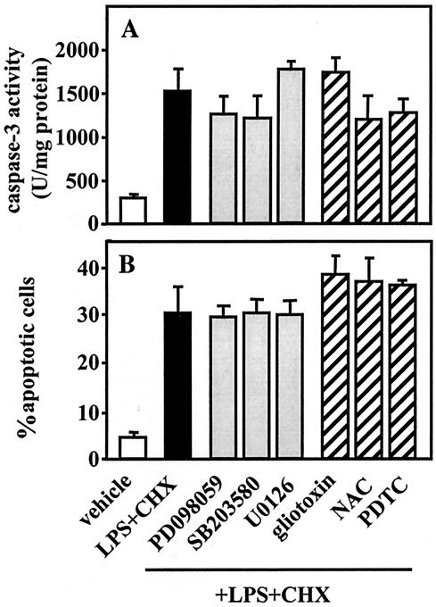
Actinobacillus actinomycetemcomitans is a major periodontopathic bacterium with multiple virulence factors, including lipopolysaccharide (LPS). Previous reports have demonstrated that LPS induced apoptosis in a murine macrophage-like cell line, J744.1, as well as in peritoneal macrophages from C3H/HeN mice in the presence of cycloheximide (CHX). However, the detailed molecular mechanisms involved in the apoptosis of macrophages induced by LPS and CHX are not well known. To clarify the possible role of LPS in the induction of macrophage apoptosis, we investigated cell death induced by LPS from A. actinomycetemcomitans and CHX in human macrophage-like U937 cells, which were differentiated by 12-O-tetradecanoylphorbol 13-acetate (TPA), and also assessed the molecular mechanisms involved in the process. We found that TPA-differentiated U937 cells usually showed resistance to LPS-induced apoptosis. However, in the presence of CHX, LPS induced release of cytochrome c without modifying steady-state levels of Bcl-2, Bcl-xL, Bax, and Bak. Treatment with LPS in the presence of CHX also led to activation of caspase-3 and apoptosis via, in part, the CD14/toll-like receptor 4 (TLR4). The induction of cytochrome c release may have been due to dephosphorylation of Akt and Bad, which were cooperatively induced by CHX and LPS. However, endogenous tumor necrosis factor alpha- and Fas-induced signals, extracellular signal-regulated kinase kinase/mitogen-activated protein kinases and I-kappa B alpha/nuclear factor-kappa B (NF-kappa B) were not required for caspase-3-dependent apoptosis. These results emphasize the possible important role of the mitochondrial apoptotic pathway leading to caspase-3 activation in LPS-induced apoptosis of human macrophages in the presence of CHX.


Figures

FIG. 1.
FIG. 1.
Effects of LPS and rhTNF-α on apoptotic cell death in TPA-differentiated U937 cells. U937 cells were pretreated with TPA (10 ng/ml) for 12 h. (A) The TPA-differentiated U937 cells (105 cells) were further treated with E. coli LPS (1 μg/ml) (▴), A. actinomycetemcomitans LPS (□), P. gingivalis LPS (▪), rhTNF-α (1 ng/ml) (•), or nothing (○) for 6 to 72 h. (B) Other TPA-differentiated U937 cells (105 cells) were treated with a 1- or 10-μg/ml of LPS from E. coli, A. actinomycetemcomitans, or P. gingivalis; rhTNF-α (1 ng/ml); or nothing for 24 h. After each treatment, apoptosis induced in the cells was evaluated by morphological assessment using Hoechst 33258 dye, as described in Materials and Methods. Values shown (percent apoptotic cells) are the means ± SD (error bars) of three separate experiments, each conducted in triplicate. Differences from the value for untreated cells (vehicle) were considered significant (**) at a P of <0.01.
FIG. 2.
FIG. 2.
LPS-induced apoptosis of TPA-differentiated U937 cells in the presence of CHX. TPA-differentiated U937 cells (105 cells [A and B] or 107 cells [C]) were treated with a 1-μg/ml concentration of E. coli LPS, A. actinomycetemcomitans LPS, or P. gingivalis LPS in the presence or absence of CHX (10 μg/ml) for 3 h. Following each treatment, apoptosis in the cells was evaluated by morphological assessment using Hoechst 33258 dye (A and B) or analysis of DNA fragmentation (C), as described in Materials and Methods. (A) Values shown (percent apoptotic cells) are the means + SD (error bars) of three separate experiments, each conducted in triplicate. Differences from the value for untreated cells were considered significant (**) at a P of <0.01. (B) Morphological features of apoptotic cells (i.e., condensed and fragmented nuclei, cell shrinkage, and formation of apoptotic bodies). White arrow indicates apoptotic cells. (C) Agarose gel detection of DNA fragmentation. Lanes: 1 vehicle; 2, CHX alone; 3, A. actinomycetemcomitans LPS alone; 4, P. gingivalis LPS alone; 5, A. actinomycetemcomitans LPS plus CHX; 6, P. gingivalis LPS plus CHX.
FIG. 3.
FIG. 3.
Effects of A. actinomycetemcomitans LPS and/or CHX on cytochrome c release, expression of Bcl-2 family proteins and phosphorylation of Akt and Bad. TPA-differentiated U937 cells (107 cells [A] or 106 cells [B and C]) were treated with A. actinomycetemcomitans LPS (1 μg/ml) in the presence or absence of CHX (10 μg/ml) for 15 min (C) or 3 h (A and B). Following each treatment, cytochrome c release (A); expression of Bcl-2, Bcl-xL, Bax, and Bak proteins (B); and phosphorylation of Bad (Ser136, Ser112) and Akt (Thr308) (C) were evaluated by Western blot analysis, as described in Materials and Methods.
FIG. 4.
FIG. 4.
Involvement of caspase-3 activation in apoptosis induced by A. actinomycetemcomitans LPS in the presence of CHX. TPA-differentiated U937 cells (107 cells) were treated with A. actinomycetemcomitans LPS (1 μg/ml) in the presence or absence of CHX (10 μg/ml) for 30 min, 1 h, and 3 h (A to C). Following each treatment, caspase-1 (A), caspase-3 (B), and caspase-8 (C) activities were measured using colorimetric assay kits according to the manufacturer's instructions. (D) TPA-differentiated U937 cells (105 cells) were also pretreated for 1 h with a 100 μM concentration of a caspase inhibitor, Z-asp-CH2-DCB, or nothing, and were subsequently cotreated with A. actinomycetemcomitans LPS (1 μg/ml) and CHX (10 μg/ml) for 3 h. Following each treatment, the effect of Z-asp-CH2-DCB on apoptosis induced by LPS and CHX (D) was evaluated by morphological assessment using Hoechst 33258-dye, as described in Materials and Methods. Values shown (units per milligram of protein [A to C] or percent apoptotic cells [D]) are the means + SD (error bars) of three separate experiments, each conducted in triplicate. Differences from the value for untreated cells (B) or cells cotreated with LPS and CHX (D) were considered significant (**) at a P of <0.01.
FIG. 5.
FIG. 5.
Participation of CD14/TLR4 in caspase-3 activity and apoptotic cell death induced by A. actinomycetemcomitans LPS in the presence of CHX. TPA-differentiated U937 cells (107 cells [A] or 105 cells [B]) were pretreated for 1 h with MY4 (5 μg/ml), control isotype-matched mouse IgG2b (5 μg/ml), HTA125 (20 μg/ml), control isotype-matched mouse IgG2a (20 μg/ml), or nothing and were subsequently cotreated with A. actinomycetemcomitans LPS (1 μg/ml) and CHX (10 μg/ml) for 3 h. Following each treatment, caspase-3 activity (A) was evaluated using colorimetric assay kits, and apoptosis (B) was evaluated by morphological assessment using Hoechst 33258 dye, as described in Materials and Methods. Values shown (units per milligram of protein [A] or percent apoptotic cells [B]) are the means + SD (error bars) of three separate experiments, each conducted in triplicate. Differences from the value for cells cotreated with LPS and CHX were considered significant (**) at a P of <0.01.
FIG. 6.
FIG. 6.
Effects of anti-FasL and anti-hTNF-α on caspase-3 activity and apoptosis induced by A. actinomycetemcomitans LPS and CHX. TPA-differentiated U937 cells (107 cells [A] or 105 cells [B]) were pretreated for 1 h with anti-FasL (10 μg/ml), anti-hTNF-α (5 μg/ml), or nothing and were subsequently cotreated with A. actinomycetemcomitans LPS (1 μg/ml) and CHX (10 μg/ml) for 3 h. Following each treatment, caspase-3 activity (A) was determined using colorimetric assay kits and apoptosis (B) by morphological assessment using Hoechst 33258 dye, as described in Materials and Methods. Values shown (units per milligram of protein [A] or percent apoptotic cells [B]) are the means + SD (error bars) of three separate experiments, each conducted in triplicate.
FIG. 7.
FIG. 7.
Activation of MAP kinases and I-κBα in TPA-differentiated U937 cells treated with A. actinomycetemcomitans LPS and/or CHX. TPA-differentiated U937 cells (106 cells) were treated with A. actinomycetemcomitans LPS (1 μg/ml) and/or CHX (10 μg/ml) for 30 min. Following each treatment, the expressions of ERK1/2, phosphorylated ERK1/2 (p-ERK1/2), p38, phosphorylated p38 (p-p38), JNK1/2, phosphorylated JNK1/2 (p-JNK1/2), and phosphorylated I-κBα (p-I-κBα) were evaluated by Western blot analysis, as described in Materials and Methods.
FIG. 8.
FIG. 8.
Effects of inhibitors of MEK/MAP kinases or NF-κB activation on caspase-3 activity and apoptosis induced by A. actinomycetemcomitans LPS and CHX. TPA-differentiated U937 cells (107 cells [A] or 105 cells [B]) were pretreated for 1 h with PD098059 (100 μM; MEK1/2/MAP kinase inhibitor), U0126 (5 μM; MEK1/2 inhibitor), SB203580 (10 μM; p38 inhibitor), inhibitors of NF-κB activation (gliotoxin [250 ng/ml], NAC [5 mM], or PDTC [100 μM]), or nothing and were subsequently cotreated with A. actinomycetemcomitans LPS (1 μg/ml) and CHX (10 μg/ml) for 3 h. Following each treatment, caspase-3 activity (A) was determined using the colorimetric assay kits and apoptosis (B) was evaluated by morphological assessment using Hoechst 33258 dye, as described in Materials and Methods. Values shown (units per milligram of protein [A] or percent apoptotic cells [B]) are the means + SD (error bars) of three separate experiments, each conducted in triplicate.

References

    1. Afford, S. C., J. Pongracz, R. A. Stockley, J. Crocker, and D. Burnett. 1992. The induction by human interleukin-6 of apoptosis in the promonocytic cell line U937 and human neutrophils. J. Biol. Chem. 267:21612-21616. - PubMed
    1. Amano, F., and H. Karahashi. 1996. A cytotoxic effect of lipopolysaccharide on a macrophage-like cell line, J774.1, in the presence of cycloheximide. J. Endotoxin Res. 3:415-423. - PubMed
    1. Bratton, S. B., G. Walker, S. M. Srinivasula, X. M. Sun, M. Butterworth, E. S. Alnemri, and G. M. Cohen. 2001. Recruitment, activation and retention of caspases-9 and -3 by Apaf-1 apoptosome and associated XIAP complexes. EMBO J. 20:998-1009. - PMC - PubMed
    1. Bremner, T. A., D. Chatterjee, Z. Han, M. F. Tsan, and J. H. Wyche. 1999. THP-1 monocytic leukemia cells express Fas ligand constitutively and kill Fas-positive Jurkat cells. Leuk. Res. 23:865-870. - PubMed
    1. Choi, K. B., F. Wong, J. M. Harlan, P. M. Chaudhary, L. Hood, and A. Karsan. 1998. Lipopolysaccharide mediates endothelial apoptosis by a FADD-dependent pathway. J. Biol. Chem. 273:20185-20188. - PubMed

Publication types

MeSH terms