. 2004 Apr;72(4):1946-55.
doi: 10.1128/IAI.72.4.1946-1955.2004.

N-linked glycosylation is required for c1 inhibitor-mediated protection from endotoxin shock in mice

Affiliations

N-linked glycosylation is required for c1 inhibitor-mediated protection from endotoxin shock in mice

Dongxu Liu et al. Infect Immun. 2004 Apr.

Abstract

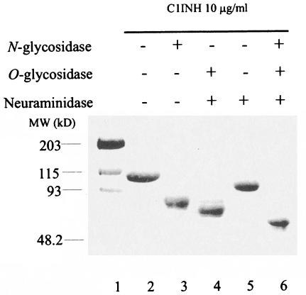
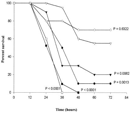
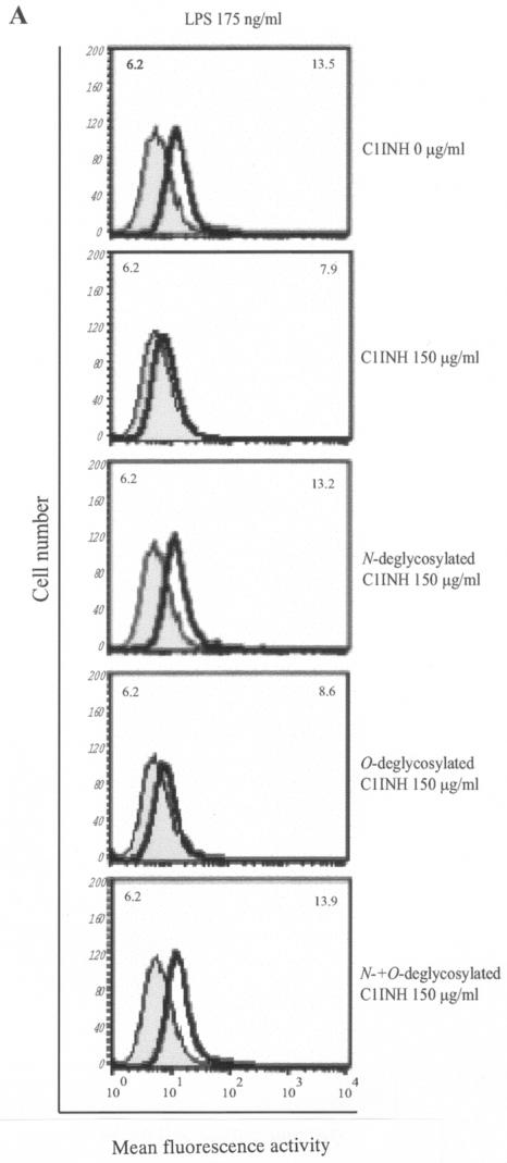
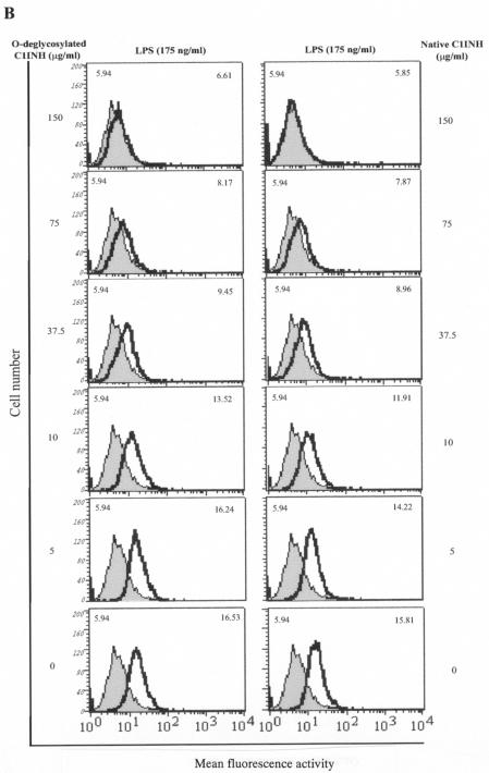
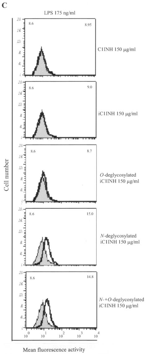
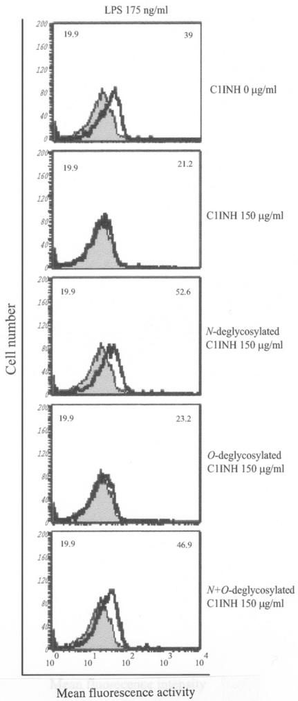
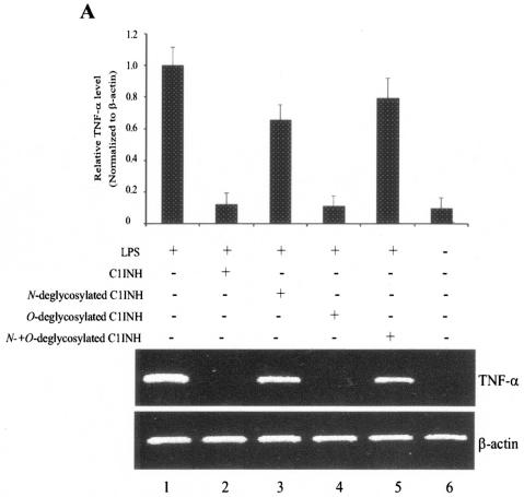
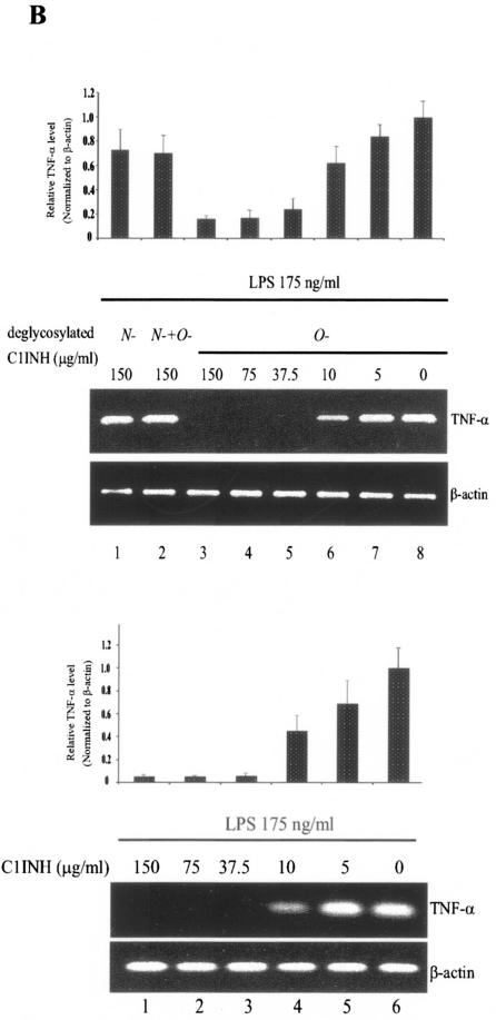
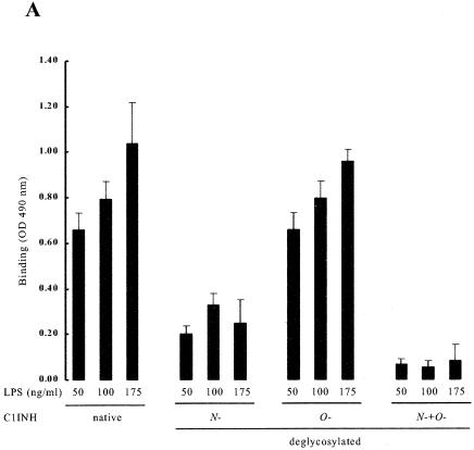
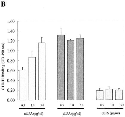
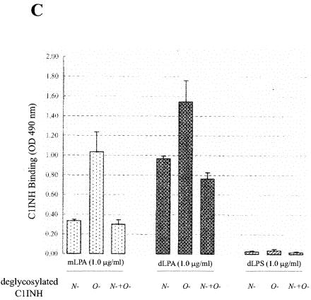
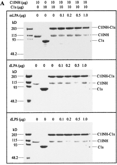
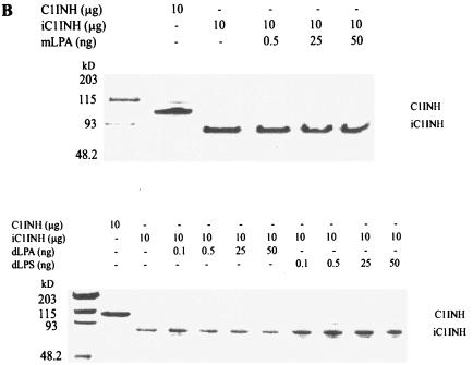
C1 inhibitor (C1INH) prevents endotoxin shock in mice via a direct interaction with lipopolysaccharide (LPS). This interaction requires the heavily glycosylated amino-terminal domain of C1INH. C1INH in which N-linked carbohydrate was removed by using N-glycosidase F was markedly less effective in protecting mice from LPS-induced lethal septic shock. N-deglycosylated C1INH also failed to suppress fluorescein isothiocyanate (FITC)-LPS binding to and LPS-induced tumor necrosis factor alpha mRNA expression by the murine macrophage-like cell line, RAW 264.7, and cells in human whole blood. In an enzyme linked immunosorbent assay, the N-deglycosylated C1INH bound to LPS very poorly. In addition, C1INH was shown to bind to diphosphoryl lipid A (dLPA) but only weakly to monophosphoryl lipid A (mLPA). As with intact LPS, binding of N-deglycosylated C1INH to dLPA and mLPA was diminished in comparison with the native protein. Removal of O-linked carbohydrate had no effect on any of these activities. Neither detoxified LPS, dLPA, nor mLPA had any effect on the rate or extent of C1INH complex formation with C1s or on cleavage of the reactive center loop by trypsin. These data demonstrate that N-linked glycosylation of C1INH is essential to mediate its interaction with the LPA moiety of LPS and to protect mice from endotoxin shock.


Figures

FIG. 1.
FIG. 1.
SDS-PAGE analysis of deglycosylated C1INH. C1INH (10 μg) was incubated with N-glycosidase F, O-glycosidase, and neuraminidase as described in Materials and Methods. The various forms of C1INH were analyzed by SDS-PAGE and stained with Coomassie brilliant blue.
FIG. 2.
FIG. 2.
Effect of deglycosylated C1INH on survival of mice in gram-negative endotoxin LPS-induced lethal endotoxemia. C57BL/6J mice were injected i.p. with a mixture of LPS (20 mg/kg) with native, plasma-derived C1INH (▵, n = 20), with N-deglycosylated C1INH (▴, n = 10), with O-deglycosylated C1INH (○, n = 10), or with both N- and O-deglycosylated C1INH (•, n = 10) (200 μg/per mouse). Control mice were injected (i.p.) with LPS (20 mg/kg) alone (▪, n = 21) or a mixture of LPS (20 mg/kg) with the glycosidase buffer (□, n = 5). The indicated P values are for each group in comparison with the group treated with native, plasma-derived C1INH.
FIG. 3.
FIG. 3.
Effect of deglycosylated C1INH on the binding of FITC-conjugated LPS to the murine macrophage cell line, RAW 264.7. LPS binding, thick line; control, shaded field; mean fluorescence intensities for the control and treated cells are indicated in the upper left and upper right corners, respectively, in each panel. (A) RAW 264.7 macrophages were incubated with FITC-LPS (175 ng/ml) in the absence or presence of C1INH, N-deglycosylated C1INH, O-deglycosylated C1INH, or both N- and O-deglycosylated C1INH (each at 150 μg/ml) in DMEM containing 10% FBS for 15 min at 37°C. (B) RAW 264.7 macrophages were incubated with FITC-LPS (175 ng/ml) in the presence of different doses of O-deglycosylated C1INH or untreated native C1INH (150, 75, 37.5, 10, or 5 μg/ml) in DMEM containing 10% FBS for 15 min at 37°C. (C) RAW 264.7 macrophages were incubated with FITC-LPS (175 ng/ml) in the presence of iC1INH (150 μg/ml) lacking N-, O-, or both N- and O-linked glycosylation. Cells were fixed with FACS solution after being washed with PBS three times and were analyzed by FACS.
FIG. 3.
FIG. 3.
Effect of deglycosylated C1INH on the binding of FITC-conjugated LPS to the murine macrophage cell line, RAW 264.7. LPS binding, thick line; control, shaded field; mean fluorescence intensities for the control and treated cells are indicated in the upper left and upper right corners, respectively, in each panel. (A) RAW 264.7 macrophages were incubated with FITC-LPS (175 ng/ml) in the absence or presence of C1INH, N-deglycosylated C1INH, O-deglycosylated C1INH, or both N- and O-deglycosylated C1INH (each at 150 μg/ml) in DMEM containing 10% FBS for 15 min at 37°C. (B) RAW 264.7 macrophages were incubated with FITC-LPS (175 ng/ml) in the presence of different doses of O-deglycosylated C1INH or untreated native C1INH (150, 75, 37.5, 10, or 5 μg/ml) in DMEM containing 10% FBS for 15 min at 37°C. (C) RAW 264.7 macrophages were incubated with FITC-LPS (175 ng/ml) in the presence of iC1INH (150 μg/ml) lacking N-, O-, or both N- and O-linked glycosylation. Cells were fixed with FACS solution after being washed with PBS three times and were analyzed by FACS.
FIG. 4.
FIG. 4.
Effect of deglycosylated C1INH on the binding of FITC-conjugated LPS to human blood cells. LPS binding, thick line; control, shaded field. Mean fluorescence intensities for the control and treated cells are indicated in the upper left and upper right corners, respectively, in each panel. The human blood cells were incubated for 15 min at 37°C with FITC-LPS (175 ng/ml) in the presence of C1INH (150 μg/ml) lacking N-, O-, or both N- and O-linked glycosylation. Cells were fixed with FACS solution after being washed with PBS three times and were analyzed by FACS.
FIG. 5.
FIG. 5.
Effect of deglycosylated C1INH on LPS-induced TNF-α mRNA expression in the murine macrophage cell line, RAW 264.7 and human blood cells. (A) total RNA from RAW 264.7 macrophages was isolated after treatment with LPS (175 ng/ml) in the presence of C1INH or with N-, O-, or both N- and O-deglycosylated C1INH (all at 150 μg/ml) for 30 min at 37°C. RT-PCR was performed with mouse TNF-α cDNA and β-actin cDNA primers. The band intensity was quantitated by using ImageQuant software (Molecular Dynamics), was normalized to the β actin level, and is expressed relative to the amount of PCR product present in the samples treated with LPS alone. (B) Total RNA from human blood cells was isolated after treatment of whole human blood with LPS (175 ng/ml) in the presence of C1INH (5, 10, 37.5, 75, or 150 μg/ml) or with N-deglycosylated C1INH (150 μg/ml), O-deglycosylated C1INH (5, 10, 37.5, 75, or 150 μg/ml), or N- and O-deglycosylated C1INH (150 μg/ml) (upper panel) for 30 min at 37°C. RT-PCR was performed with human TNF-α cDNA and β-actin cDNA primers. PCR products were quantitated as in panel A.
FIG. 5.
FIG. 5.
Effect of deglycosylated C1INH on LPS-induced TNF-α mRNA expression in the murine macrophage cell line, RAW 264.7 and human blood cells. (A) total RNA from RAW 264.7 macrophages was isolated after treatment with LPS (175 ng/ml) in the presence of C1INH or with N-, O-, or both N- and O-deglycosylated C1INH (all at 150 μg/ml) for 30 min at 37°C. RT-PCR was performed with mouse TNF-α cDNA and β-actin cDNA primers. The band intensity was quantitated by using ImageQuant software (Molecular Dynamics), was normalized to the β actin level, and is expressed relative to the amount of PCR product present in the samples treated with LPS alone. (B) Total RNA from human blood cells was isolated after treatment of whole human blood with LPS (175 ng/ml) in the presence of C1INH (5, 10, 37.5, 75, or 150 μg/ml) or with N-deglycosylated C1INH (150 μg/ml), O-deglycosylated C1INH (5, 10, 37.5, 75, or 150 μg/ml), or N- and O-deglycosylated C1INH (150 μg/ml) (upper panel) for 30 min at 37°C. RT-PCR was performed with human TNF-α cDNA and β-actin cDNA primers. PCR products were quantitated as in panel A.
FIG. 6.
FIG. 6.
Analysis of the binding of deglycosylated C1INH to LPS and LPA. The interaction of C1INH or deglycosylated C1INH with immobilized LPS, mLPA, dLPA, and dLPS was analyzed by ELISA. (A) Binding of deglycosylated C1INH (150 μg/ml) to LPS (50, 100, or 175 ng/ml) in comparison with native C1INH (150 μg/ml); (B) binding of C1INH (150 μg/ml) to mLPA (0.5, 1.0, or 5.0 μg/ml), dLPA (0.5, 1.0, or 5.0 μg/ml), and dLPS (0.5, 1.0, and 5.0 μg/ml); (C) binding of deglycosylated C1INH (150 μg/ml) to mLPA (1.0 μg/ml), dLPA (1.0 μg/ml), or dLPS (1.0 μg/ml).
FIG. 6.
FIG. 6.
Analysis of the binding of deglycosylated C1INH to LPS and LPA. The interaction of C1INH or deglycosylated C1INH with immobilized LPS, mLPA, dLPA, and dLPS was analyzed by ELISA. (A) Binding of deglycosylated C1INH (150 μg/ml) to LPS (50, 100, or 175 ng/ml) in comparison with native C1INH (150 μg/ml); (B) binding of C1INH (150 μg/ml) to mLPA (0.5, 1.0, or 5.0 μg/ml), dLPA (0.5, 1.0, or 5.0 μg/ml), and dLPS (0.5, 1.0, and 5.0 μg/ml); (C) binding of deglycosylated C1INH (150 μg/ml) to mLPA (1.0 μg/ml), dLPA (1.0 μg/ml), or dLPS (1.0 μg/ml).
FIG. 7.
FIG. 7.
Effects of mLPA, dLPA, and dLPS on the formation of C1INH-C1s complexes and C1INH cleavage by trypsin. (A) mLPA (0.1, 0.2, 0.5, or 1.0 μg), dLPA (0.1, 0.2, 0.5, or 1.0 μg), and dLPS (0.1, 0.2, 0.5, or 1.0 μg) have no effect on the rate or extent of C1INH-C1s (10 μg: 10 μg) complex formation, as assessed by SDS-PAGE stained with Coomassie brilliant blue. (B) mLPA (0.5, 25, or 50 ng), dLPA (0.1, 0.5, 25, or 50 ng), and dLPS (0.1, 0.5, 25, or 50 ng) have no effect on the formation of cleaved C1INH (10 μg) by trypsin, as assessed by SDS-PAGE stained with Coomassie brilliant blue.
FIG. 7.
FIG. 7.
Effects of mLPA, dLPA, and dLPS on the formation of C1INH-C1s complexes and C1INH cleavage by trypsin. (A) mLPA (0.1, 0.2, 0.5, or 1.0 μg), dLPA (0.1, 0.2, 0.5, or 1.0 μg), and dLPS (0.1, 0.2, 0.5, or 1.0 μg) have no effect on the rate or extent of C1INH-C1s (10 μg: 10 μg) complex formation, as assessed by SDS-PAGE stained with Coomassie brilliant blue. (B) mLPA (0.5, 25, or 50 ng), dLPA (0.1, 0.5, 25, or 50 ng), and dLPS (0.1, 0.5, 25, or 50 ng) have no effect on the formation of cleaved C1INH (10 μg) by trypsin, as assessed by SDS-PAGE stained with Coomassie brilliant blue.

References

    1. Appelmelk, B. J., Y.-Q. An, M. Geerts, B. G. Thijs, H. A. De Boer, D. M. MacLaren, J. de Graaff, and J. H. Juijens. 1994. Lactoferrin is a lipid A-binding proten. Infect. Immun. 62:2628-2632. - PMC - PubMed
    1. Astiz, M. E., R. C. Rackow, J. G. Still, S. T. Howell, A. Cato, K. B. Von Eschen, J. T. Ulrich, J. A. Rudbach, G. McMahon, R. Vargas, and W. Stern. 1995. Pretreatment of normal humans with monophosphoryl lipid A induces tolerance to endotoxin: a prospective, double-blind, randomized, controlled trial. Crit. Care Med. 23:9-17. - PubMed
    1. Battafarano, R. J., P. S. Dahlberg, C. A. Ratz, J. W. Johnston, B. H. Gray, J. R. Haseman, K. H. Mayo, and D. L. Dunn. 1995. Peptide derivatives of three distinct lipopolysaccharide binding proteins inhibit lipopolysaccharide-induced tumor necrosis factor-alpha secretion in vitro. Surgery 118:318-324. - PubMed
    1. Bock, S. C., K. Skriver, E. Nielsen, H. C. Thogersen, B. Wiman, V. H. Donaldson, R. L. Eddy, J. Marrinan, E. Radziejewska, R. Huber, T. B. Shows, and S. Magnussen. 1986. Human C1 inhibitor: primary structure, cDNA cloning, and chromosomal localization. Biochemistry 25:4292-4301. - PubMed
    1. Bone, R. C. 1993. gram-negative sepsis: a dilemma of modern medicine. Clin. Microbiol. Rev. 6:57-68. - PMC - PubMed

Publication types

MeSH terms

Substances