. 2004 Apr;72(4):2002-13.
doi: 10.1128/IAI.72.4.2002-2013.2004.

Lon protease activity causes down-regulation of Salmonella pathogenicity island 1 invasion gene expression after infection of epithelial cells

Affiliations

Lon protease activity causes down-regulation of Salmonella pathogenicity island 1 invasion gene expression after infection of epithelial cells

Jennifer D Boddicker et al. Infect Immun. 2004 Apr.

Abstract

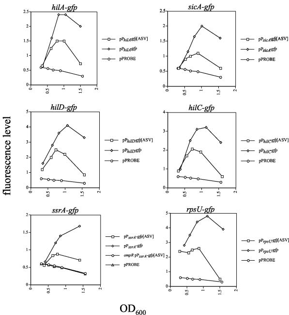
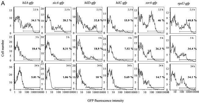
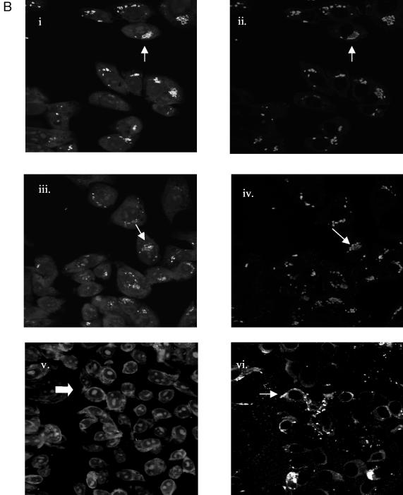
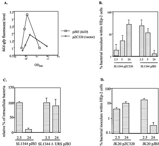
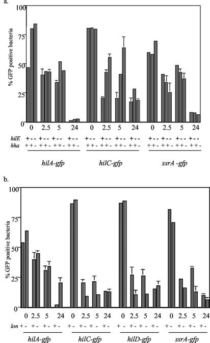
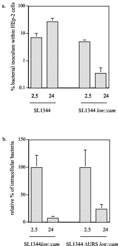
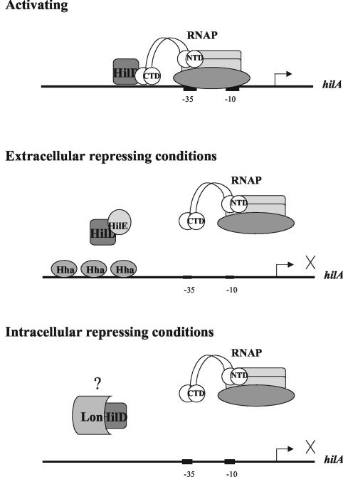
Salmonella enterica serovar Typhimurium causes self-limiting gastroenteritis in humans and a typhoid-like disease in mice that serves as a model for typhoid infections in humans. A critical step in Salmonella pathogenesis is the invasion of enterocytes and M cells of the small intestine via expression of a type III secretion system, encoded on Salmonella pathogenicity island 1 (SPI-1), that secretes effector proteins into host cells, leading to engulfment of the bacteria within large membrane ruffles. The in vitro regulation of invasion genes has been the subject of much scientific investigation. Transcription of the hilA gene, which encodes an OmpR/ToxR-type transcriptional activator of downstream invasion genes, is increased during growth under high-osmolarity and low-oxygen conditions, which presumably mimic the environment found within the small intestine. Several negative regulators of invasion gene expression have been identified, including HilE, Hha, and Lon protease. Mutations within the respective genes increase the expression of hilA when the bacteria are grown under environmental conditions that are not favorable for hilA expression and invasion. In this study, the intracellular expression of invasion genes was examined, after bacterial invasion of HEp-2 epithelial cells, using Salmonella strains containing plasmid-encoded short-half-life green fluorescent protein reporters of hilA, hilD, hilC, or sicA expression. Interestingly, the expression of SPI-1 genes was down-regulated after invasion, and this was important for the intracellular survival of the bacteria. In addition, the effects of mutations in genes encoding negative regulators of invasion on intracellular hilA expression were examined. Our results indicate that Lon protease is important for down-regulation of hilA expression and intracellular survival after the invasion of epithelial cells.


Figures

FIG. 1.
FIG. 1.
Fluorescence levels from Salmonella containing GFP reporters at various times during a growth curve. The fluorescence levels shown represent one experiment that was repeated at least two times with similar results.
FIG. 2.
FIG. 2.
Expression of SPI-1 invasion genes decreases after infection of HEp-2 epithelial cells. (A) FACS analysis was performed after lysing of infected epithelial cells and stainingof intracellular Salmonella (see Materials and Methods) 2.5, 5, and 24 h after inoculation of bacteria onto HEp-2 cell monolayers. The percentage of GFP-positive bacteria and the length of infection are indicated on each histogram. The horizontal lines represent the gate used to determine the percentage of GFP-positive bacteria in each sample. (B) Confocal microscopy was performed on cell monolayers infected with Salmonella containing pPhilA-gfp[ASV] 2.5, 5, and 24 h after infection. After infection, samples were fixed, permeabilized, and stained with ethidium bromide, anti-Salmonella antiserum, and a secondary antibody conjugated to Cy 5. The top two micrographs show the same microscopic field 2.5 h after infection. Likewise, the middle and bottom micrographs show the same fields 5 and 24 h after infection, respectively. The micrographs on the left show the GFP fluorescence of bacteria within epithelial cells stained with ethidium bromide. The micrographs on the right show bacteria within the same fields as on the left stained with Salmonella-specific antibody and Cy 5 as a positive control for the presence of bacteria. The arrowheads point to bacteria that are expressing GFP and that are stained with Cy 5 in adjacent micrographs of the same field. The arrow in the lower left micrograph points to bacteria that are not expressing GFP (as no green fluorescence was detected); however, the diffuse (red) fluorescence that is visible is likely due to staining of the bacterial nucleic acids by ethidium bromide; the arrowhead in the lower right micrograph points to bacteria in the same area of the microscopic field that are stained with Cy 5.
FIG. 2.
FIG. 2.
Expression of SPI-1 invasion genes decreases after infection of HEp-2 epithelial cells. (A) FACS analysis was performed after lysing of infected epithelial cells and stainingof intracellular Salmonella (see Materials and Methods) 2.5, 5, and 24 h after inoculation of bacteria onto HEp-2 cell monolayers. The percentage of GFP-positive bacteria and the length of infection are indicated on each histogram. The horizontal lines represent the gate used to determine the percentage of GFP-positive bacteria in each sample. (B) Confocal microscopy was performed on cell monolayers infected with Salmonella containing pPhilA-gfp[ASV] 2.5, 5, and 24 h after infection. After infection, samples were fixed, permeabilized, and stained with ethidium bromide, anti-Salmonella antiserum, and a secondary antibody conjugated to Cy 5. The top two micrographs show the same microscopic field 2.5 h after infection. Likewise, the middle and bottom micrographs show the same fields 5 and 24 h after infection, respectively. The micrographs on the left show the GFP fluorescence of bacteria within epithelial cells stained with ethidium bromide. The micrographs on the right show bacteria within the same fields as on the left stained with Salmonella-specific antibody and Cy 5 as a positive control for the presence of bacteria. The arrowheads point to bacteria that are expressing GFP and that are stained with Cy 5 in adjacent micrographs of the same field. The arrow in the lower left micrograph points to bacteria that are not expressing GFP (as no green fluorescence was detected); however, the diffuse (red) fluorescence that is visible is likely due to staining of the bacterial nucleic acids by ethidium bromide; the arrowhead in the lower right micrograph points to bacteria in the same area of the microscopic field that are stained with Cy 5.
FIG. 3.
FIG. 3.
Plasmid expression of hilD increases bacterial invasion but decreases intracellular growth and survival. (A) Expression of hilA was estimated by determining the fluorescence level from Salmonella strain SL1344 pPhilA-gfp[ASV] carrying pJB3, which expresses hilD from the lac promoter, or the parent vector pZC320 at various times during a growth curve. The data shown are from one experiment that is representative of two separate experiments performed with similar results. (B) An invasion assay was performed with SL1344 containing pZC320 or pJB3 to determine invasion and intracellular survival of the bacteria from 2.5 to 24 h after infection. (C) Invasion and intracellular survival of SL1344(pJB3) and SL1344 ΔURS(pJB3) were determined. Since SL1344 ΔURS is noninvasive, the SL1344 ΔURS(pJB3) strain was coinfected with wt Salmonella that did not contain pJB3. The percentage of the original inoculum of each strain containing pJB3 that was recovered on LB-ampicillin plates after 2.5 h of infection was set to 100% for each strain, and the number of intracellular bacteria containing pJB3 at 24 h is reported as a percentage of that shown at 2.5 h. (D) Invasion and intracellular survival of the SPI-2 ssaV::cam mutant strain, JK20, carrying pZC320 or pJB3 after 2.5 or 24 h of infection. The invasion assay data are the mean plus the standard deviation of one experiment performed in triplicate that is representative of two or more independent experiments.
FIG. 4.
FIG. 4.
Effects of mutations in negative regulators on the expression of GFP reporters after infection of HEp-2 epithelial cells. FACS analysis was performed 2.5, 5, or 24 h after infection of epithelial cells with Salmonella hilE or hilE hha (a) and lon (b) mutant strains containing unstable (tagged) hilA, hilD, hilC, or ssrA-gfp reporters. The data presented are the mean plus the standard deviation of one experiment performed in duplicate that is representative of several independent experiments that had similar results. +, present; −, absent.
FIG. 5.
FIG. 5.
Effects of a lon mutation on invasion and intracellular survival of Salmonella. Invasion and intracellular survival of Salmonella strain SL1344 containing a lon::cam mutation 2.5 and 24 h after infection were determined and compared to those of wt Salmonella. The data shown are the mean plus the standard deviation of one experiment performed in triplicate that is representative of two separate experiments with similar results.
FIG. 6.
FIG. 6.
Model of hilA regulation under conditions that are activating for invasion and conditions that are repressive for invasion. CTD, C-terminal domain; NTD, N-terminal domain.

References

    1. Akbar, S., L. M. Schechter, C. P. Lostroh, and C. A. Lee. 2003. AraC/XylS family members, HilD and HilC, directly activate virulence gene expression independently of HilA in Salmonella typhimurium. Mol. Microbiol. 47:715-728. - PubMed
    1. Altier, C., M. Suyemoto, and S. D. Lawhon. 2000. Regulation of Salmonella enterica serovar Typhimurium invasion genes bycsrA. Infect. Immun. 68:6790-6797. - PMC - PubMed
    1. Altier, C., M. Suyemoto, A. I. Ruiz, K. D. Burnham, and R. Maurer. 2000. Characterization of two novel regulatory genes affecting Salmonella invasion gene expression. Mol. Microbiol. 35:635-646. - PubMed
    1. Andersen, J. B., C. Sternberg, L. K. Poulsen, S. P. Bjorn, M. Givskov, and S. Molin. 1998. New unstable variants of green fluorescent protein for studies of transient gene expression in bacteria. Appl. Environ. Microbiol. 64:2240-2246. - PMC - PubMed
    1. Bajaj, V., C. Hwang, and C. A. Lee. 1995. hilA is a novel ompR/toxR family member that activates the expression of Salmonella typhimurium invasion genes. Mol. Microbiol. 18:715-727. - PubMed

Publication types

MeSH terms

LinkOut - more resources