. 2004 Apr;72(4):2067-74.
doi: 10.1128/IAI.72.4.2067-2074.2004.

Mycobacterium tuberculosis lipomannan induces apoptosis and interleukin-12 production in macrophages

Affiliations

Mycobacterium tuberculosis lipomannan induces apoptosis and interleukin-12 production in macrophages

D N Dao et al. Infect Immun. 2004 Apr.

Abstract

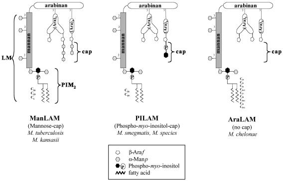
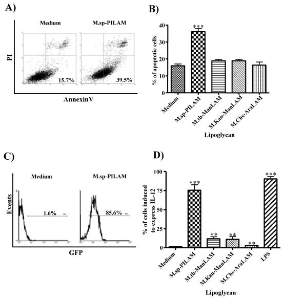
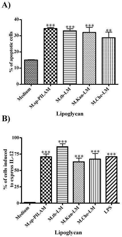
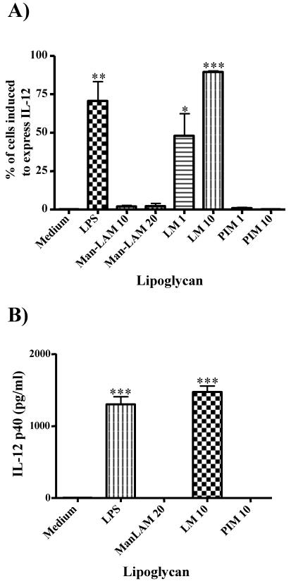
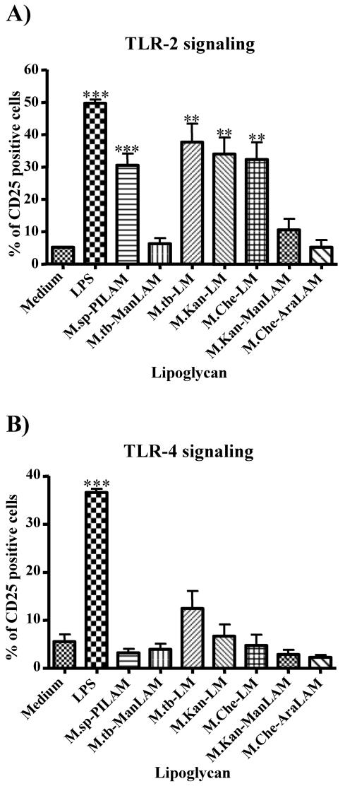
The mycobacterial cell wall component lipoarabinomannan (LAM) has been described as a virulence factor of Mycobacterium tuberculosis, and modification of the terminal arabinan residues of this compound with mannose caps (producing mannosyl-capped LAM [ManLAM]) in M. tuberculosis or with phosphoinositol caps (producing phosphoinositol-capped LAM [PILAM]) in Mycobacterium smegmatis has been implicated in various functions associated with these lipoglycans. A structure-function analysis was performed by using LAMs and their biosynthetic precursor lipomannans (LMs) isolated from different mycobacterial species on the basis of their capacity to induce the production of interleukin-12 (IL-12) and/or apoptosis of macrophage cell lines. Independent of the mycobacterial species, ManLAMs did not induce IL-12 gene expression or apoptosis of macrophages, whereas PILAMs induced IL-12 secretion and apoptosis. Interestingly, uncapped LAM purified from Mycobacterium chelonae did not induce IL-12 secretion or apoptosis. Furthermore, LMs, independent of their mycobacterial origins, were potent inducers of IL-12 and apoptosis. The precursor of LM, phosphatidyl-myo-inositol dimannoside, had no activity, suggesting that the mannan core of LM was required for the activity of LM. The specific interaction of LM with Toll-like receptor 2 (TLR-2) but not with TLR-4 suggested that these responses were mediated via the TLR-2 signaling pathway. Our experiments revealed an important immunostimulatory activity of the biosynthetic LAM precursor LM. The ratio of LAM to LM in the cell wall of mycobacteria may be an important determinant of virulence, and enzymes that modify LM could provide targets for development of antituberculosis drugs and for derivation of attenuated strains of M. tuberculosis.


Figures

FIG. 1.
FIG. 1.
Structures of the three representative families of LAM molecules isolated from different mycobacterial species and used in this study.
FIG. 2.
FIG. 2.
PILAM induces apoptosis and IL-12 gene expression in macrophages. (A) Differentiated human THP-1 cells either were not treated (left panel) or were incubated with PILAM (20 μg/ml) for 24 h and then stained with AnnexinV-Alexa488 and propidium iodide and analyzed by flow cytometry to determine the induction of apoptosis. (B) Percentage of apoptotic THP-1 cells treated with different LAM molecules which are representative of the three LAM families (ManLAM, PILAM, and AraLAM) and were isolated from various mycobacterial species. The percentage of apoptotic cells was determined as the number of AnnexinV-Alexa488-positive and propidium iodide-negative cells in a population of 10,000 cells analyzed by flow cytometry. M.sp.-PILAM, Mycobacterium sp. PILAM; M.tb-ManLAM, M. tuberculosis ManLAM; M.Kan-ManLAM, M. kansasii ManLAM; M.Che-AraLAM, M. chelonae AraLAM. (C) RAW cells transfected with an IL-12p40 gene promoter-GFP gene fusion plasmid (RAW/pIL-12-GFP) either were not treated (left panel) or were treated with 20 μg of PILAM per ml (right panel) for 16 h. Activation of the IL-12p40 promoter was determined by flow cytometry analysis of 5,000 events per condition, and the percentage of GFP-positive cells is indicated. (D) Induction of IL-12 expression by different lipoglycans analyzed as described above. The percentages of cells induced to express IL-12 are indicated. For panels B and D the means and standard deviations for three to five experiments are shown, and statistical analyses were performed by using the unpaired t test to compare treated cells to untreated cells; three asterisks indicate that the P value is <0.001, and two asterisks indicate that the P value is <0.01.
FIG. 3.
FIG. 3.
LM induces apoptosis and IL-12 gene expression in macrophages. (A) Differentiated THP-1 cells were incubated with either PILAM (20 μg/ml) or with LMs isolated from M. tuberculosis, M. kansasii, and M. chelonae (10 μg/ml) for 24 h. The percentage of apoptosis was determined by flow cytometry analysis of 10,000 events after cells were stained with AnnexinV and propidium iodide. (B) RAW/pIL-12-GFP cells were incubated with wither PILAM (20 μg/ml), LMs (10 μg/ml), or LPS (100 ng/ml) for 16 h, and flow cytometry was used to determine the induction of GFP-positive cells in 5,000 events. The results are the means of three independent experiments. A statistical analysis was performed as described in the legend to Fig. 2; three asterisks indicate that the P value is <0.001, and two asterisks indicate that the P value is <0.01. M.sp.-PILAM, Mycobacterium sp. PILAM; M.tb-LM, M. tuberculosis LM; M.Kan-LM, M. kansasii LM; M.Che-LM, M. chelonae LM.
FIG. 4.
FIG. 4.
PIM2 does not induce IL-12 gene expression in macrophages. (A) RAW/p-IL-12-GFP cells were incubated with either LPS (100 ng/ml), ManLAM (10 or 20 μg/ml), LM (1 or 10 μg/ml), or PIM2 (PIM) (1 or 10 μg/ml) (all isolated from M. tuberculosis) for 16 h, and IL-12 gene induction was measured by flow cytometry analysis as described in the legend to Fig. 2. (B) Bone marrow-derived murine macrophages were incubated with lipoglycans as described above for 24 h, and the secretion of IL-12 into the supernatant was analyzed by an ELISA. The results are the means of three independent experiments, and a statistical analysis was performed as described in the legend to Fig. 2; three asterisks indicate that the P value is <0.001, two asterisks indicate that the P value is <0.01, and one asterisk indicates that the P value is <0.05.
FIG. 5.
FIG. 5.
LM activates cells in a TLR-2-dependent manner. CHO/CD14/TLR-2 (A) and CHO/CD14/TLR-4 (B) reporter cell lines were incubated either with LAM (20 μg/ml), with LMs (10 μg/ml) isolated from various mycobacterial species, or with LPS (1 μg/ml) for 16 h. Cellular activation was measured by determining the expression of CD25 at the cell surface by using anti-CD25 monoclonal antibody and flow cytometry analysis. The percentage of CD25-positive cells was determined, and the averages of three independent experiments are shown. A statistical analysis was performed as described in the legend to Fig. 2; three asterisks indicate that the P value is <0.001, and two asterisks indicate that the P value is <0.01. M.sp.-PILAM, Mycobacterium sp. PILAM; M.tb-ManLAM, M. tuberculosis ManLAM; M.tb-LM, M. tuberculosis LM; M.Kan-LM, M. kansasii LM; M.Che-LM, M. chelonae LM; M.Kan-ManLAM, M. kansasii ManLAM; M.Che-AraLAM, M. chelonae AraLAM.

References

    1. Aliprantis, A. O., R. B. Yang, M. R. Mark, S. Suggett, B. Devaux, J. D. Radolf, G. R. Klimpel, P. Godowski, and A. Zychlinsky. 1999. Cell activation and apoptosis by bacterial lipoproteins through toll-like receptor-2. Science 285:736-739. - PubMed
    1. Altare, F., A. Durandy, D. Lammas, J. F. Emile, S. Lamhamedi, F. Le Deist, P. Drysdale, E. Jouanguy, R. Doffinger, F. Bernaudin, O. Jeppsson, J. A. Gollob, E. Meinl, A. W. Segal, A. Fischer, D. Kumararatne, and J. L. Casanova. 1998. Impairment of mycobacterial immunity in human interleukin-12 receptor deficiency. Science 280:1432-1435. - PubMed
    1. Brennan, P. J. 2003. Structure, function, and biogenesis of the cell wall of Mycobacterium tuberculosis. Tuberculosis (Edinb.) 83:91-97. - PubMed
    1. Brightbill, H. D., D. H. Libraty, S. R. Krutzik, R. B. Yang, J. T. Belisle, J. R. Bleharski, M. Maitland, M. V. Norgard, S. E. Plevy, S. T. Smale, P. J. Brennan, B. R. Bloom, P. J. Godowski, and R. L. Modlin. 1999. Host defense mechanisms triggered by microbial lipoproteins through toll-like receptors. Science 285:732-736. - PubMed
    1. Celada, A., P. W. Gray, E. Rinderknecht, and R. D. Schreiber. 1984. Evidence for a gamma-interferon receptor that regulates macrophage tumoricidal activity. J. Exp. Med. 160:55-74. - PMC - PubMed

Publication types

LinkOut - more resources