. 2004 Apr;72(4):2101-10.
doi: 10.1128/IAI.72.4.2101-2110.2004.

Inhibition of major histocompatibility complex II expression and antigen processing in murine alveolar macrophages by Mycobacterium bovis BCG and the 19-kilodalton mycobacterial lipoprotein

Affiliations

Inhibition of major histocompatibility complex II expression and antigen processing in murine alveolar macrophages by Mycobacterium bovis BCG and the 19-kilodalton mycobacterial lipoprotein

Scott A Fulton et al. Infect Immun. 2004 Apr.

Abstract

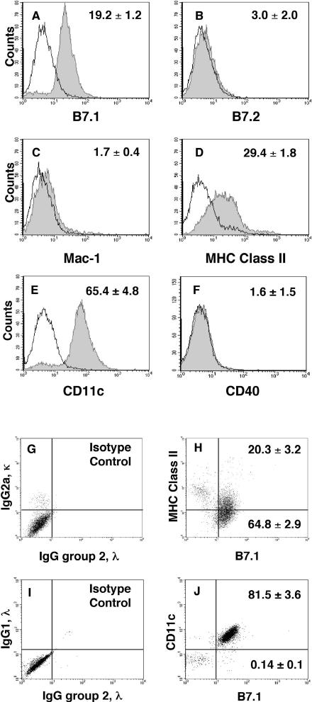
Alveolar macrophages constitute a primary defense against Mycobacterium tuberculosis, but they are unable to control M. tuberculosis without acquired T-cell immunity. This study determined the antigen-presenting cell function of murine alveolar macrophages and the ability of the model mycobacterium, Mycobacterium bovis BCG, to modulate it. The majority (80 to 85%) of alveolar macrophages expressed both CD80 (B7.1) and CD11c, and 20 to 30% coexpressed major histocompatibility complex II (MHC-II). Gamma interferon (IFN-gamma) enhanced MHC-II but not B7.1 expression. Naive or IFN-gamma-treated alveolar macrophages did not express CD86 (B7.2), CD11b, Mac-3, CD40, or F4/80. M. bovis BCG and the 19-kDa mycobacterial lipoprotein inhibited IFN-gamma-regulated MHC-II expression on alveolar macrophages, and inhibition was dependent on Toll-like receptor 2. The inhibition of MHC-II expression by the 19-kDa lipoprotein was associated with decreased presentation of soluble antigen to T cells. Thus, susceptibility to tuberculosis may result from the ability of mycobacteria to interfere with MHC-II expression and antigen presentation by alveolar macrophages.


Figures

FIG. 1.
FIG. 1.
Resident alveolar macrophages express CD80 (B7.1), CD11c, and MHC-II. (A to F) Alveolar macrophages were isolated from 8- to 10-week-old female C57BL/6 mice and stained as described in Materials and Methods. A total of 5,000 events per sample was analyzed by flow cytometry. Histograms show both specific staining (shaded) and isotype control staining (unshaded) for CD80 (B7.1), CD86 (B7.2), CD11b (Mac-1), CD11c, CD40, and MHC-II. Data shown on the panels are the mean specific MFIs (± SEM) from five independent experiments demonstrating the expression of B7.1, MHC-II, and CD11c (for FcRIII/II, MFI = 16.9 ± 7.1; data not shown). B7.2, Mac-1, and CD40 were not detected. (G to J) Dot blots show coexpression of MHC-II and CD11c on B7.1-expressing alveolar macrophages. The mean percentages (± SEM) of total cells expressing B7.1 and coexpressing MHC-II and CD11c from five independent experiments are shown on the panels inserts. Staining with isotype control antibodies was less than 2 to 3% of the total events detected in any single quadrant.
FIG. 2.
FIG. 2.
IFN-γ enhances expression of MHC-II on alveolar macrophages. (A to F) Alveolar macrophages were isolated from mice and cultured at the indicated times in the presence (shaded) or absence (dark black line; unshaded) of murine IFN-γ (2 ng/ml). Cells were stained for MHC-II and B7.1 as described, and histograms from a representative experiment (n = 5) are shown. Specific staining in the absence and presence of IFN-γ are shown and compared to isotype control staining. In panel G, the mean specific geoMFI (± SEM) for MHC-II from five experiments is shown. The Wilcoxon rank sum test was used to compare MHC-II expression between control and IFN-γ-stimulated alveolar macrophages. Statistically significant differences in specific geoMFI are designated by an asterisk (P ≤ 0.05).
FIG. 3.
FIG. 3.
Enhanced coexpression of MHC-II on IFN-γ-treated B7.1+ alveolar macrophages. (A to F) Alveolar macrophages were isolated, cultured at the indicated times with IFN-γ (2 ng/ml) or without IFN-γ and then stained for B7.1 and MHC-II. A total of 5,000 to 7,500 events were collected for two-color analysis and representative dot blots are shown. Quadrant settings were used to calculate the distribution of cell types within the entire (ungated) alveolar macrophages population. The levels of expression of MHC-II and B7.1 from six different experiments are summarized and shown on panels (mean % total ± SEM) for upper- and lower-right quadrants. Isotype control staining was less than 2 to 3% in each quadrant.
FIG. 4.
FIG. 4.
Phagocytosis of M. bovis BCG by alveolar macrophages. Alveolar macrophages were incubated overnight with fluos-BCG at a low MOI (3 to 5) and analyzed by flow cytometry. (A) A representative histogram shows phagocytosis of fluos-BCG. (B) MHC-II and B7.1 expression on fluos-BCG-positive cells (region R1, panel A) from a representative experiment reveals similar uptake of bacteria by both MHC-II+ and MHC-II B7.1+ alveolar macrophages.
FIG. 5.
FIG. 5.
M. bovis BCG infection and a 19-kDa mycobacterial lipoprotein inhibit IFN-γ-inducible MHC-II expression on alveolar macrophages. (A to D) Alveolar macrophages were cultured with or without BCG at an MOI of 3 to 5 and with or without the 19-kDa lipoprotein (250 ng/ml) for 24 h. IFN-γ (2 ng/ml) was added after infection or pretreatment with the 19-kDa lipoprotein, and alveolar macrophages were cultured for an additional 24, 48, and 72 h in the presence of IFN-γ. The expression of MHC-II was measured by flow cytometry, and histograms representative of four independent experiments are shown. IFN-γ-induced MHC-II expression on BCG-infected or 19-kDa-lipoprotein-treated macrophages (black lines) is compared to control treated cells (shaded) after 48 and 72 h of IFN-γ exposure. Isotype control staining for both treatment groups is shown with overlapping thin lines. (E to F) Alveolar macrophages were isolated, cultured overnight, and then treated without (medium) or with the 19-kDa lipoprotein (500 ng/ml) for 24 h. After 24 h, fresh 19-kDa lipoprotein was added along with IFN-γ (2 ng/ml), and cells were incubated for an additional 48 h. CIITA and MHC-II mRNA expression was measured by real-time PCR and normalized to GAPDH expression. Data from a representative experiment (n = 3) are shown. Decreases in CIITA and MHC-II mRNA expression were analyzed by using a paired Student's t test, and significant differences are designated with asterisks (P ≤ 0.05).
FIG. 6.
FIG. 6.
The 19-kDa mycobacterial lipoprotein inhibits antigen processing and presentation in alveolar macrophages. Alveolar macrophages were harvested, plated in duplicate in 96-well flat bottom plates (65,000 cells/well), and incubated overnight. Adherent cells were washed and incubated for 24 h with or without 19-kDa lipoprotein. Next, cells received IFN-γ (2 ng/ml) in fresh medium (controls) or medium supplemented with fresh 19-kDa lipoprotein. After 48 h, cells were washed extensively and pulsed for 2 h with ovalbumin, and antigen presentation was measured 24 h later as described. Ovalbumin-specific DOBW responses were measured, and mean (± standard deviation) DOBW responses from a representative experiment (n = 3) are shown. Percent inhibition was calculated as the reduction in the OD550 measured by the CTLL assay for IL-2 (see Materials and Methods). Statistically significant inhibition (P ≤ 0.05) of ovalbumin processing was measured at each antigen dose by using a paired Student's t test.
FIG. 7.
FIG. 7.
Inhibition of MHC-II expression and antigen processing by the 19-kDa lipoprotein is dependent on TLR-2. (A to B) Alveolar macrophages were isolated from age-matched C57BL/6 wild-type and C57BL/6 TLR-2−/− mice. After overnight incubation with (dark lines) or without (shaded) the 19-kDa lipoprotein (250 ng/ml), alveolar macrophages were treated for 48 h with IFN-γ (2 ng/ml) and then analyzed for surface MHC-II expression. Histograms representative of three different experiments are shown. (C) Alveolar macrophages were isolated from wild-type C57BL/6 and TLR-2 −/− mice, pretreated with the 19-kDa lipoprotein or medium, and then incubated for 48 h with IFN-γ with or without fresh 19-kDa lipoprotein or medium. Next, cells were pulsed with ovalbumin, and DOBW responses were analyzed as described in the legend of Fig. 6. Mean (± standard deviation) DOBW responses for wild-type alveolar macrophages and TLR-2 −/− alveolar macrophages pretreated with medium or 19-kDa lipoprotein from a representative experiment (n = 3) are shown. By a paired Student's t test, statistically significant inhibition (P ≤ 0.05) was detected at each ovalbumin dose in wild-type cells pretreated with the 19-kDa lipoprotein. Antigen processing and presentation were not inhibited in TLR-2 −/− cells pretreated with the 19-kDa lipoprotein.

References

    1. Beatty, W. L., E. R. Rhoades, H. J. Ullrich, D. Chatterjee, J. E. Heuser, and D. G. Russell. 2000. Trafficking and release of mycobacterial lipids from infected macrophages. Traffic 1:235-247. - PubMed
    1. Brightbill, H. D., D. H. Libraty, S. R. Krutzik, R. B. Yang, J. T. Belisle, J. R. Bleharski, M. Maitland, M. V. Norgard, S. E. Plevy, S. T. Smale, P. J. Brennan, B. R. Bloom, P. J. Godowski, and R. L. Modlin. 1999. Host defense mechanisms triggered by microbial lipoproteins through toll-like receptors. Science 285:732-736. - PubMed
    1. Chensue, S. W., K. Warmington, J. Ruth, P. Lincoln, M. C. Kuo, and S. L. Kunkel. 1994. Cytokine responses during mycobacterial and schistosomal antigen-induced pulmonary granuloma formation. Production of Th1 and Th2 cytokines and relative contribution of tumor necrosis factor. Am. J. Pathol. 145:1105-1113. - PMC - PubMed
    1. Constant, S. L., K. S. Lee, and K. Bottomly. 2000. Site of antigen delivery can influence T cell priming: pulmonary environment promotes preferential Th2-type differentiation. Eur. J. Immunol. 30:840-847. - PubMed
    1. Cooper, A. M., J. E. Callahan, M. Keen, J. T. Belisle, and I. M. Orme. 1997. Expression of memory immunity in the lung following re-exposure to Mycobacterium tuberculosis. Tuber. Lung Dis. 78:67-73. - PubMed

Publication types