. 2004 Apr;72(4):2229-39.
doi: 10.1128/IAI.72.4.2229-2239.2004.

Role of extracellular phospholipases and mononuclear phagocytes in dissemination of cryptococcosis in a murine model

Affiliations

Role of extracellular phospholipases and mononuclear phagocytes in dissemination of cryptococcosis in a murine model

Rosemary Santangelo et al. Infect Immun. 2004 Apr.

Abstract

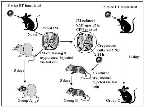
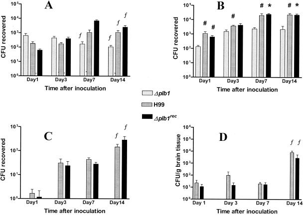
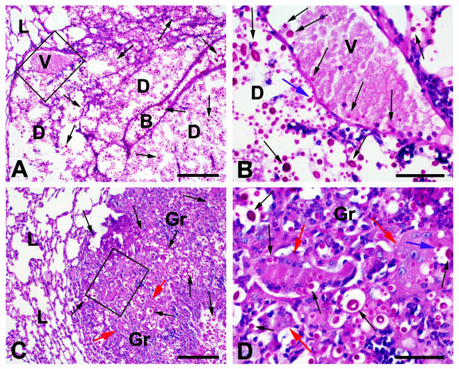
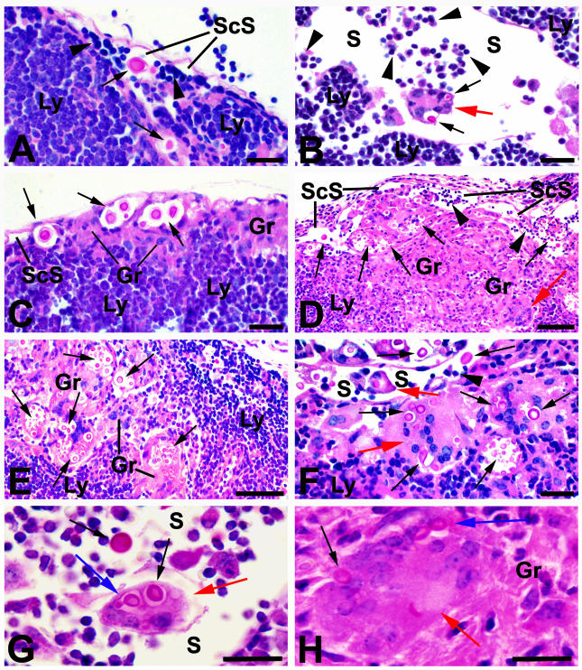
Secreted phospholipase B (PLB) activity promotes the survival and replication of Cryptococcus neoformans in macrophages in vitro. We therefore investigated the role of mononuclear phagocytes and cryptococcal PLB in the dissemination of infection in a mouse model, using C. neoformans var. grubii wild-type strain H99, a PLB1 deletion mutant (Delta plb1), and a reconstituted strain (Delta plb1(rec)). PLB facilitated the entry of endotracheally administered cryptococci into lung IM. PLB was also required for lymphatic spread from the lung to regional lymph nodes and for entry into the blood. Langhans-type giant cells containing budding cryptococci were seen free in the lymphatic sinuses of hilar nodes of H99- and Delta plb1(rec)-infected mice, suggesting that they may have a role in the dissemination of cryptococcal infection. The transfer of infected lung macrophages to recipient mice by tail vein injections demonstrated that these cells can facilitate hematogenous dissemination of cryptococci to the brain, independent of cryptococcal PLB secretion. PLB activities of cryptococci isolated from lung macrophages or infected brains were not persistently increased. We conclude that mononuclear phagocytes are a vehicle for cryptococcal dissemination and that PLB activity is necessary for the initiation of interstitial pulmonary infections and for dissemination from the lung via the lymphatics and blood. PLB is not, however, essential for the establishment of neurological infections when cryptococci are presented within, or after passage through, mononuclear phagocytes.


Figures

FIG. 1.
FIG. 1.
Experimental protocol comparing the effects on virulence and phospholipase activity of inoculation with cryptococci inside macrophages or cultured from them. ET, endotracheal; IM, interstitial lung macrophages. BL-1a and Δplb1 strains were tested separately with this protocol.
FIG. 2.
FIG. 2.
Kinetics of appearance of cryptococcal CFU in AM (A), IM (B), PBM (C), and brain tissues (D). Organs and blood were harvested from mice after endotracheal inoculation with 3 × 10⁷ cells of the H99, Δplb1rec, and Δplb1 cryptococcal strains. Note that a log scale was used to report CFU. Data represent the means ± standard errors of the means of three experiments (two for day 14) using three mice for each strain per time point. Statistics were calculated by combining data from all experiments. No CFU were recovered from blood monocytes or brain tissues from mice inoculated with Δplb1. Significant differences from Δplb1 are noted as follows: #, P < 0.05; *, P < 0.005 by the paired two-tail t test; #*, P < 0.05 by the Mann-Whitney U test, except for H99 on day 7, for which P = 0.06. f, significantly different from day 1 (P < 0.05 by the unpaired two-tail t test and the Mann-Whitney U test).
FIG. 3.
FIG. 3.
PAS-stained paraffin sections of lung tissue harvested from mice 14 days after infection with H99. Cryptococci were evident as strongly PAS-positive round cells (black arrows), often surrounded by a capsule and occasionally budding (blue arrows). (A) Although the alveolar structure of the lung (L) could be identified in many sites, this was lost in areas containing large numbers of extracellular cryptococci, which apparently caused tissue destruction (D). Major structures such as bronchioles (B) were often retained. (B) At a higher magnification of areas showing tissue destruction, occasional cryptococci within blood vessels (V) were noted. (C) Areas of granulomatous reaction (Gr) containing organisms within epithelioid macrophages and Langhans-type giant cells were also seen (red arrows). (D) At a higher magnification, budding cryptococci (blue arrows) were noted in some giant cells. Bars = 150 μm for panels A and C and 60 μm for panels B and D.
FIG. 4.
FIG. 4.
PAS-stained sections of hilar and cervical lymph nodes collected from mice 14 days after infection with strain H99. (A) The lymph node stroma (Ly), heavily infiltrated with lymphocytes, is readily apparent and is separated from subcapsular lymphatic sinuses (ScS) containing recirculating lymphocytes (arrowheads) by a layer of endothelium. Cryptococci (black arrows) were often present free within the subcapsular and other lymphatic spaces (S). (B) Cryptococci were also found in Langhans-type giant cells (red arrows), some of which were free within the lymphatic spaces (S) in panels B, F, and G. (C) Small granulomas (Gr) comprised of epithelioid macrophages with phagocytosed cryptococci were present in subcapsular sites in some areas. In others, these granulomatous lesions occupied large portions of the peripheral lymph node tissues (D) as well as more central zones of nodes (E). Langhans-type giant cells (red arrows) containing cryptococci were also found among the epithelioid macrophages in these lesions (D, F, and H). Budding cryptococci (blue arrows) were present in Langhans-type giant cells (red arrows), free within lymphatic sinuses (G), and within granulomas (H). Bars = 50 μm for panels A, B, C, and F; 100 μm for panels D and E; and 25 μm for panels G and H.
FIG. 5.
FIG. 5.
PAS-stained paraffin sections of brain tissue collected from mice after 14 days of infection with H99. Cryptococci appear as strongly PAS-positive cells (black arrows), often surrounded by a capsule and occasionally budding (blue arrows). (A and B) Brain tissue (Br) contained foci of cryptococci; organisms were also found in capillaries (Cp). (C) Venules (V) containing cryptococci were present within the brain tissue, as well as on the surface (D). Bars = 50 μm.

References

    1. Casadevall, A., and J. R. Perfect. 1998. Cryptococcus neoformans. ASM Press, Washington, D.C.
    1. Chen, S. C. A., M. Muller, J. Z. Zhou, L. C. Wright, and T. C. Sorrell. 1997. Phospholipase activity in Cryptococcus neoformans: a new virulence factor? J. Infect. Dis. 175:414-420. - PubMed
    1. Chen, S. C. A., L. C. Wright, R. T. Santangelo, M. Muller, V. R. Moran, P. W. Kuchel, and T. C. Sorrell. 1997. Identification of extracellular phospholipase B, lysophospholipase, and acyltransferase produced by Cryptococcus neoformans. Infect. Immun. 65:405-411. - PMC - PubMed
    1. Chrétien, F., O. Lortholary, I. Kansau, S. Neuville, F. Gray, and F. Dromer. 2002. Pathogenesis of cerebral Cryptococcus neoformans infection after fungemia. J. Infect. Dis. 186:522-530. - PubMed
    1. Cox, G. M., H. C. Mc Dade, S. C. Chen, S. C. Tucker, M. Gottfredsson, L. C. Wright, T. C. Sorrell, S. D. Leidich, A. Casadevall, M. A. Ghannoum, and J. R. Perfect. 2001. Extracellular phospholipase activity is a virulence factor for Cryptococcus neoformans. Mol. Microbiol. 39:166-175. - PubMed

Publication types

MeSH terms

LinkOut - more resources