. 2004 Apr;72(4):2240-7.
doi: 10.1128/IAI.72.4.2240-2247.2004.

Probiotic bifidobacteria protect mice from lethal infection with Shiga toxin-producing Escherichia coli O157:H7

Affiliations

Probiotic bifidobacteria protect mice from lethal infection with Shiga toxin-producing Escherichia coli O157:H7

Takashi Asahara et al. Infect Immun. 2004 Apr.

Abstract

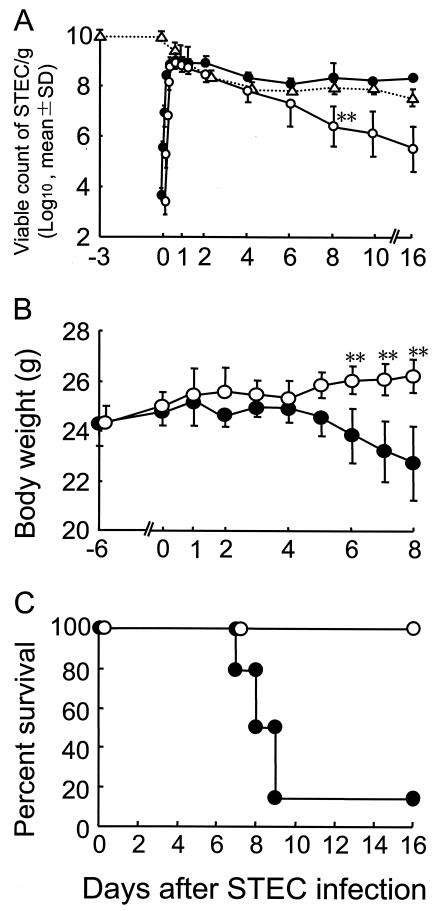
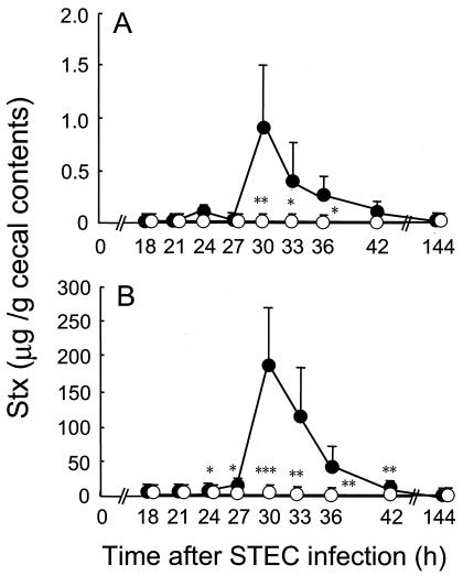
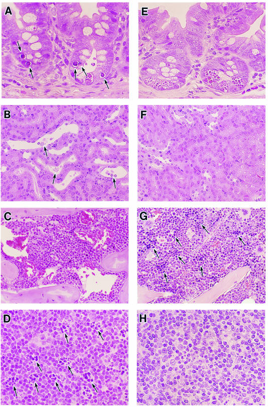
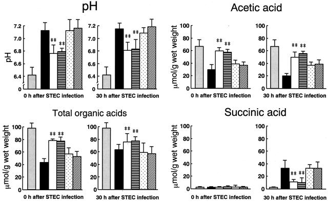
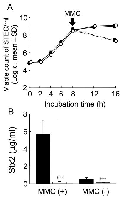
The anti-infectious activity of probiotic Bifidobacteria against Shiga toxin-producing Escherichia coli (STEC) O157:H7 was examined in a fatal mouse STEC infection model. Stable colonization of the murine intestines was achieved by the oral administration of Bifidobacterium breve strain Yakult (naturally resistant to streptomycin sulfate) as long as the mice were treated with streptomycin in their drinking water (5 mg/ml). The pathogenicity of STEC infection, characterized by marked body weight loss and subsequent death, observed in the infected controls was dramatically inhibited in the B. breve-colonized group. Moreover, Stx production by STEC cells in the intestine was almost completely inhibited in the B. breve-colonized group. A comparison of anti-STEC activity among several Bifidobacterium strains with natural resistance to streptomycin revealed that strains such as Bifidobacterium bifidum ATCC 15696 and Bifidobacterium catenulatum ATCC 27539(T) did not confer an anti-infectious activity, despite achieving high population levels similar to those of effective strains, such as B. breve strain Yakult and Bifidobacterium pseudocatenulatum DSM 20439. The effective strains produced a high concentration of acetic acid (56 mM) and lowered the pH of the intestine (to pH 6.75) compared to the infected control group (acetic acid concentration, 28 mM; pH, 7.15); these effects were thought to be related to the anti-infectious activity of these strains because the combination of a high concentration of acetic acid and a low pH was found to inhibit Stx production during STEC growth in vitro.


Figures

FIG. 1.
FIG. 1.
Inhibition of lethal intestinal STEC infection by B. breve colonization in SM-treated mice. SM sulfate at a concentration of 5 mg/ml in drinking water was given to 28 mice from day −6 until day 16. B. breve strain Yakult (1 × 10⁸ to 3 × 10⁸ CFU/mouse/day) in 0.1 ml of saline was administered to half of the mice once a day from day −5 to −3, and the other half of the mice were administered saline on the same schedule as that for the B. breve treatment. Mice were infected orally with STEC (5 × 10³ CFU) on day 0 and then treated with MMC at an inoculum dose of 0.25 mg/kg of body weight three times at 18, 21, and 24 h after the STEC infection. (A) Feces for bacteriological analysis were obtained from 6 randomly selected mice in each group on days 0 (at 3, 6, 9, 12, 15, and 18 h), 1, 3, 4, 7, 10, and 16 after the STEC infection, with the exception of the control group on days 10 to 16 (n = 2). Viable counts of STEC and B. breve were examined as described in the text. Symbols: •, number of STEC organisms in the STEC-infected control mice; ○, number of STEC organisms in the B. breve-treated mice; ▵, number of B. breve organisms in B. breve-treated mice. (B) All 14 mice in each group were weighed every day until day 8. Symbols: •, STEC-infected control; ○, B. breve-treated mice. (C) The STEC-infected control mice (•) and B. breve-treated mice (○) were observed for survival for 14 days after the challenge infection. **, a significant difference was observed between the B. breve-treated and the untreated control groups (P < 0.01).
FIG. 2.
FIG. 2.
Inhibition of MMC-induced production of Shiga toxins by B. breve colonization. Mice were infected with STEC and treated with MMC as described in the legend to Fig. 1 and then dissected at the indicated periods after STEC infection to examine Stx production. The concentrations of Stx1 (A) and Stx2 (B) in the intestinal contents were determined by RPLA test as described in the text. The results were expressed as the means and standard deviations of the results from 6 mice. Significant differences in Stx concentration were observed between the B. breve-treated and the untreated control groups (*, P < 0.05; **, P < 0.01; ***, P < 0.001).
FIG. 3.
FIG. 3.
Histopathological analysis. Hematoxylin and eosin staining of the ileum (A and E), kidney (B and F), bone marrow (C and G), and mesenteric lymph node (D and H) from a mouse in the STEC-infected control group (STEC inoculum, 3.8 × 10³ CFU) (A to D) and a mouse in the B. breve-treated group (E to H). Organs were obtained on day 2 (A and E) or 7 (B to D and F to H) after STEC infection. Black arrows: panel A, changes suggestive of apoptosis; panel B, necrotic tubular endothelial cells with distention; panel D, changes suggestive of apoptotic bodies; panel G, erythroblasts. Magnifications in both groups: ileum, ×520; kidney, ×520; bone marrow, ×260; mesenteric lymph node, ×520.
FIG. 4.
FIG. 4.
Changes in intestinal pH and concentrations of organic acids after STEC infection in SM-treated mice. Mice were treated as shown in Table 2. Cecal contents were obtained from mice both at the time of STEC infection (0 h) and 30 h after STEC infection. pH and organic acid concentrations were determined as described in the text. Results are expressed as the means and standard deviations of the results from 6 mice. Columns: grey, nontreated healthy mice; black, SM-treated mice; white, B. breve strain Yakult-treated mice; hatched, B. pseudocatenulatum DSM20439-treated mice; slashed, B. bifidum ATCC 15696-treated mice; vertically lined, B. catenulatum ATCC 27539T-treated mice. **, significant differences are shown for the Bifidobacterium-treated groups versus the untreated control group (P < 0.01).
FIG. 5.
FIG. 5.
Inhibition of Stx production but not STEC proliferation at higher AA concentrations and lower pH values in vitro. The pH and concentration of AA were adjusted in the growth medium so that the conditions were the same as those in the control cecum (•) (pH 7.15; AA concentration, 28 mM) or the B. breve-colonized cecum (○) (pH 6.75; AA concentration, 56 mM). STEC was added to each medium at a final concentration of 105 CFU/ml and cultivated at 37°C for 8 h. Cultures were then divided into two groups, and either 20 μl of fresh medium or MMC at a final concentration of 1 μg/ml in 20 μl of medium was added to each group, and the tubes were incubated for an additional 8 h. (A) Viable bacterial counts were determined at the indicated periods during incubation. The straight line and the dotted line show growth without (−) and with (+) MMC, respectively. (B) Stx2 concentrations were determined after incubation for 16 h. Columns: black, control, white, B. breve. Results are expressed as the means and standard deviations of the results from triplicate cultures. ***, significant differences are shown for growth under the B. breve colonization conditions versus growth under control conditions (P < 0.001).

References

    1. Abe, H., I. Tatsuno, T. Tobe, A. Okutani, and C. Sasakawa. 2002. Bicarbonate ion stimulates the expression of locus of enterocyte effacement-encoded genes in enterohemorrhagic Escherichia coli O157:H7. Infect. Immun. 70:3500-3509. - PMC - PubMed
    1. Aiba, Y., H. Ishikawa, K. Shimizu, S. Noda, Y. Kitada, M. Sasaki, and Y. Koga. 2002. Role of internalization in the pathogenicity of Shiga toxin-producing Escherichia coli infection in gnotobiotic murine model. Microbiol. Immunol. 46:723-731. - PubMed
    1. Asahara, T., K. Shimizu, K. Nomoto, M. Watanuki, and R. Tanaka. 2001. Antibacterial effect of fermented milk containing Bifidobacterium breve, Bifidobacterium bifidum and Lactobacillus acidophilus against indigenous Escherichia coli infection in mice. Microb. Ecol. Health Dis. 13:16-24.
    1. Asahara, T., K. Nomoto, K. Shimizu, M. Watanuki, and R. Tanaka. 2001. Increased resistance of mice to Salmonella enterica serovar Typhimurium infection by synbiotic administration of Bifidobacteria and transgalactosylated oligosaccharides. J. Appl. Microbiol. 91:985-996. - PubMed
    1. Benjamin, M. M., and A. R. Datta. 1995. Acid tolerance of enterohemorrhagic Escherichia coli. Appl. Environ. Microbiol. 61:1669-1672. - PMC - PubMed

MeSH terms