. 2004 Apr;72(4):2338-49.
doi: 10.1128/IAI.72.4.2338-2349.2004.

Cytokine and inducible nitric oxide synthase mRNA expression during experimental murine cryptococcal meningoencephalitis

Affiliations

Cytokine and inducible nitric oxide synthase mRNA expression during experimental murine cryptococcal meningoencephalitis

Claudia M L Maffei et al. Infect Immun. 2004 Apr.

Abstract

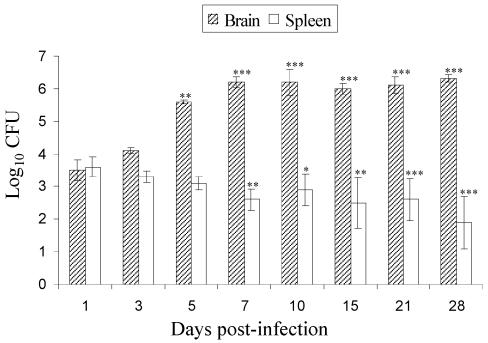
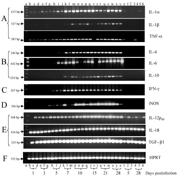
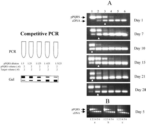
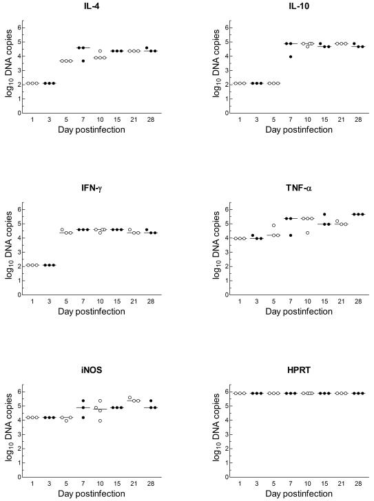
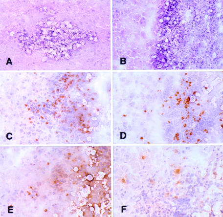
The immune events that take place in the central nervous system (CNS) during cryptococcal infection are incompletely understood. We used competitive reverse transcription-PCR to delineate the time course of the local expression of mRNAs encoding a variety of cytokines and inducible nitric oxide synthase (iNOS) during progressive murine cryptococcal meningoencephalitis and assessed the CNS inflammatory response using immunohistochemistry. Interleukin 18 (IL-18), transforming growth factor beta1, and IL-12p(40) mRNAs were constitutively expressed in the brains of infected and uninfected mice; IL-2 mRNA was not detected at any time. Increased levels of transcripts corresponding to IL-1 alpha, tumor necrosis factor alpha (TNF-alpha), and iNOS were detected as early as day 1 postinfection, with TNF-alpha rising by approximately 30-fold and iNOS increasing by approximately 5-fold by day 7. Each remained at these levels thereafter. IL-4, IL-6, and gamma interferon transcripts were detected on day 5, and IL-1 beta and IL-10 transcripts were detected beginning on day 7. Once detected, each remained at a relatively constant level through 28 days of infection. This cytokine profile does not suggest a polarized Th1 or Th2 response. Immunohistochemistry did not reveal inflammatory infiltrates before day 7, despite the presence of cryptococci. Intraparenchymal abscesses with inflammatory cells in their peripheries were found beginning on day 10. The infiltrates were comprised primarily of cells expressing CD4, CD8, or CD11b; low numbers of cells expressing CD45R/B220 were also present. The persistence of Cryptococcus observed in the CNS may result from an ineffective immune response, perhaps owing to an insufficient anticryptococcal effector function of endogenous glial cells resulting from competing pro- and anti-inflammatory cytokines. These data detail the immune response in the brain and could be important for the future design of specific immunomodulatory therapies for this important opportunistic infection.


Figures

FIG. 1.
FIG. 1.
Yeast burdens in brains and spleens of BALB/c mice after intravenous infection with 4 × 10⁴ cells of C. neoformans (isolate 9759). The data are expressed as log10 geometric mean CFU per organ ± 95% confidence interval (n = 5 mice/time). The differences between brain and spleen at each time compared to day 1 postinfection were statistically significant as indicated (*, P < 0.05; **, P < 0.01; ***, P < 0.001).
FIG. 2.
FIG. 2.
Temporal expression of cytokine, iNOS, and HPRT mRNAs in brains of uninfected BALB/c mice (lanes 1 to 6) and BALB/c mice infected with 4 × 10⁴ cells of C. neoformans (lanes b to z). Total RNA was extracted from half of each brain from uninfected mice on days 5 and 28 and from infected mice on days 1, 3, 5, 7, 10, 15, 21, and 28. The RNA samples were obtained from three or four mice per time point. RT-PCR was performed, and the PCR products were electrophoresed on 1.5% agarose gels containing 0.5 μg of ethidium bromide per ml and observed with a UV transilluminator. Lane a, molecular size marker (100-bp ladder). (A) Proinflammatory cytokines; (B) Th2 cytokines; (C) Th1 cytokines; (D) inducible enzyme; (E) constitutive cytokines; (F) internal control.
FIG. 3.
FIG. 3.
Competitive PCR. (Left panel) Schematic of a typical PCR experiment (described in Materials and Methods). (A) Titration of iNOS mRNA transcripts in the brains of infected BALB/c mice sampled at different times postinfection. A constant amount of experimental cDNA was titrated against sequential dilutions of linear pPQRS plasmid competitor during the PCR amplification. Lane 1, 3.8 × 10⁵ copies of plasmid DNA/μl; lane 2, 7.7 × 10⁴ copies of plasmid DNA/μl; lane 3, 1.5 × 10⁴ copies of plasmid DNA/μl; lane 4, 3.1 × 10³ copies of plasmid DNA/μl; lane 5, 6.2 × 10² copies of plasmid DNA/μl; lane 6, 1.2 × 10² copies of plasmid DNA/μl. The arrowheads indicate the points of equivalence in copy numbers of the two different PCR products. The sizes of the PCR products were 390 bp for pPQRS iNOS and 306 bp for iNOS. (B) Example of the reproducibility of the pPQRS methodology for the expression of iNOS transcripts in three mice (a, b, and c) at the same time postinfection. The lanes are the same as for panel A.
FIG. 4.
FIG. 4.
Quantification of DNA copies of cytokines (IFN-γ, TNF-α, IL-4, and IL-10), iNOS, and HPRT during the course of cerebral cryptococcal infection in BALB/c mice, determined by competitive PCR as described in Materials and Methods. The symbols represent individual mice, and the bars represent the medians for the time points examined. On days 1 and 3, IFN-γ, IL-4, and IL-10 were not detected, and IL-10 was not detected on day 5. The statistical significance of differences during the evolution of infection and the correlation among the cytokines are discussed in the text. Values of 2 indicate that no product was detected, and the lower limit of sensitivity of the assay was ∼100 copies.
FIG. 5.
FIG. 5.
(A and B) Cryosections of typical intraparenchymal cryptococcal abscesses from day 10 (A) and day 21 (B) postinfection (hematoxylin and eosin staining). (C to F) Adjacent serial cryosections (immunoperoxidase with hematoxylin counterstain) demonstrating the inflammatory response in a cryptococcal abscess from day 28 postinfection. Intact brain parenchyma is on the left side of the field in each panel. (C) Anti-CD4; (D) anti-CD8; (E) anti-CD11b; (F) anti-CD45R/B220. Numerous CD4+ and CD8+ lymphoid cells and CD11b+ cells are shown in the abscess. There are fewer B cells in the infiltrate in panel F. Magnifications: ×206 (A), ×171 (B), and ×294 (C to F).

References

    1. Aguirre, K., E. A. Havell, G. W. Gibson, and L. L. Johnson. 1995. Role of tumor necrosis factor and gamma interferon in acquired resistance to Cryptococcus neoformans in the central nervous system of mice. Infect. Immun. 63:1725-1731. - PMC - PubMed
    1. Almeida, G. M., R. M. Andrade, and C. A. Bento. 2001. The capsular polysaccharides of Cryptococcus neoformans activate normal CD4(+) T cells in a dominant Th2 pattern. J. Immunol. 167:5845-5851. - PubMed
    1. Ashman, R. B., E. M. Bolitho, and A. Fulurija. 1995. Cytokine mRNA in brain tissue from mice that show strain-dependent differences in the severity of lesions induced by systemic infection with Candida albicans yeast. J. Infect. Dis. 172:823-830. - PubMed
    1. Barluzzi, R., A. Brozzetti, D. Delfino, F. Bistoni, and E. Blasi. 1998. Role of the capsule in microglial cell-Cryptococcus neoformans interaction: impairment of antifungal activity but not of secretory functions. Med. Mycol. 36:189-197. - PubMed
    1. Barluzzi, R., A. Brozzetti, G. Mariucci, M. Tantucci, R. G. Neglia, F. Bistoni, and E. Blasi. 2000. Establishment of protective immunity against cerebral cryptococcosis by means of an avirulent, nonmelanogenic Cryptococcus neoformans strain. J. Neuroimmunol. 109:75-86. - PubMed

Publication types

MeSH terms