

Review
doi: 10.1038/nri1310.
Immune surveillance in the skin: mechanisms and clinical consequences
Affiliations
- PMID: 15039758
- PMCID: PMC7097017
- DOI: 10.1038/nri1310
Item in Clipboard
Review
Immune surveillance in the skin: mechanisms and clinical consequences
Nat Rev Immunol.
2004 Mar.
Abstract
The skin, as the primary interface between the body and the environment, provides the first line of defence against a broad array of microbial pathogens and trauma. In addition to its properties as a physical barrier, the skin has many active defence mechanisms. In this review, we discuss the interaction between the innate and adaptive immune systems in the skin as a model for immune function at epithelial-cell interfaces with the environment. How these mechanisms account for the robust nature of cutaneous immune surveillance and how their dysregulation drives the pathogenesis of inflammatory skin disorders and skin-based tumours are the subjects of this review.
Conflict of interest statement
The authors declare no competing financial interests.
Figures
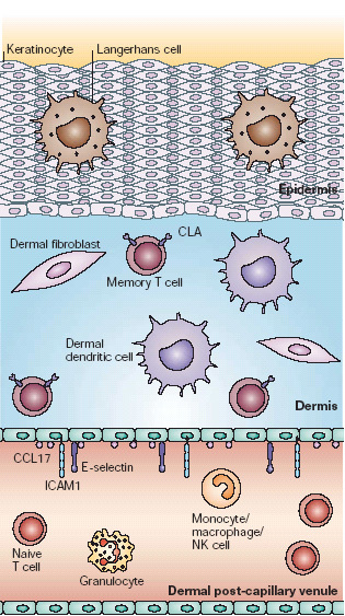
Human skin is composed of three distinct compartments relevant to its immune functions. First, the epidermis is composed of keratinized epithelial cells and functions as both a physical barrier and an early warning system. Immune cells resident in the epidermis include specialized dendritic cells (DCs) known as Langerhans cells and intraepithelial lymphocytes. Second, the dermis is mainly composed of connective tissue produced by dermal fibroblasts. Immune system cells resident in non-inflamed dermis include dermal DCs, mast cells and a small number of cutaneous lymphocyte antigen (CLA)-positive memory T cells. Third, dermal post-capillary venules constitutively express low levels of E-selectin, CC-chemokine ligand 17 (CCL17) and intercellular adhesion molecule 1 (ICAM1). These support the margination and baseline emigration of CLA+ memory T cells into non-inflamed skin. CLA− T cells, including both naive cells and memory/effector cells that are targeted to other tissues, as well as granulocytes and other immune cells, lack the appropriate receptors to attach to dermal vessels and emigrate into non-inflamed skin.
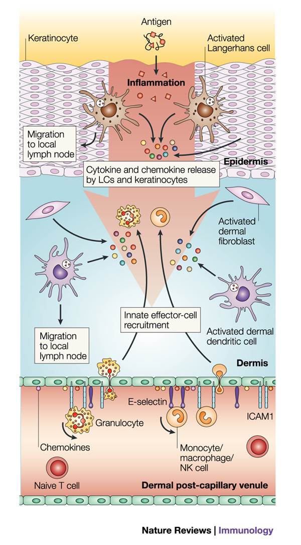
Epithelial-cell injury or pathogen invasion leads to the release of primary cytokines and the activation of both skin cells (keratinocytes and fibroblasts) and resident innate immune cells (Langerhans cells (LCs), dermal dendritic cells (DCs) and mast cells), stimulating downstream activation cascades. Activated Langerhans cells and dermal DCs are stimulated to mature and emigrate from the tissue to the draining lymph node, carrying antigen for presentation to naive and memory T cells. The cytokines and chemokines produced in response to this activation cascade act on the local endothelia through nuclear factor-κB (NF-κB)-mediated pathways to upregulate the expression of adhesion molecules, including E-selectin, P-selectin and intercellular adhesion molecule 1 (ICAM1), and direct the recruitment of additional innate immune components according to the specific signals that are generated — for example, neutrophils, eosinophils and natural killer (NK) cells. CCL17, CC-chemokine ligand 17.
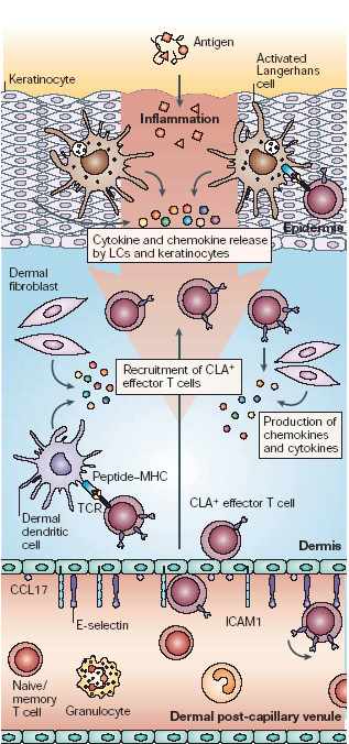
Circulating cutaneous lymphocyte antigen (CLA)-positive T cells represent a library of memory T cells with T-cell receptors (TCRs) specific for antigens previously encountered in the skin. Cytokines released by keratinocytes, fibroblasts and resident antigen-presenting cells stimulate the upregulation of expression of E-selectin and intercellular adhesion molecule 1 (ICAM1) through nuclear factor-κB (NF-κB)-mediated activation pathways. Production and presentation of T-cell-specific chemokines, such as CC-chemokine ligand 17 (CCL17), CCL22 and CCL27, on the local endothelium results in the recruitment of CLA+ T cells in an antigen non-specific manner. T cells entering the tissue that encounter their specific antigen presented by local macrophages or dendritic cells will be activated to proliferate and carry out their specific functions. Those that do not encounter their cognate antigen, which might be most of the cells that are recruited, will enter the lymphatics and return to the general circulation. LC, Langerhans cell.
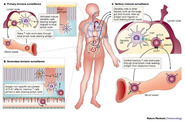
a | Primary immune surveillance is the process by which the innate immune response ensures the effective engagement of the adaptive immune response. Antigens encountered in the skin are carried by activated dendritic cells through the afferent lymphatics to the draining lymph nodes, and presented to naive and central memory T cells circulating through the node. This increases the likelihood of encountering T cells that express the appropriate T-cell receptor (TCR). T cells that encounter their cognate antigen proliferate and differentiate into effector cells expressing homing receptors for the tissue served by that node. b | Secondary immune surveillance provides a mechanism for ensuring rapid and effective local adaptive immune responses to previously encountered antigens. Tissue inflammation results in the upregulation of expression of adhesion molecules and presentation of specific chemokines on the local endothelium. Effector memory T cells that express the appropriate counter-receptors are recruited in an antigen non-specific manner. Those cells that encounter their cognate antigen presented by local antigen-presenting cells (APCs) participate in the local inflammatory response, whereas those that do not return to the general circulation. c | Tertiary immune surveillance represents a mechanism by which the immune system can hedge its bets, providing enhanced adaptive immune responses to antigens encountered in tissues distinct from those in which they were previously encountered. Central memory T cells produced in skin-draining lymph nodes express L-selectin and CC-chemokine receptor 7 (CCR7), which allows them to recirculate through lymph nodes throughout the body, where they can provide enhanced responses to antigen encountered through a different environmental interface. CLA, cutaneous lymphocyte antigen.
References
-
- Leung DY. Atopic dermatitis: immunobiology and treatment with immune modulators. Clin. Exp. Immunol. 1997;107(Suppl. 1):25–30. - PubMed
Publication types
MeSH terms
Substances
LinkOut - more resources
Full Text Sources
Other Literature Sources
Medical
