. 2004 Apr 6;101(14):5105-10.
doi: 10.1073/pnas.0304823101. Epub 2004 Mar 23.

Galanin acts as a neuroprotective factor to the hippocampus

Affiliations

Galanin acts as a neuroprotective factor to the hippocampus

Caroline R Elliott-Hunt et al. Proc Natl Acad Sci U S A. .

Abstract

The expression of the neuropeptide galanin is markedly up-regulated in many areas of the central and peripheral nervous system after injury. We have recently demonstrated that peripheral sensory neurons depend on galanin for neurite extension after injury, mediated by activation of the second galanin receptor subtype (GALR2). We therefore hypothesized that galanin might also act in a similar manner in the CNS, reducing cell death in hippocampal models of excitotoxicity. Here we report that galanin acts an endogenous neuroprotective factor to the hippocampus in a number of in vivo and in vitro models of injury. Kainate-induced hippocampal cell death was greater in both the CA1 and CA3 regions of galanin knockout animals than in WT controls. Similarly, exposure to glutamate or staurosporine induced significantly more neuronal cell death in galanin knockout organotypic and dispersed primary hippocampal cultures than in WT controls. Conversely, less cell death was observed in the hippocampus of galanin overexpressing transgenic animals after kainate injection and in organotypic cultures after exposure to staurosporine. Further, exogenous galanin or the previously described high-affinity GALR2 agonist, both reduced cell death when coadministered with glutamate or staurosporine in WT cultures. These results demonstrate that galanin acts an endogenous neuroprotective factor to the hippocampus and imply that a galanin agonist might have therapeutic uses in some forms of brain injury.


Figures

Fig. 1.
Fig. 1.
Immunohistochemistry for galanin in WT (A and C) and line 46 galanin overexpressing (OE) animals (B and D)at ×20 (Upper), and CA1 hippocampal subfield at ×40 (Lower). (Bar = 100 μm.) The OE animals have considerably more galanin expression throughout the CA subfields and dentate gyrus. At higher power, it is evident that the majority of galanin overexpression in the CA1 region is in the terminal fields rather than in cell bodies.
Fig. 2.
Fig. 2.
Effects of i.p. administration of 20 mg/kg kainate on hippocampal cell death in vivo. Galanin knockout (KO) animals had significantly more neuronal cell death than strain-matched WT controls, whereas galanin overexpressing (OE) animals had less cell death than strain-matched WT controls. Cell death was measured by counting the numbers of terminal deoxynucleotidyl transferase-mediated fluorescein-dUTP nick end labeling-positive cells in every sixth section of the CA1 and CA3 regions of the hippocampus. Each point represents the mean ± SEM (n = 3). **, P < 0.01; ***, P < 0.001.
Fig. 3.
Fig. 3.
Immunohistochemistry staining for galanin in WT dispersed primary hippocampal neurons and slice organotypic cultures. (A) One-half of all dispersed neurons are positive for galanin after 9 days in culture (Upper Left), corresponding bright-field image (Upper Right), and overlay (Lower). (B) High levels of galanin staining are noted throughout WT organotypic cultures. Because of the thickness of the slice culture (≈250 μm after 2 wk in culture), the neurons in the periphery of the field are not in focus.
Fig. 4.
Fig. 4.
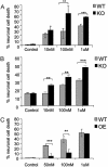
Responses of hippocampal cultures after incubation with 10 nM to 1 μM staurosporine (St). (A) Organotypic cultures from galanin knockout (KO) cultures had significantly more neuronal cell death than WT controls at each concentration of staurosporine in the CA1 subfield. Cell death was visualized by propidium iodide staining. Results are expressed as a percentage of the area expressing fluorescence as compared to the untreated “control” cultures. Each point represents the mean ± SEM, n = 4. **, P < 0.01; ***, P < 0.001. (B) Primary hippocampal cultures were incubated for with 10 nM to 1 μM staurosporine. KO cultures had significantly more neuronal cell death than WT cultures at each concentration of staurosporine. Each point represents the mean ± SEM. n = 4. **, P < 0.01; ***, P < 0.001. (Bar = 100 μm.) (C) Organotypic cultures from galanin overexpressing (OE) mice were incubated with 50 nM to 1 μM staurosporine. OE cultures had significantly less neuronal cell death than WT cultures at 100 nM and 50 nM staurosporine. Each point represents the mean ± SEM. n = 4, **, P < 0.01, ***, P < 0.001.
Fig. 5.
Fig. 5.
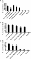
(A) WT organotypic hippocampal cultures were incubated with the indicated concentrations of galanin and AR-M1896 during treatment with 100 nM staurosporine. Galanin significantly reduced staurosporine-induced cell death. Each point represents the mean ± SEM. n = 4; **, P < 0.01. (B) WT organotypic hippocampal cultures were incubated with the indicated concentrations of galanin during treatment with 4 mM l-glutamic acid. Galanin significantly reduced glutamate-induced cell death over the range 10 nM-1 μM. Each point represents the mean ± SEM. n = 4; *, P < 0.05; **, P < 0.01. (C) Dispersed primary hippocampal neurons were incubated with the indicated concentrations of galanin or AR-M1896 during treatment with staurosporine. AR-M1896 and galanin significantly reduced staurosporine-induced cell death in WT cultures. Each point represents the mean ± SEM (n = 4). n = 3; **, P < 0.01.

Similar articles

Cited by

References

    1. Tatemoto, K., Rokaeus, A., Jornvall, H., McDonald, T. J. & Mutt, V. (1983) FEBS Lett. 164, 124-128. - PubMed
    1. Fisone, G., Wu, C. F., Consolo, S., Nordstrom, O., Brynne, N., Bartfai, T., Melander, T. & Hokfelt, T. (1987) Proc. Natl. Acad. Sci. USA 84, 7339-7343. - PMC - PubMed
    1. Misane, I., Razani, H., Wang, F. H., Jansson, A., Fuxe, K. & Ogren, S. O. (1998) Eur. J. Neurosci. 10, 1230-1240. - PubMed
    1. Pieribone, V. A., Xu, Z. Q., Zhang, X., Grillner, S., Bartfai, T. & Hokfelt, T. (1995) Neuroscience 64, 861-874. - PubMed
    1. Hokfelt, T., Xu, Z. Q., Shi, T. J., Holmberg, K. & Zhang, X. (1998) Ann. N.Y. Acad. Sci. 863, 252-263. - PubMed

Publication types