Comparative Study
. 2004 Mar 24;24(12):2886-97.
doi: 10.1523/JNEUROSCI.4161-03.2004.

Novel neuronal phenotypes from neural progenitor cells

Affiliations
Comparative Study

Novel neuronal phenotypes from neural progenitor cells

Eleni A Markakis et al. J Neurosci. .

Abstract

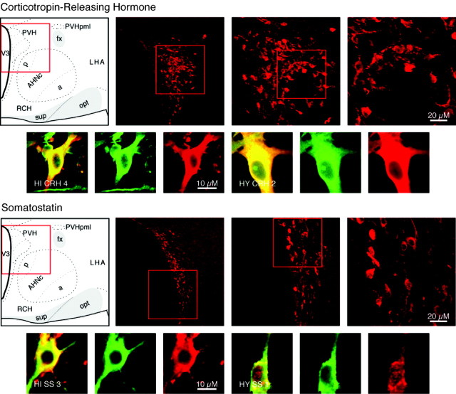
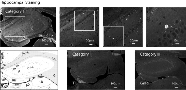
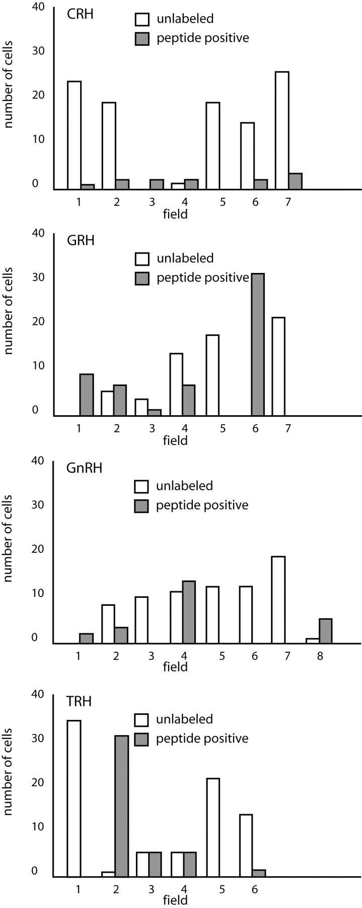
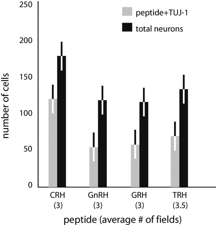
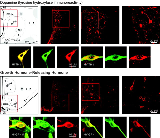
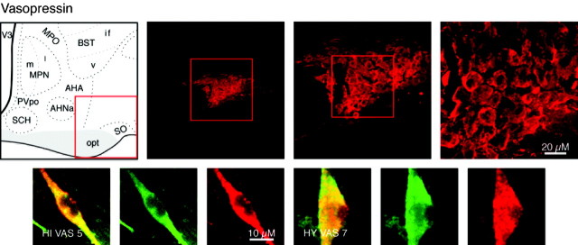
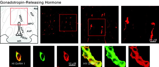
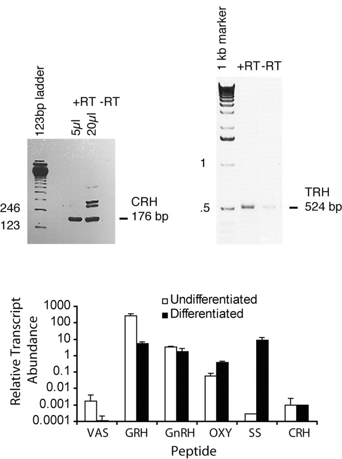
We report the first isolation of progenitor cells from the hypothalamus, a derivative of the embryonic basal plate that does not exhibit neurogenesis postnatally. Neurons derived from hypothalamic progenitor cells were compared with those derived from progenitor cultures of hippocampus, an embryonic alar plate derivative that continues to support neurogenesis in vivo into adulthood. Aside from their different embryonic origins and their different neurogenic potential in vivo, these brain regions were chosen because they are populated with cells of three different categories: Category I cells are generated in both hippocampus and hypothalamus, Category II cells are generated in the hypothalamus but are absent from the hippocampus, and Category III is a cell type generated in the olfactory placode that migrates into the hypothalamus during development. Stem-like cells isolated from other brain regions, with the ability to generate neurons and glia, produce neurons of several phenotypes including gabaergic, dopaminergic, and cholinergic lineages. In the present study, we extended our observations into neuroendocrine phenotypes. The cultured neural precursors from 7-week-old rat hypothalamus readily generated neuropeptide-expressing neurons. Hippocampal and hypothalamic progenitor cultures converged to indistinguishable populations and produced neurons of all three categories, confirming that even short-term culture confers or selects for immature progenitors with enough plasticity to elaborate neuronal phenotypes usually inhibited in vivo by the local microenvironment. The range of phenotypes generated from neuronal precursors in vitro now includes the peptides found in the neuroendocrine system: corticotropin-releasing hormone, growth hormone-releasing hormone, gonadotropin-releasing hormone, oxytocin, somatostatin, thyrotropin-releasing hormone, and vasopressin.


Figures

Figure 1.
Figure 1.
Category I neuropeptides CRH and SS. Confocal photomicrographs in the top rows are rat brain sections stained with the antibody to CRH and SS, respectively. At left is an atlas graphic (Swanson, 1998) indicating the approximate location within the hypothalamus. In each image, the red box serves to orient the location of higher magnification images that follow. In the second row are two sets of three confocal photomicrographs of immunocytochemistry for the neuropeptide (red) and TUJ-1 (green) double-labeled (merged) cells detected in differentiated adult progenitor cultures of hippocampal (HI) and hypothalamic (HY) origins. AHNa,c,p, Anterior hypothalamic nucleus anterior, central, posterior parts; fx, columns of the fornix; LHA, lateral hypothalamic area; opt, optic tract; PVHpml, paraventricular nucleus hypothalamus posterior magnocellular part lateral zone; RCH, retrochiasmatic area; sup, supraoptic commisures; V3, third ventricle.
Figure 2.
Figure 2.
Hippocampal staining. Confocal photomicrographs and montages of immunohistochemistry for neuroendocrine peptides of each generative category in the hippocampus. In the top row, the Category I peptide CRH is shown. At left is a photo-montage of two low-power confocal images. In each image, the box serves to orient subsequent higher-power images. At lower left is an atlas graphic (Swanson, 1998) indicating the approximate level of the hippocampus in all images. Category II and III neurons producing TH and GnRH, respectively, were absent in the hippocampus, as shown in their photo-montages. In all three images, background was enhanced to show tissue details. AD, Anterodorsal nucleus thalamus; alv, alveus; CA3so,sp,sr, field CA3 stratum oriens, pyramidal layer, stratum radiatum; cing, cingulum bundle; cc, corpus calosum; df, dorsal fornix; DG, dentate gyrus; fi, fimbria; LD, lateral dorsal nucleus thalamus; RSPV, retrosplenial area ventral part; sm, stria medularis; V3, third ventricle; VIP, velum interpositum.
Figure 3.
Figure 3.
Undifferentiated hypothalamic field counts. This graphic representation of the numbers of peptide-positive neurons producing CRH, GnRH, GRH, or TRH detected in undifferentiated hypothalamic cell cultures per field shows the distribution of these cells that seem to be found in clusters.
Figure 4.
Figure 4.
Differentiated hypothalamic neuron numbers. Average numbers of peptide and TUJ-1 double-labeled neurons versus total neurons detected in differentiated hypothalamic cultures are shown by peptide for CRH, GnRH, GRH, or TRH. Bars represent SD from the two counts. Three fields were tallied per count, with the exception of TRH, in which 3.5 fields were scored.
Figure 5.
Figure 5.
Category II neuropeptides DA and GRH. Confocal photomicrographs in the top rows are rat brain sections stained with the antibody to TH and GRH, respectively. At left is an atlas graphic (Swanson, 1998) indicating the approximate location within the hypothalamus. In each image, the red box serves to orient the location of higher magnification images that follow. In the second row are two sets of three confocal photomicrographs of immunocytochemistry for the neuropeptide (red) and TUJ-1 (green) double-labeled (merged) cells detected in differentiated adult progenitor cultures of hippocampal (HI) and hypothalamic (HY) origins. AHNa,c, Anterior hypothalamic nucleus anterior, central parts; DMHa,p,v, dorsomedial nucleus hypothalamus anterior, posterior, ventral parts; fx, columns of the fornix; LHA, lateral hypothalamic area; MEex, median eminence external lamina; NC, nucleus circularis; opt, optic tract; PVHap, paraventricular nucleus hypothalamus anterior, parvicellular part; RCH, retrochiasmatic area; RE, nucleus reuniens; SCH, suprachiasmatic nucleus; sup, supraoptic commisures; TU, tuberal area; V3, third ventricle; VMHc,vl, ventromedial nucleus hypothalamus central, ventrolateral parts.
Figure 6.
Figure 6.
Category II neuropeptides TRH and OXY. Confocal photomicrographs in the top rows are rat brain sections stained with the antibody to TRH and OXY, respectively. At left is an atlas graphic (Swanson, 1998) indicating the approximate location within the hypothalamus. In each image, the red box serves to orient the location of higher magnification images that follow. In the second row are two sets of three confocal photomicrographs of immunocytochemistry for the neuropeptide (red) and TUJ-1 (green) double-labeled (merged) cells detected in differentiated adult progenitor cultures of hippocampal (HI) and hypothalamic (HY) origins. AHA, Anterior hypothalamic area; AHNa,p,c anterior hypothalamic nucleus anterior, central, posterior parts; BSTif,v, bed nucleus stria terminalis posterior division interfasicular nucleus, anterior division ventral nucleus; fx, columns of the fornix; LHA, lateral hypothalamic area; MPO, medial preoptic area; MPNm,l, medial preoptic nucleus medial, lateral parts; opt, optic tract; PVpo, preoptic periventricular nucleus; SCH, suprachiasmatic nucleus; SO, supraoptic nucleus; TU, tuberal area; V3, third ventricle.
Figure 7.
Figure 7.
Category II neuropeptide VAS. Confocal photomicrographs in the top row are rat brain sections stained with the antibody to VAS. At left is an atlas graphic (Swanson, 1998) indicating the approximate location within the hypothalamus. In each image, the red box serves to orient the location of higher magnification images that follow. In the second row are two sets of three confocal photomicrographs of VAS (red) and TUJ-1 (green) double-labeled (merged) cells detected in differentiated adult progenitor cultures of hippocampal (HIVAS5) and hypothalamic (HYVAS7) origins. AHA, Anterior hypothalamic area; AHNa, anterior hypothalamic nucleus anterior part; BSTif,v, bed nucleus stria terminalis posterior division interfasicular nucleus, anterior division ventral nucleus; MPO, medial preoptic area; MPNm,l, medial preoptic nucleus medial part, lateral part; opt, optic tract; PVpo, preoptic periventricular nucleus; SCH, suprachiasmatic nucleus; SO, supraoptic nucleus; V3, third ventricle.
Figure 8.
Figure 8.
Category III neuropeptide GnRH. Confocal photomicrographs in the top row are rat brain sections stained with the antibody to GnRH. At left is an atlas graphic (Swanson, 1998) indicating the approximate location within the hypothalamus. In each image, the red box serves to orient the location of higher magnification images that follow. In the second row are two sets of three confocal photomicrographs of GnRH (red) and TUJ-1 (green) double-labeled (merged) cells detected in differentiated adult progenitor cultures of hippocampal (HIGnRH1) and hypothalamic (HYGnRH1) origins. aco, Anterior commissure olfactory limb; ADP, anterodorsal preoptic nucleus; AVP, anteroventral preoptic nucleus; AVPV, anteroventral periventricular nucleus hypothalamus; MEPO, median preoptic nucleus; MPO, medial preoptic area; och, optic chiasm; PSCH, suprachiasmatic preoptic nucleus; V3p, third ventricle preoptic recess.
Figure 9.
Figure 9.
PCR. Nonquantitative RT-PCR gels confirm the presence of CRH mRNA in undifferentiated hypothalamic cultures and TRH mRNA in differentiated hypothalamic cultures. Q-PCR graph shows that the abundance of each peptide transcript was determined by quantitative PCR relative to a reference sample of RNA isolated from the intact rat neuraxis (brain and spinal cord). The reference sample contains authentic transcripts from all neuronal and glial cell types of the brain, including neurendocrine peptide transcripts. Although these data do not indicate absolute abundance of each transcript in the cultures, these do indicate that authentic transcripts for neural peptides exist within the culture at levels that fall within the linear detection range of the assay and within the range detected in normal rat brain tissues.

References

    1. Alvarez-Bolado G, Swanson LW (1996) Developmental brain maps: structure of the embryonic rat brain. Amsterdam: Elsevier.
    1. Alvarez-Buylla A (1990) Mechanism of neurogenesis in adult avian brain. Experientia 46: 948–955. - PubMed
    1. Barry J, Dubois MP, Puolain P (1973) LRF producing cells of the mammalian hypothalamus. Z Zellforsch 146: 351–366. - PubMed
    1. Bennett-Clarke C, Joseph SA (1982) Immunocytochemical distribution of LHRH neurons and processes in the rat: hypothalamic and extrahypothalamic locations. Cell Tissue Res 221: 493–504. - PubMed
    1. Bloch B, Brazeau P, Ling N, Bohlen P, Esch F, Wherenberg WB, Benoit R, Bloom F, Guillemin R (1983) Immunohistochemical detection of growth hormone-releasing factor in brain. Nature 301: 607–608. - PubMed

Publication types

MeSH terms