. 2004 Mar 24;24(12):2963-73.
doi: 10.1523/JNEUROSCI.0155-04.2004.

Nuclear factor-(kappa)B modulates the p53 response in neurons exposed to DNA damage

Affiliations

Nuclear factor-(kappa)B modulates the p53 response in neurons exposed to DNA damage

Hossein Aleyasin et al. J Neurosci. .

Abstract

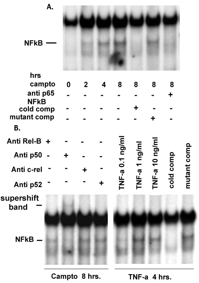
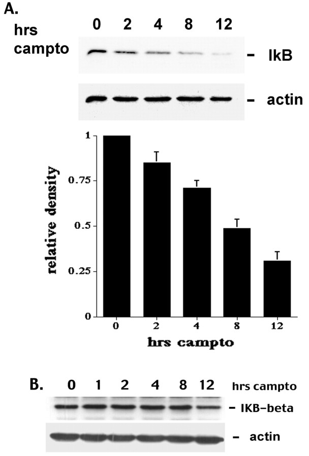
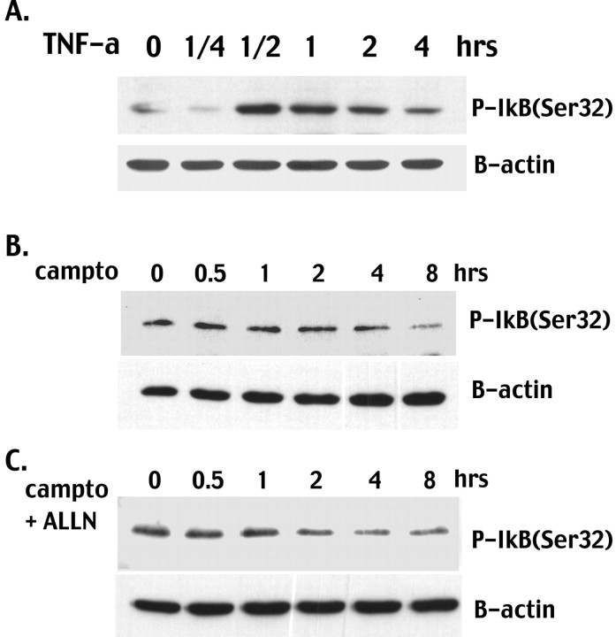
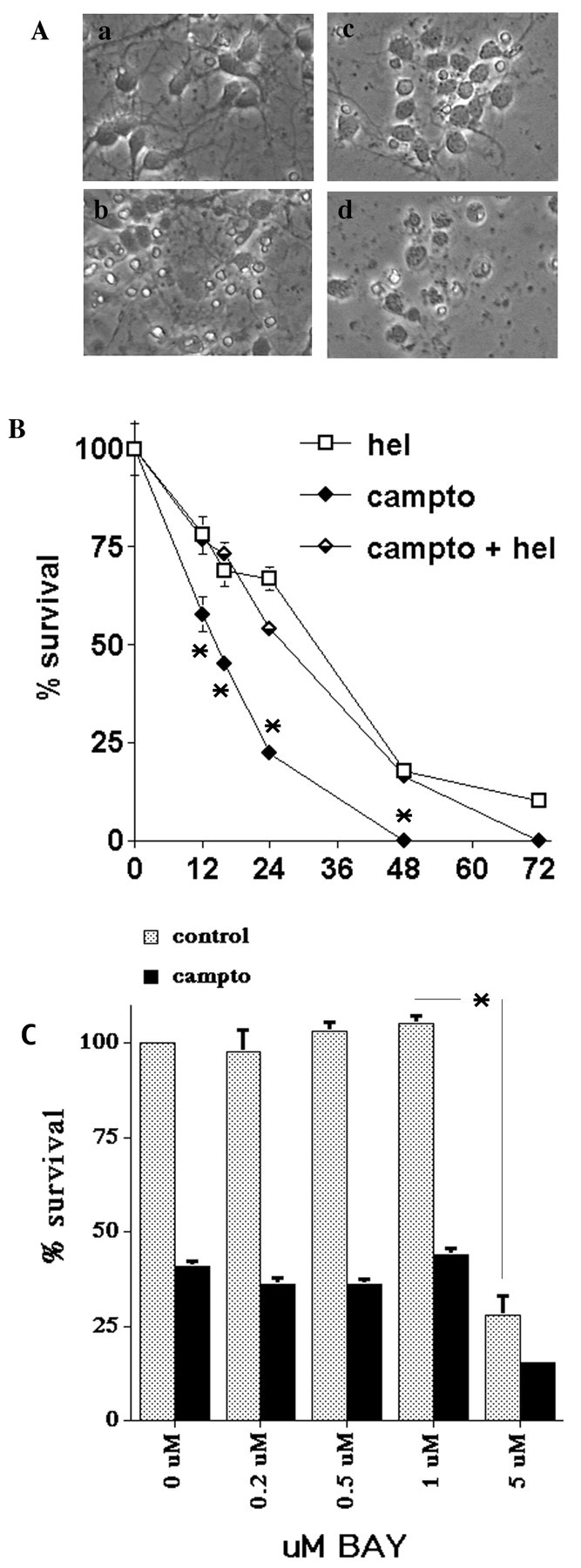
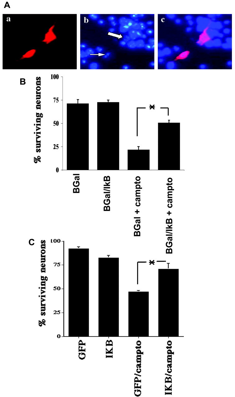
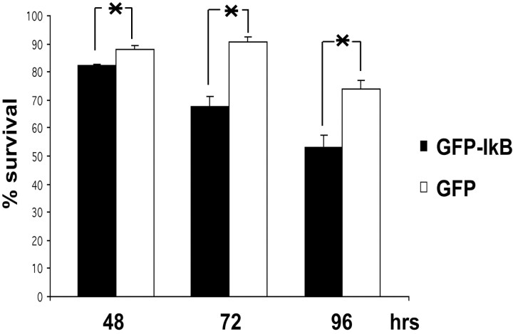
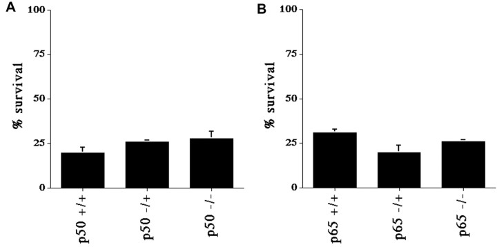
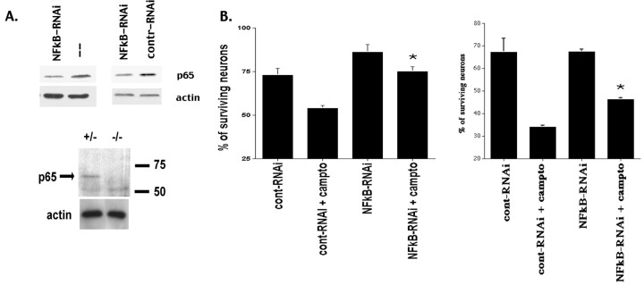
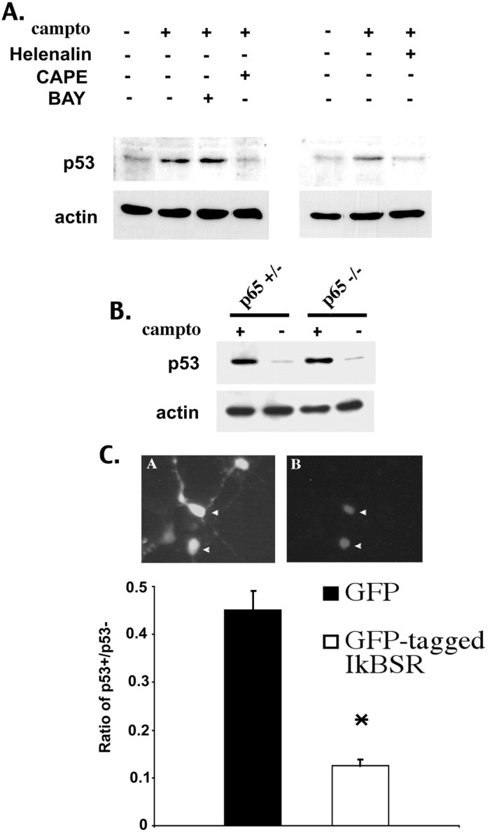
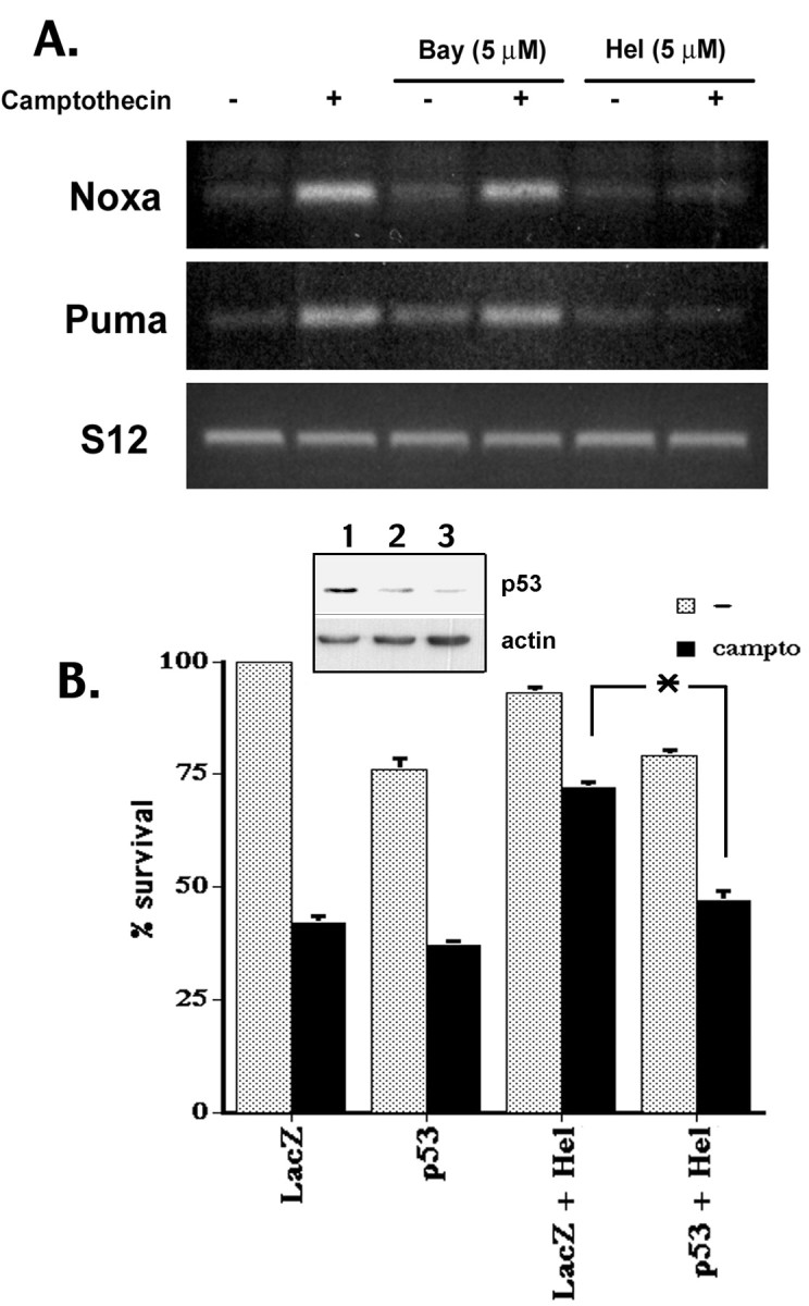
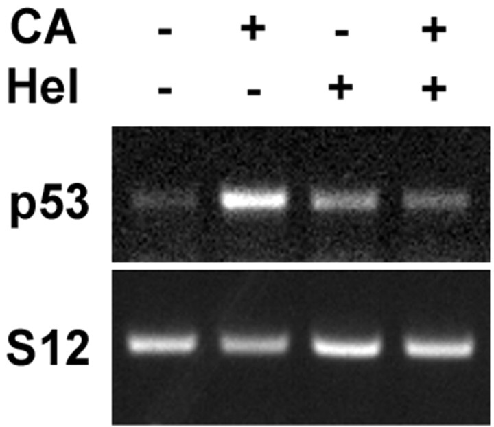
Previous studies have shown that DNA damage-evoked death of primary cortical neurons occurs in a p53 and cyclin-dependent kinase-dependent (CDK) manner. The manner by which these signals modulate death is unclear. Nuclear factor-kappaB (NF-kappaB) is a group of transcription factors that potentially interact with these pathways. Presently, we show that NF-kappaB is activated shortly after induction of DNA damage in a manner independent of the classic IkappaB kinase (IKK) activation pathway, CDKs, ATM, and p53. Acute inhibition of NF-kappaB via expression of a stable IkappaB mutant, downregulation of the p65 NF-kappaB subunit by RNA interference (RNAi), or pharmacological NF-kappaB inhibitors significantly protected against DNA damage-induced neuronal death. NF-kappaB inhibition also reduced p53 transcripts and p53 activity as measured by the p53-inducible messages, Puma and Noxa, implicating the p53 tumor suppressor in the mechanism of NF-kappaB-mediated neuronal death. Importantly, p53 expression still induces death in the presence of NF-kappaB inhibition, indicating that p53 acts downstream of NF-kappaB. Interestingly, neurons cultured from p65 or p50 NF-kappaB-deficient mice were not resistant to death and did not show diminished p53 activity, suggesting compensatory processes attributable to germline deficiencies, which allow p53 activation still to occur. In contrast to acute NF-kappaB inhibition, prolonged NF-kappaB inhibition caused neuronal death in the absence of DNA damage. These results uniquely define a signaling paradigm by which NF-kappaB serves both an acute p53-dependent pro-apoptotic function in the presence of DNA damage and an anti-apoptotic function in untreated normal neurons.


Figures

Figure 1.
Figure 1.
Camptothecin treatment induces NF-κB DNA binding. Nuclear protein extracts of cultured cortical neurons subject to camptothecin for the indicated times were subjected to EMSA. A, NF-κB-related band appeared as early as 2 hr and reached its maximum intensity at ∼8 hr. The induced band was reactive with the antibody to NF-κB family member p65. B, The camptothecin-induced NF-κB band was also reactive to the antibody to p50, but not Rel-B, c-Rel, or p52. TNF-α-treated cortical neurons also were included as a positive control. Cold comp refers to the addition of 125-fold of excess amounts of unlabeled probe. Mutant comp refers to the addition of a mutant form of the NF-κB-specific probe that is unable to compete with the consensus radiolabeled probe.
Figure 2.
Figure 2.
Camptothecin-induced activation of NF-κB is accompanied by IκB degradation. A, Total cell lysates of cultured cortical neurons at indicated times were analyzed for IκB-α by Western blot analysis. Densitometric analysis of IκB degradation from three independent experiments is provided. All data are normalized to loading controls. Error bars are represented as ±SEM. B, Western blot analyses of cortical neurons for IκB-β levels after camptothecin treatment.
Figure 3.
Figure 3.
Lack of increase in IκB-α phosphorylation in cortical neurons after DNA damage. A, Positive control for Ser32 phosphorylation; fibroblasts were treated with TNF (10 μm) for the indicated times and analyzed by Western blot analyses, using a phospho-epitope-specific IκB-α antibody. Total cell lysates of cultured cortical neurons also were treated with camptothecin alone (B) or camptothecin and the proteasomal inhibitor ALLN (C; 50 μm) for the indicated times and analyzed for Ser32 IκB-α phosphorylation as above.
Figure 4.
Figure 4.
Effects of pharmacological inhibitors of the NF-κB pathway. Cultured cortical neurons were cotreated with camptothecin (10 μm) and the indicated concentrations of the p65 inhibitor helenalin (hel) or the IKK inhibitor BAY 11-7082. A, Phase-contrast photomicrographs of neuronal cultures untreated (a), treated with camptothecin (b), and cotreated with 5 μm (c) or 10 μm (d) helenalin for 12 hr. B, Quantitation of neuronal survival with helenalin (2.5 μm) as assessed by nuclear counts under the conditions as indicated. Similar results were obtained for 5 μm helenalin; however, the time course of toxicity was accelerated. Each data point is the mean ± SEM from three independent cultures. *p < 0.05 when campto versus campto + hel are compared at each time point or as indicated. C, Quantitation of neuronal survival with BAY 11-7082 (12 hr) as assessed by nuclear counts under the conditions as indicated.
Figure 5.
Figure 5.
Inhibition of NF-κB by induction of IκBSR protects primary cortical neurons against camptothecin-induced cell death. A, B, Primary cortical neurons were cotransfected with β-gal+ empty pcDNA3 vector (βGal) or β-gal+IκBSR expressing pcDNA3 (βGal+IκBSR) and treated with or without camptothecin (campto). Aa, Cultured cortical neurons were immunostained by anti-β-gal antibody (as described in Materials and Methods). Ab, Nuclei were stained with Hoechst, a nuclear-specific fluorescent dye. Ac, Merged micrograph of a and b. Condensed or fragmented nuclei were scored as dead (shown by thin arrow) versus intact nuclei (shown by thick arrow). B, Quantitation of survival assay among indicated groups of cotransfected primary cortical neurons after 14 hr of treatment with camptothecin. C, Identical experiment as in A and B with the exception that IκBSR was expressed via recombinant adenovirus. GFP, Green fluorescent protein; IκB, GFP-tagged IκBSR. Each data point is the mean ± SEM of three independent cultures. *p < 0.05 (t test).
Figure 6.
Figure 6.
Chronic expression of IκBSR results in neuronal death. Embryonic cortical neurons were infected with either GFP or GFP-tagged IκBSR expressing adenoviruses at the time of plating (MOI = 250). At 48, 72, and 96 hr after plating the neuronal cultures were fixed, and GFP-containing neurons were assessed for survival by analyses of nuclear integrity. Each data point is the mean ± SEM of three independent cultures. *p < 0.05 (t test).
Figure 7.
Figure 7.
p50 or p65 deficiency is not protective against camptothecin-induced neuronal death. Cortical neurons were obtained from embryos derived from a double heterozygote breeding of either p50 (A) or p65 (B). Cultures were exposed to camptothecin for 14 hr and analyzed by nuclear counts. Each data point is the mean ± SEM from three independent embryos.
Figure 8.
Figure 8.
NF-κB p65 suppression by RNA interference (RNAi) protects cortical neurons against camptothecin-induced neurotoxicity. Cortical neurons were transfected with short-interfering RNA (siRNA) oligonucleotides as described in Materials and Methods. A, Top, The levels of p65 protein were reduced in neurons that had been transfected with p65 RNAi (NF-κB-RNAi) as compared with nontransfected cells (–) or cells that had been transfected with nonspecific scrambled RNAi (contr-RNAi; 48 hr after transfection). β-Actin blot was provided as a loading control. Similar results were obtained by using both transfection strategies as described in Materials and Methods. A, Bottom, The specificity of the p65 antibody is demonstrated by using cortical cultures obtained from p65-deficient and wild-type littermate controls. B, Survival of cortical neurons, transfected with p65 NF-κB or control nonspecific RNAi and treated with camptothecin or vehicle for 12 hr, was assessed by nuclear morphology. Two independent experiments with different transfection methods are shown. Right, GeneSilencer; left, Lipofectamine 2000 (see Materials and Methods). Each data point is the mean ± SEM of three separate cultures. *p < 0.05 (t test) when comparing cont-RNAi + campto versus NF-κB-RNAi + campto.
Figure 9.
Figure 9.
The effect of NF-κB suppression on p53 activation after camptothecin treatment. A, Cultured cortical neurons were treated for 8 hr with camptothecin (10 μm) or cotreated with indicated pharmacological inhibitors of NF-κB. Total protein extracts were probed for p53 in a Western blot analysis. B, Cortical neurons, which had been harvested from 14.5 d littermate embryos of indicated p65 genotypes, were subjected to camptothecin or vehicle treatment for 8 hr. Protein extracts were probed for p53. β-Actin was used as a loading control. C, Expression of IκBSR inhibits p53 induction. Cortical neurons were infected with GFP or GFP-tagged IκBSR-expressing adenoviruses (MOI = 250) at the time of plating. At 24 hr after plating they were treated with camptothecin (10 μm) for 8 hr to induce p53. Neurons were fixed and immunostained for p53. Neurons that expressed GFP (left panel) and nuclear p53 (right panel) were scored as positive (indicated with arrowheads). Neurons that expressed GFP but displayed no p53 signal were scored as negative. Bar graph representing quantitation of p53-positive neurons in cells expressing GFP control or GFP-IκBSR is indicated below the panels. Each data point is the mean ± SEM of three independent experiments. *p < 0.05 (t test).
Figure 10.
Figure 10.
A, The p65 inhibitor helenalin (Hel), but not the IKK inhibitor BAY 11-7082 (Bay), inhibits the p53-inducible genes Noxa and Puma. Cortical neurons were treated with camptothecin with and without cotreatment as indicated for 12 hr. Noxa and Puma levels then were analyzed by RT-PCR as described in Results. S12 (a ribosomal protein used as a loading control) also was analyzed as a negative control, which did not change during camptothecin treatment. B, Adenoviral-mediated expression of p53 still induces death even in the presence of the NF-κB inhibitor helenalin (Hel; 5 μm). Neurons were infected with adenovirus expressing lacZ control or p53 as indicated in Materials and Methods. Camptothecin treatment was initiated 24 hr after infection. Survival was assessed at 12 hr after camptothecin treatment in the presence or absence of helenalin. Inset demonstrates the overexpression of p53. Lane 1, Infection with p53-expressing virus; lane 2, infection with lacZ control; lane 3, no infection. *p < 0.05 (t test).
Figure 11.
Figure 11.
NF-κB inhibition blocks induction of p53 transcripts induced by DNA damage. Cortical neurons were treated with camptothecin with and without cotreatment with helenalin (Hel; 5 μm) as indicated for 2 hr. Then p53 transcription levels were analyzed by RT-PCR as described in Results. S12 (a ribosomal protein used as a loading control) also was analyzed as a negative control, which did not change during camptothecin treatment.

References

    1. Alam ZI, Jenner A, Daniel SE, Lees AJ, Cairns N, Marsden CD, Jenner P, Halliwell B (1997) Oxidative DNA damage in the parkinsonian brain: an apparent selective increase in 8-hydroxyguanine levels in substantia nigra. J Neurochem 69: 1196–1203. - PubMed
    1. Bales KR, Du Y, Dodel RC, Yan GM, Hamilton-Byrd E, Paul SM (1998) The NF-kappaB/Rel family of proteins mediates Aβ-induced neurotoxicity and glial activation. Brain Res Mol Brain Res 57: 63–72. - PubMed
    1. Barkett M, Gilmore TD (1999) Control of apoptosis by Rel/NF-kappaB transcription factors. Oncogene 18: 6910–6924. - PubMed
    1. Barnes DE, Stamp G, Rosewell I, Denzel A, Lindahl T (1998) Targeted disruption of the gene encoding DNA ligase IV leads to lethality in embryonic mice. Curr Biol 8: 1395–1398. - PubMed
    1. Benoit V, Hellin AC, Huygen S, Gielen J, Bours V, Merville MP (2000) Additive effect between NF-kappaB subunits and p53 protein for transcriptional activation of human p53 promoter. Oncogene 19: 4787–4794. - PubMed

Publication types

MeSH terms