. 2004 Mar 24;24(12):3013-22.
doi: 10.1523/JNEUROSCI.5416-03.2004.

Group III metabotropic glutamate receptors maintain tonic inhibition of excitatory synaptic input to hypocretin/orexin neurons

Affiliations

Group III metabotropic glutamate receptors maintain tonic inhibition of excitatory synaptic input to hypocretin/orexin neurons

Claudio Acuna-Goycolea et al. J Neurosci. .

Abstract

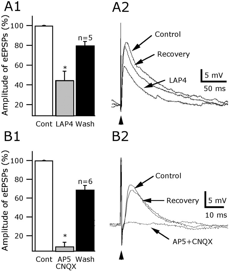
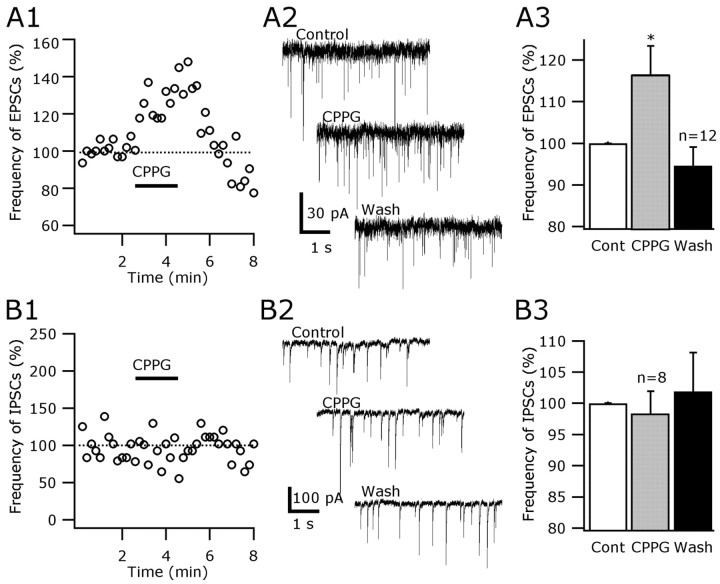
Hypocretin/orexin neurons play an important role in hypothalamic arousal. Synaptic glutamate input to hypocretin neurons regulates cell firing. We studied the actions of group III metabotropic glutamate receptors (mGluRs) in modulating the activity of hypocretin neurons using whole-cell voltage- and current-clamp recording in mouse whole hypothalamic slices or minislices consisting only of the lateral hypothalamus. Selective green fluorescent protein expression was used to detect live hypocretin neurons. The mGluR agonist l-(+)-2-amino-4-phosphonobutyric acid (l-AP-4) inhibited synaptic input to hypocretin neurons in a dose-dependent manner; both spontaneous glutamate and GABA-mediated synaptic currents were reduced in frequency. l-AP-4 also reduced the amplitude of postsynaptic potentials evoked by a stimulating electrode placed medial or lateral to the recorded cell. No postsynaptic effect of l-AP-4 was found relative to membrane potential, input resistance, or AMPA-evoked currents. l-AP-4 appeared to act by a presynaptic mechanism and reduced the frequency of both glutamate- and GABA-mediated miniature events recorded in the presence of tetrodotoxin, with no change in amplitude. (RS)-phosphonopentanoic acid (CPPG), a group III mGluR antagonist, suppressed the actions of l-AP-4. Of substantial interest, CPPG by itself increased synaptic activity recorded in hypocretin neurons, suggesting an ongoing inhibitory tone attributable to activation of group III mGluRs. Glutamatergic interneurons have been suggested to play a role in a positive feedback recruitment of hypocretin on hypocretin neurons. l-AP-4 blocked hypocretin-mediated increases in EPSCs and attenuated the hypocretin-mediated increase in spike frequency. Together, these data suggest that tonically active inhibitory mGluRs are expressed on local hypocretin-sensitive glutamate neurons within the lateral hypothalamus that modulate the output of the hypocretin arousal system.


Figures

Figure 1.
Figure 1.
Group III metabotropic glutamate receptors inhibit spontaneous excitatory synaptic activity in hypocretin neurons. A, Time course of the inhibitory l-AP-4 (100 μm) effects on the sPSCs. B, Raw traces showing the reversible effect of l-AP-4 on the postsynaptic current frequency. C, Dose–response relationships for l-AP-4 inhibition of EPSC frequency. D, Parallel experiments in the presence of 30 μm BIC show a reduction of EPSCs (*p < 0.01; n = 6). The group III mGluR antagonist CPPG (200 μm) substantially reduced the l-AP-4-mediated inhibition of EPSC frequency (*p <0.01; n=6). Glutamate receptor antagonists (50 μm AP-5 and 10 μm CNQX) blocked the EPSCs, suggesting glutamate as the active transmitter (data not shown).
Figure 2.
Figure 2.
mGluR attenuation of evoked glutamate-mediated excitatory potentials. EPSPs were evoked by electrical stimulation (arrowhead) in the lateral hypothalamus, medial or lateral to the recorded cell. A1, The amplitude of evoked potentials was depressed by 55.2 ± 9% (mean ± SEM) in 100 μm l-AP-4 and subsequently recovered (*p<0.001). A2, Representative traces demonstrating the inhibitory effect of l-AP-4 (100 μm) on excitatory evoked transmission in hypocretin cells. The membrane potential of the cells was held at –65 mV. B1, B2, These evoked EPSPs were attributable to glutamate release, because they were blocked by AP-5 (50 μm) and CNQX (10 μm) (*p < 0.001; n = 6).
Figure 3.
Figure 3.
l-AP-4 depressed local synaptic glutamate release in the lateral hypothalamus. A, Schematic representation of the lateral hypothalamic region dissected out to study the local excitatory inputs to hypocretin cells. ARH, Arcuate nucleus of the hypothalamus; cpd, cerebral peduncle; DMH, dorsomedial hypothalamus; fx, fornix; LHA, lateral hypothalamic area; ME, median eminence; opt, optic tract; VMH, ventromedial hypothalamus; V3, third ventricle; ZI, zonaincerta. B, The group III mGluR agonist l-AP-4 (100 μm) reversibly depressed the frequency of spontaneous EPSCs. C, Trace 1 shows the excitatory synaptic response, but not direct response, to glutamate micro application 1 mm away from the recorded cell. Trace 2 shows that, in the presence of TTX, the glutamate microdrop failed to increase the EPSC frequency. Trace 3 shows the glutamate microdrop experiment after ionotropic glutamate receptors blockade. No increase in the EPSC frequency was detected. In trace 4 is shown the slow inward current evoked by glutamate directly applied to the recorded cell. D, TTX at 0.5 μm changed (left shift) the cumulative fractions of the EPSC amplitude. Arrowhead shows the point at which all events in TTX can be accounted for (maximum of 27 pA). E, l-AP-4 (100 μm) reduced the frequency of large spike-dependent EPSCs (amplitude, ≥28 pA) by 40.5 ± 9.3% (*p < 0.05; n = 5).
Figure 4.
Figure 4.
Presynaptic mechanism: group III mGluRs attenuate miniature EPSC frequency. A, mEPSCs were recorded from hypocretin cells held at –60 mV in the presence of 0.5 μm TTX. l-AP-4 (100 μm) induced a reversible decrease in the frequency of mEPSCs. B, Summary bar graph of data illustrating a significant effect of l-AP-4 on the frequency (mean ± SEM) of mEPSCs (*p < 0.005; n = 8; ANOVA). C, Cumulative histogram of a typical cell showing the lack of l-AP-4 effect on mEPSC amplitude (p = 0.71; n = 5; Kolmogorov–Smirnov test). D, I–V relationships in a typical cell. l-AP-4 did not alter the input resistance of hypocretin cells (n = 8; p = 0.86; ANOVA). E, l-AP-4 did not alter the response of postsynaptic AMPA receptors, as shown in the representative traces in hypocretin neurons before (left), during (middle), and after (right) bath application of 100 μm l-AP-4. These results support the view that l-AP-4 presynaptically modulates excitatory transmission in hypocretin cells.
Figure 5.
Figure 5.
Presynaptic mGluR inhibition of GABA-mediated synaptic currents. A, Using whole-cell voltage clamp and the glutamate receptor antagonists AP-5 (50 μm) and CNQX (10 μm) in the bath and KCl in the pipette, spontaneous IPSCs were recorded. l-AP-4 (100 μm) decreased the frequency of inhibitory currents in these typical traces. B, Bar graph shows the mean ± SEM IPSC frequency before, during, and after l-AP-4 (*p < 0.001; n = 6; ANOVA). C, In the presence of 0.5 μm TTX in the bath, the effect of l-AP-4 on mIPSCs was examined. l-AP-4 decreased the frequency (D) (*p < 0.001; n = 6; ANOVA) but did not alter the amplitude (E) (p > 0.05; n = 7; Kolmogorov–Smirnov test) of mIPSCs, suggesting a presynaptic effect on inhibitory transmission. F, Bar graph shows that, in the presence of the group III mGluR antagonist CPPG (200 μm), l-AP-4 actions were suppressed.
Figure 6.
Figure 6.
Normal mGluR inhibitory tone blocked by group III antagonists. A1, CPPG (200 μm), a selective group III mGluR antagonist, increased the spontaneous EPSCs in hypocretin neurons. BIC at 30 μm was used in all experiments. A2, Representative traces showing the CPPG (200 μm) effect on the frequency of the EPSCs. A3, The data from 12 neurons were combined in a bar graph. The frequency of EPSCs was increased by 15.5 ± 6.9% (mean ± SEM) during CPPG administration and returned to the baseline after washout. This CPPG effect on the frequency of EPSCs was statistically significant (*p < 0.05; n = 12). B1, B2, CPPG (200 μm) did not increase the frequency of the inhibitory currents in hypocretin cells (50 μm AP-5 and 10 μm CNQX were bath applied in all experiments). B3, The IPSC frequency was 98.3 ± 3.6% in CCPG compared with control levels (100%) (n = 8; p = 0.79; ANOVA). These results suggest that group III mGluRs maintain a tonic inhibition of glutamatergic, but not GABAergic, synaptic inputs in hypocretin cells.
Figure 7.
Figure 7.
l-AP-4 depresses hypocretin-mediated excitation of hypocretin cells. A, B, Group III mGluR activation depresses the hypocretin-mediated increase in EPSCs and firing rate. A, C, Hcrt-2 (2 μm) reversibly increased the frequency of EPSCs (traces 1–3). This effect was statistically significant (*p < 0.05; n = 5). In the presence of l-AP-4 in the bath (traces 4, 5), no change in the frequency of EPSCs was induced by hypocretin (p = 0.82; n = 5). C, The effects of Hcrt-2 in the absence or presence of 100 μm l-AP-4 are compared (*p < 0.05; n = 5; ANOVA and Bonferroni post hoc test). B, D, l-AP-4 depressed the hypocretin-mediated spike frequency increase. B, Representative traces showing the excitatory effect of Hcrt-2 (2 μm) in normal ACSF (traces 1–3) and ACSF containing l-AP-4 (traces 4, 5). D, Bar graph shows that l-AP-4 caused a significant reduction of the hypocretin-induced increase in spike frequency (*p < 0.05; n = 7; ANOVA followed by a Bonferroni comparison). n.s., Not significant.

References

    1. Abrahamson EE, Moore RY (2001) The posterior hypothalamic area: chemoarchitecture and afferent connections. Brain Res 889: 1–22. - PubMed
    1. Awad-Granko H, Conn PJ (2001) Activation of groups I or III metabotropic glutamate receptors inhibits excitatory transmission in the rat subthalamic nucleus. Neuropharmacology 41: 32–34. - PubMed
    1. Awatramani GB, Slaughter MM (2001) Intensity-dependent, rapid activation of presynaptic metabotropic glutamate receptors at a central synapse. J Neurosci 21: 741–749. - PMC - PubMed
    1. Belousov AB, van den Pol AN (1997) Local synaptic release of glutamate from neurons in the rat hypothalamic arcuate nucleus. J Physiol (Lond) 499: 747–761. - PMC - PubMed
    1. Bergles DE, Jahr CE (1997) Synaptic activation of glutamate transporters in hippocampal astrocytes. Neuron 19: 1297–1308. - PubMed

Publication types

MeSH terms

LinkOut - more resources