. 2004 Mar 24;24(12):3023-30.
doi: 10.1523/JNEUROSCI.5745-03.2004.

Sensory neuron signaling to the brain: properties of transmitter release from olfactory nerve terminals

Affiliations

Sensory neuron signaling to the brain: properties of transmitter release from olfactory nerve terminals

Gabe J Murphy et al. J Neurosci. .

Abstract

Olfactory receptor neurons (ORNs) convey sensory information directly to the CNS via conventional glutamatergic synaptic contacts in olfactory bulb glomeruli. To better understand the process by which information contained in the odorant-evoked firing of ORNs is transmitted to the brain, we examined the properties of glutamate release from olfactory nerve (ON) terminals in slices of the rat olfactory bulb. We show that marked paired pulse depression is the same in simultaneously recorded periglomerular and tufted neurons, and that this form of short-term plasticity is attributable to a reduction of glutamate release from ON terminals. We used the progressive blockade of NMDA receptor (NMDAR) EPSCs by MK-801 [(5R,10S)-(+)-5-methyl-10,11-dihydro-5H-dibenzo[a,d]cyclohepten-5-10-imine hydrogen maleate] and stationary fluctuation analysis of AMPA receptor (AMPAR) EPSCs to determine the probability of release (P(r)) of ON terminals; both approaches indicated that P(r) is unusually high (>/=0.8). The low-affinity glutamate receptor antagonists gamma-d-glutamylglycine and l-amino-5-phosphonovaleric acid blocked ON-evoked AMPAR- and NMDAR-mediated EPSCs, respectively, to the same extent under conditions of low and high P(r), suggesting that multivesicular release is not a feature of ON terminals. Although release from most synapses exhibits a highly nonlinear dependence on extracellular Ca(2+), we find that the relationship between glutamate release and extracellular Ca(2+) at ON terminals is nearly linear. Our results suggest that ON terminals have specialized features that may contribute to the reliable transmission of sensory information from nose to brain.


Figures
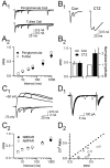
Figure 1.
Figure 1.
ON terminals show strong presynaptic paired pulse depression. A1, The average response of a simultaneously recorded PG and tufted cell to 5, 10, 25, and 50 msec paired pulse stimulation. A2, Average PPR (EPSC2 /EPSC1) over a range of interstimulus intervals (n = 7 pairs). B1, ON-evoked EPSC (Vh = –70 mV) before (Con) and after (CTZ) application of cyclothiazide (100 μm). B2, Average effect of CTZ on PPR (left axis) and EPSC1 amplitude (right axis; n = 5). C1, AMPAR- and NMDAR-mediated paired pulse depression in a representative PG neuron. C2, Average AMPAR- and NMDAR-mediated PPR are not significantly different (n = 7 cells; p > 0.2). D1, Fifteen consecutive AMPAR-mediated responses in a PG neuron evoked by paired pulse stimulation of the ON layer. D2, CV2 ratio (EPSC1 CV2/EPSC2 CV2) versus PPR (25–100 msec ISI) in 11 experiments.
Figure 2.
Figure 2.
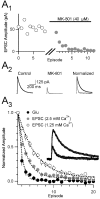
Progressive blockade of ON-evoked NMDAR EPSCs by MK-801 indicates Pr from ON terminals is high. A1, NMDAR EPSC amplitude versus episode number for a typical experiment. After establishing a stable ON-evoked NMDAR-mediated response (open circles), stimulation was halted, and MK-801 was introduced. In the presence of MK-801, the peak NMDAR EPSC amplitude (gray circles) declined rapidly when stimulation was resumed. A2, Average response to ON stimulation before (control) and average of the first five responses in MK-801. A3, NMDAR-mediated currents evoked by brief (15 msec) pressure application of glutamate (Glu; 100 μm) and ON stimulation in normal aCSF (2.5 mm Ca2+; gray circles) or low Ca2+ (1.25 mm; open circles) are inhibited progressively in the presence of MK-801 with a monoexponental time course (τglu = 0.89 episodes; τEPSC 2.5 = 2.12 episodes; τEPSC 1.25 = 4.7 episodes). Inset, ON-evoked NMDAR-mediated currents under control conditions and in MK-801. White dots represent fits to the data generated by the kinetic model (see Materials and Methods).
Figure 3.
Figure 3.
Nonstationary fluctuation analysis of ON-evoked AMPAR-mediated EPSCs indicates Pr from ON terminals is high. A1, Ten consecutive ON-evoked EPSCs under control conditions and in the presence of two different concentrations of Cd2+. A2, Variance–mean plot for the representative experiment in A1. The estimated Pr in this experiment is 0.96. B, The predicted (small closed circle) and measured (small open circle) quantal size for each experiment. The average predicted (large filled circle) and measured (large open circle) quantal sizes were not significantly different (p = 0.66; left axis). Pr for each of the six individual experiments (small open circles) and the average estimated for the population (large open circle) are plotted on the right axis.
Figure 4.
Figure 4.
ON-evoked EPSCs are equally sensitive to low-affinity glutamate receptor antagonists under conditions of high or low Pr. A, γ-DGG (2 mm) blocks AMPAR-mediated EPSCs to the same extent, whether Pr is high (2.5 mm Ca2+) or low (0.5 mm Ca2+). Traces from two representative experiments are shown above; the average fractional block of EPSC1 and EPSC2 by γ-DGG when Pr is high (n = 12) and low (n = 10) shown below was indistinguishable (p > 0.6). B, l-APV (1 mm) inhibits NMDAR-mediated EPSCs evoked by ON stimulation to the same degree when Pr is high and low (n = 5; p > 0.5).
Figure 5.
Figure 5.
ON transmitter release exhibits unusual Ca2+ cooperativity. A1, ON-evoked glomerular fEPSPs from a representative experiment in normal (2.5 mm) and reduced Ca2+. A2, Normalized amplitude of ON-evoked glomerular fEPSPs (filled circles) and AMPAR-mediated EPSCs (open circles) as a function of extracellular Ca2+. Each point represents the average of three to six experiments. Predicted relationships for data with an identical K0.5 (1.19) and cooperativity (n) values of 2 and 4 are shown as dashed lines. B1, LOT-evoked fEPSPs in piriform cortex from a representative experiment in normal (2.5) and reduced Ca2+ aCSF. B2, Normalized amplitude of LOT-evoked fEPSPs; predicted relationships for n = 2 and 4 are shown as in A2, with K0.5 = 1.28. All experiments in this figure were performed at room temperature.

References

    1. Aroniadou-Anderjaska V, Zhou FM, Priest CA, Ennis M, Shipley MT (2000) Tonic and synaptically evoked presynaptic inhibition of sensory input to the rat olfactory bulb via GABA(B) heteroreceptors. J Neurophysiol 84: 1194–1203. - PubMed
    1. Augustine GJ, Charlton MP, Smith SJ (1985) Calcium entry and transmitter release at voltage-clamped nerve terminals of squid. J Physiol (Lond) 367: 163–181. - PMC - PubMed
    1. Bellingham MC, Walmsley B (1999) A novel presynaptic inhibitory mechanism underlies paired pulse depression at a fast central synapse. Neuron 23: 159–170. - PubMed
    1. Berton F, Iborra C, Boudier JA, Seagar MJ, Marqueze B (1997) Developmental regulation of synaptotagmin I, II, III, and IV mRNAs in the rat CNS. J Neurosci 17: 1206–1216. - PMC - PubMed
    1. Borst JG, Sakmann B (1996) Calcium influx and transmitter release in a fast CNS synapse. Nature 383: 431–434. - PubMed

Publication types

MeSH terms