. 2004 Mar 24;24(12):3040-50.
doi: 10.1523/JNEUROSCI.4934-03.2004.

Protein kinase A activation promotes plasma membrane insertion of DCC from an intracellular pool: A novel mechanism regulating commissural axon extension

Affiliations

Protein kinase A activation promotes plasma membrane insertion of DCC from an intracellular pool: A novel mechanism regulating commissural axon extension

Jean-François Bouchard et al. J Neurosci. .

Abstract

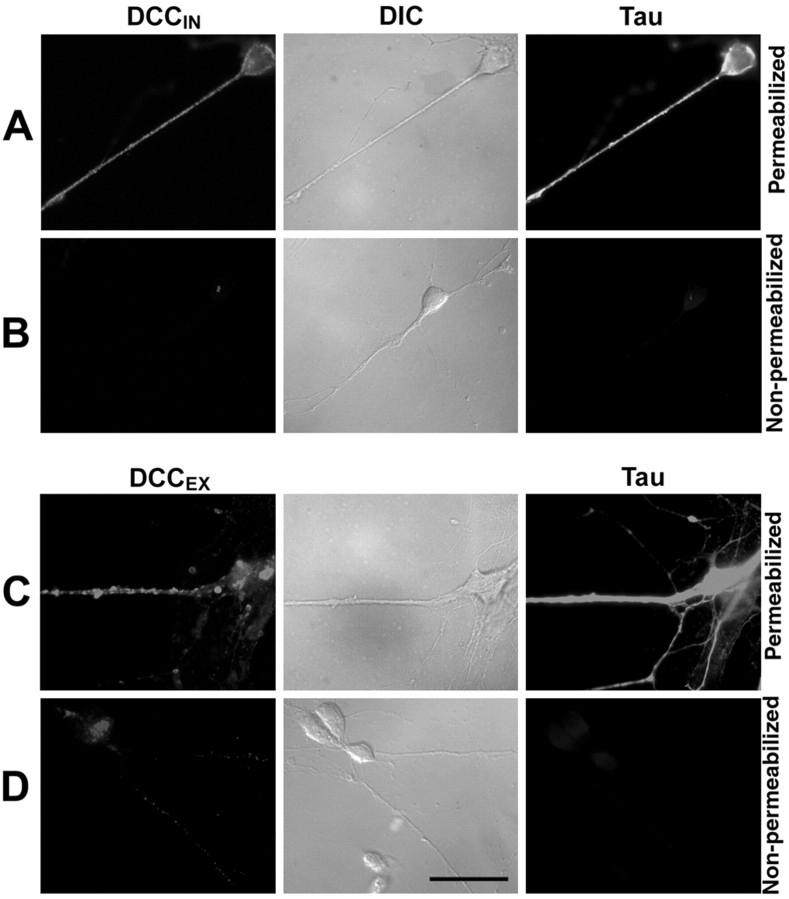
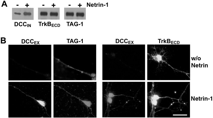
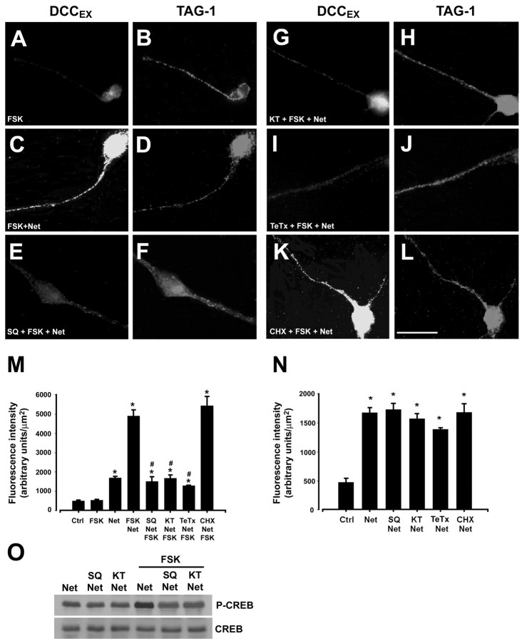
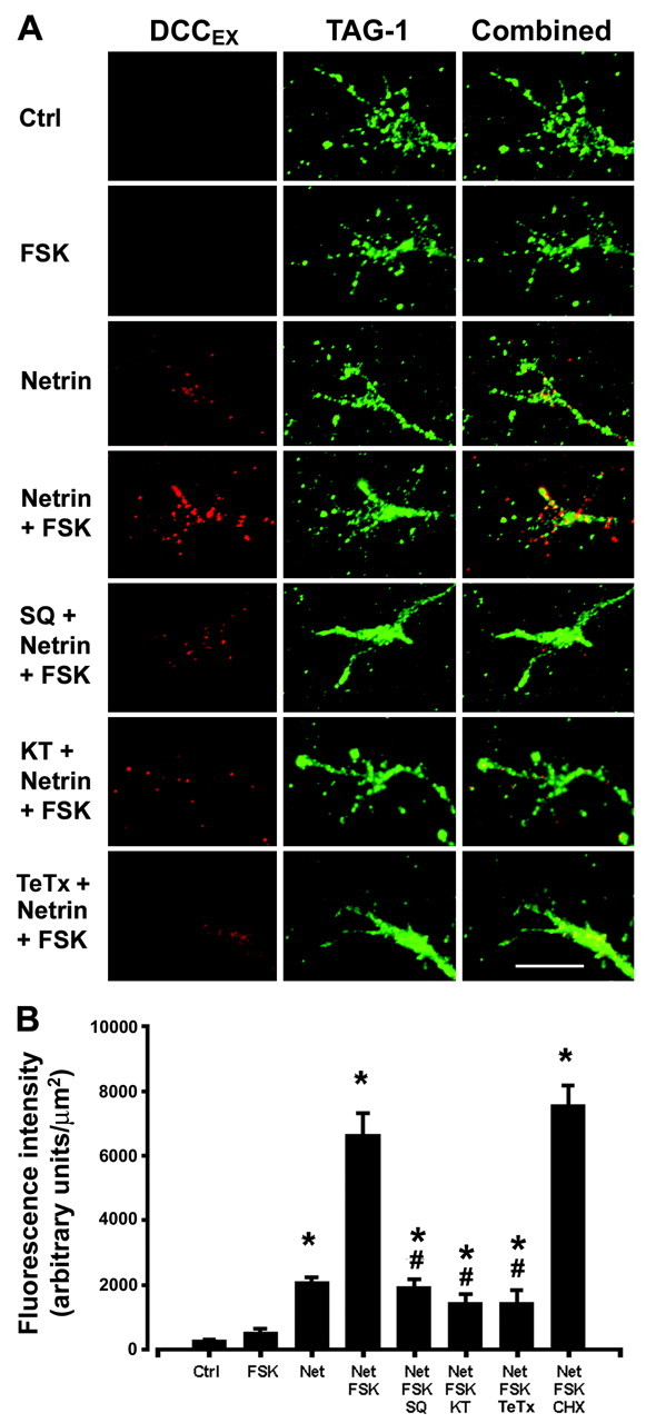
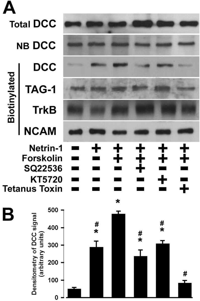
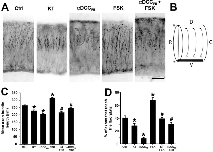
Protein kinase A (PKA) exerts a profound influence on axon extension during development and regeneration; however, the molecular mechanisms underlying these effects of PKA are not understood. Here, we show that DCC (deleted in colorectal cancer), a receptor for the axon guidance cue netrin-1, is distributed both at the plasma membrane and in a pre-existing intracellular vesicular pool in embryonic rat spinal commissural neurons. We hypothesized that the intracellular pool of DCC could be mobilized to the plasma membrane and enhance the response to netrin-1. Consistent with this, we show that application of netrin-1 causes a modest increase in cell surface DCC, without increasing the intracellular concentration of cAMP or activating PKA. Intriguingly, activation of PKA enhances the effect of netrin-1 on DCC mobilization and increases axon extension in response to netrin-1. PKA-dependent mobilization of DCC to the plasma membrane is selective, because the distributions of transient axonal glycoprotein-1, neural cell adhesion molecule, and trkB are not altered by PKA in these cells. Inhibiting adenylate cyclase, PKA, or exocytosis blocks DCC translocation on PKA activation. These findings indicate that netrin-1 increases the amount of cell surface DCC, that PKA potentiates the mobilization of DCC to the neuronal plasma membrane from an intracellular vesicular store, and that translocation of DCC to the cell surface increases axon outgrowth in response to netrin-1.


Figures

Figure 1.
Figure 1.
Embryonic spinal commissural neurons in vitro. A, Neurons derived from dissociated dorsal E13 rat spinal cord and cultured for 6 d in vitro. Cells were fixed, permeabilized, and immunostained for TAG-1 (red). Nuclei were stained with Hoechst 33258 dye (blue). Scale bar, 100 μm. B, Confocal microscopy revealed a punctate distribution of DCC immunoreactivity in the cytoplasm and outlining the surface of two commissural neurons cultured as in A (anti-DCCIN, Cy3 secondary). Scale bar, 5 μm.
Figure 2.
Figure 2.
Plasma membrane and intracellular DCC in commissural neurons. Dissociated commissural neurons grown 6 d in vitro (A–D). After fixation, cells in A and C were permeabilized with 0.1% Tween. Cells in B and D were not permeabilized. Cells were labeled with either anti-DCCIN (A, B), anti-DCCEX (C, D), or anti-Tau (A–D). A–D, Middle, Differential interference contrast (DIC) optics. Magnification, 100×. Scale bar, 25 μm.
Figure 3.
Figure 3.
Netrin-1 increases DCC immunoreactivity at the cell surface. Dissociated commissural neurons were grown for 6 d in vitro before treatment for 15 min with 50 ng/ml netrin-1 or vehicle. A, Cell surface proteins were biotinylated and then isolated using streptavidin–agarose beads. The biotinylated proteins were analyzed by Western blot with antibodies directed against DCC, trkB, or TAG-1. B, Cells were fixed but not permeabilized, then immunostained with anti-DCCEX, anti-TAG-1, or anti-trkBECD, all against extracellular epitopes. Magnification, 100×. Scale bar, 25 μm.
Figure 4.
Figure 4.
DCC distribution is regulated by PKA and netrin-1. Dissociated commissural neurons, 6 d in vitro (A–L). Cells in C–L were treated with 50 ng/ml netrin-1; cells in A and B were not. Cells were exposed for 15 min to 10 μm FSK (A–L), 1 mm SQ 22536 (E,F), 200 nm KT5720 (G,H), 1.6 nm TeTx(I,J), or 100 μm CHX(K,L). After treatment, cultures were fixed without permeabilization and immunostained with anti-DCCEX (A, C, E, G, I, K) or anti-TAG-1 (B, D, F, H, J, L), both recognized extracellular epitopes. A–L, Cy2- or Cy3-conjugated secondary antibodies. Magnification, 100×. Scale bar, 25 μm. M, Quantification of DCC fluorescence intensity (mean ± SEM; n = 6–14 per condition). *p < 0.01 versus control or application of 10 μm FSK alone; #p < 0.01 versus 50 ng/ml netrin-1 in combination with 10 μm FSK. N, Effect of a 15 min application of netrin-1 alone on DCC immunofluorescence intensity. *p < 0.05 versus control. O, Western blot analysis of total cell extracts for phospho-CREB (P-CREB) and total CREB (∼43 kDa).
Figure 5.
Figure 5.
Distribution of DCC in growth cones is regulated by PKA and netrin-1. A, Dissociated commissural neurons were cultured for 2 d in vitro before treatment for 15 min with or without 50 ng/ml netrin-1. FSK (10 μm) was then added for 15 min in combination with 1 mm SQ22536, 200 nm KT5720, or 1.6 nm TeTx. Cells were fixed without permeabilization and immunostained with anti-DCCEX or anti-TAG-1 (Cy3- or Alexa 488-conjugated secondary antibodies). Magnification, 100×. Scale bar, 10 μm. B, Quantification of DCC fluorescence intensity. Values represent the mean ± SEM (n = 6–8 per condition). *p < 0.01 versus control or 10 μm FSK alone; #p < 0.01 versus netrin-1 (50 ng/ml) plus FSK (10 μm).
Figure 6.
Figure 6.
Netrin-1 does not activate PKA in embryonic rat spinal commissural neurons. Dissociated commissural neurons were cultured for 2 d in vitro (B) or 6 d in vitro (A) before treatment for 5 or 15 min with 50 or 200 ng/ml netrin-1 or 10 μm FSK. A, Western blot analysis of total cell extracts for phospho-CREB (P-CREB) and total CREB (∼43 kDa). The histograms illustrate quantification of the optical density of CREB phosphorylation. In B, cells were fixed, permeabilized, and immunostained with anti-cAMP (Alexa 546-conjugated secondary antibody). Magnification, 100×. Scale bar, 10 μm. The histograms illustrate quantification of cAMP fluorescence intensities. Values represent the mean ± SEM (n = 3 and 16 per condition, respectively, for A and B). *p < 0.01 versus control.
Figure 7.
Figure 7.
Netrin-1 and FSK increase cell surface DCC. Dissociated commissural neurons were cultured for 6 d in vitro before treatment for 15 min with or without 50 ng/ml netrin-1. FSK (10 μm) was then added for 15 min in combination with 1 mm SQ22536, 200 nm KT5720, or 1.6 nm TeTx. Cell surface proteins were biotinylated and isolated. A, Total, nonbiotinylated, and biotinylated proteins were analyzed by Western blot with antibodies directed against either DCC (∼180 kDa), TAG-1 (∼135 kDa), trkB (∼145 kDa), or NCAM (∼200 kDa). B, Quantification of the optical density of biotinylated DCC immunoreactivity. Values are the mean ± SEM (n = 4 per condition). *p < 0.01 versus control; #p < 0.01 versus 50 ng/ml netrin-1 in combination with 10 μm FSK.
Figure 8.
Figure 8.
PKA activation enhances netrin-1-dependent commissural axon outgrowth. A–I, E13 rat dorsal spinal cord axon outgrowth assays: control (A); 10 μm FSK (B); 50 ng/ml netrin-1 (C); 50 ng/ml netrin-1 plus 10 μm FSK (D); 50 ng/ml netrin-1, 10 μm FSK, and 1 mm SQ 22536 (E); 50 ng/ml netrin-1, 10 μm FSK, and 200 nm KT5720 (F); 50 ng/ml netrin-1, 10 μm FSK, and 1.6 nm TeTx (G); 50 ng/ml netrin-1, 10 μm FSK, and 30 ng/ml anti-DCCFB (H); 50 ng/ml netrin-1, 10 μm FSK, and 10 μg/ml anti-DCCFB (I). Magnification, 20×. Scale bar, 100 μm. J, Quantification of axon outgrowth shown in A–I. *p < 0.01 versus control; #p < 0.01 versus 50 ng/ml netrin-1 plus 10 μm FSK. K, Quantification of the effect of increasing concentrations of anti-DCCFB in the presence of netrin-1 and FSK. *p < 0.01 versus 50 ng/ml netrin-1 plus 10 μm FSK. J and K show the mean total axon bundle length per explant ± SEM for between 4 and 26 explants per condition.
Figure 9.
Figure 9.
PKA regulates axon extension to the ventral midline of the embryonic spinal cord in a DCC-dependent manner. A, Brachial segments of E11 rat dorsal spinal cords were embedded in collagen and cultured for 40 hr in the following conditions: control; 200 nm KT5720; 10 μg/ml anti-DCCFB; 10 μm FSK; 10 μg/ml anti-DCCFB + 10 μm FSK. TAG-1 immunoflorescence (black and white reversed image). Magnification, 10×. Scale bar, 100 μm. B, Diagram of an E11 spinal cord explant illustrating commissural neuron cell bodies in the dorsal (D) spinal cord extending an axon ventrally (V) to the floor plate (FP). RP, Roof plate; R, rostral; C, caudal. C, Quantification of axon bundle length (mean ± SEM for 183–548 axons per condition). D, Quantification of the percentage of axons reaching the floor plate (mean ± SEM for 6–10 explants per condition). *p < 0.01 versus control; #p < 0.01 versus 10 μm FSK.

References

    1. Ackerman SL, Kozak LP, Przyborski SA, Rund LA, Boyer BB, Knowles BB (1997) The mouse rostral cerebellar malformation gene encodes an UNC-5-like protein. Nature 386: 838–842. - PubMed
    1. Barker PA, Shooter EM (1994) Disruption of NGF binding to the low affinity neurotrophin receptor p75LNTR reduces NGF binding to TrkA on PC12 cells. Neuron 13: 203–215. - PubMed
    1. Barres BA, Chun LL, Corey DP (1989) Calcium current in cortical astrocytes: induction by cAMP and neurotransmitters and permissive effect of serum factors. J Neurosci 9: 3169–3175. - PMC - PubMed
    1. Brittis PA, Lu Q, Flanagan JG (2002) Axonal protein synthesis provides a mechanism for localized regulation at an intermediate target. Cell 110: 223–235. - PubMed
    1. Brummendorf T, Lemmon V (2001) Immunoglobulin superfamily receptors: cis-interactions, intracellular adapters and alternative splicing regulate adhesion. Curr Opin Cell Biol 13: 611–618. - PubMed

Publication types

MeSH terms