. 2004 Apr 6;101(14):4810-4.
doi: 10.1073/pnas.0401106101. Epub 2004 Mar 24.

Dynamics of coilin in Cajal bodies of the Xenopus germinal vesicle

Affiliations

Dynamics of coilin in Cajal bodies of the Xenopus germinal vesicle

Svetlana Deryusheva et al. Proc Natl Acad Sci U S A. .

Abstract

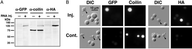
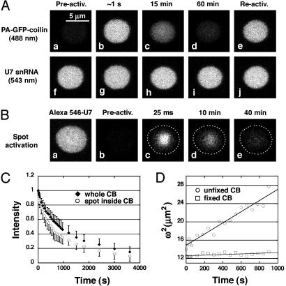
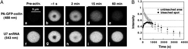
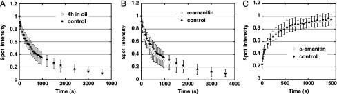
Cajal bodies (CBs) are complex organelles found in the nuclei of a wide variety of organisms, including vertebrates, invertebrates, plants, and yeast. In most cell types CBs are <1 microm in diameter, severely limiting the range of experimental observations that can be made on them. By contrast, CBs in the amphibian oocyte nucleus (also called the germinal vesicle) are 2-10 microm in diameter. We have taken advantage of this large size to carry out kinetic studies on coilin, a protein that is specifically enriched in CBs. We labeled coilin with photoactivatable green fluorescent protein and analyzed the movement of the protein by confocal microscopy in unfixed germinal vesicles isolated in oil. We showed that coilin leaves the CB relatively slowly (minutes rather than seconds) with kinetics similar to earlier measurements on its entrance. We also showed that coilin diffuses very slowly within the CB, consistent with its being in a large macromolecular complex. Finally, we found that the movement of coilin is not directly affected by the transcriptional state of the nucleus or ongoing nucleocytoplasmic exchange. These data on the kinetics of coilin reinforce the conclusion that CB components are in a constant state of flux, consistent with models that postulate an active role for CBs in nuclear physiology.


Figures

Fig. 1.
Fig. 1.
PA-GFP-coilin is correctly translated in the oocyte cytoplasm, imported into the GV, and targeted to CBs. (A) Western blot of GV proteins from oocytes that had been injected with transcripts of PA-GFP-coilin. The newly translated protein is detectable as a band at ≈105 kDa with antibodies against GFP, coilin, or the HA tag. Endogenous coilin in both control and injected oocytes appears as a band at ≈75 kDa with anti-coilin antibody. (B) Nuclear organelles from control oocytes and oocytes injected with transcripts of PA-GFP-coilin. In the injected oocytes, CBs are labeled with antibodies against GFP, coilin, or the HA tag, demonstrating that PA-GFP-coilin is targeted to CBs. In control oocytes, endogenous coilin is detectable in CBs only with the antibody against coilin. Arrowheads point to CBs.
Fig. 2.
Fig. 2.
Photoactivated PA-GFP-coilin disappears from CBs over a time period of ≈1h. (A) (a) Before activation, the CB is not detectable at 488 nm. (b) After photoactivation of PA-GFP-coilin with light of 405 nm, the CB fluoresces strongly at 488 nm. (c and d) Over time, the fluorescence disappears. (e) Fluorescence in the CB can be reactivated at the end of the observations, demonstrating that unactivated PA-GFP-coilin from the nucleoplasm has replaced activated PA-GFP-coilin in the CB. (fj) The fluorescence of Alexa 546-labeled U7 snRNA in the same CB remains unchanged during the experiment. (B) An experiment similar to that in A, except that only a small spot inside the CB was photoactivated. (a and b) Before photoactivation Alexa-546-U7 snRNA in the CB fluoresces at 543 nm (a), but PA-GFP-coilin in the same CB is not detectable at 488 nm (b). (ce) After photoactivation, PA-GFP-coilin disappears from the CB over a period of ≈1 h. (C) Quantitative data showing loss of PA-GFP-coilin from CBs as a function of time after photoactivation. The kinetics are similar for activation of the whole CB (filled diamonds) and of a spot within the CB (open circles). (D) Quantitative data showing very slow diffusion of PA-GFP-coilin within a CB. The increasing diameter (ω) of a photoactivated spot within a CB was determined for a period of 15 min after photoactivation. From the plot of ω2 versus t, one can calculate a diffusion coefficient (D) from the relationship D = ω2/8t (23). A spot within a fixed CB showed no increase in diameter.
Fig. 3.
Fig. 3.
Behavior of a photobleached spot within a photoactivated CB. (A) Before photoactivation, the CB is not detectable at 488 nm (PA-GFP-coilin) (a), but fluoresces strongly at 543 nm (Alexa 546-U7 snRNA) (f). When illuminated with the full intensity of the 405-nm laser for 1 s, PA-GFP-coilin is photoactivated throughout the CB and a spot is photobleached where the beam is most intense (b). A spot is also bleached in the Alexa-546-U7 snRNA (g). Over time, the PA-GFP-coilin disappears, whereas the Alexa-546-U7 snRNA undergoes conventional FRAP (ce and hj). (B) Quantitative data showing typical loss of intensity for PA-GFP-coilin in the photoactivated (unbleached) area of the CB (open circles). Within the bleached spot, the intensity increases for a few minutes and then decreases with kinetics similar to the unbleached area (filled diamonds).
Fig. 4.
Fig. 4.
The kinetics of coilin in the CB are independent of ongoing nucleocytoplasmic exchange or transcription. (A) PA-GFP-coilin leaves a photoactivated spot in a CB at the same rate in freshly isolated GVs (filled diamonds) as in isolated GVs held in oil for 4 h before observation (open circles). (B) PA-GFP-coilin leaves a photoactivated spot in a CB at the same rate in control GVs (filled diamonds) as in GVs from transcriptionally inactive oocytes (α-amanitin injected) (open circles). (C) FRAP kinetics of GFP-coilin are the same in control (filled diamonds) and transcriptionally inactive oocytes (α-amanitin injected) (open circles).

References

    1. Ramón y Cajal, S. (1903) Trab. Lab. Invest Biol. (Madrid) 2, 129–221.
    1. Ogg, S. C. & Lamond, A. I. (2002) J. Cell Biol. 159, 17–21. - PMC - PubMed
    1. Gall, J. G. (2000) Annu. Rev. Cell Dev. Biol. 16, 273–300. - PubMed
    1. Gall, J. G. (2003) Nat. Rev. Mol. Cell. Biol. 4, 975–980. - PubMed
    1. Andrade, L. E. C., Chan, E. K. L., Raška, I., Peebles, C. L., Roos, G. & Tan, E. M. (1991) J. Exp. Med. 173, 1407–1419. - PMC - PubMed

Publication types

LinkOut - more resources