. 2004 Apr 6;101(14):5024-9.
doi: 10.1073/pnas.0308386101. Epub 2004 Mar 25.

The spatial orientation of Helicobacter pylori in the gastric mucus

Affiliations

The spatial orientation of Helicobacter pylori in the gastric mucus

Sören Schreiber et al. Proc Natl Acad Sci U S A. .

Abstract

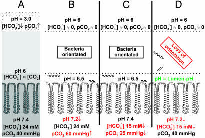
The highly motile human pathogen Helicobacter pylori lives deep in the gastric mucus layer. To identify which chemical gradient guides the bacteria within the mucus layer, combinations of luminal perfusion, dialysis, and ventilation were used to modify or invert transmucus gradients in anaesthetized Helicobacter-infected mice and Mongolian gerbils. Neither changes in lumen or arterial pH nor inversion of bicarbonate/CO2 or urea/ammonium gradients disturbed Helicobacter orientation. However, elimination of the mucus pH gradient by simultaneous reduction of arterial pH and bicarbonate concentration perturbed orientation, causing the bacteria to spread over the entire mucus layer. H. pylori thus uses the gastric mucus pH gradient for chemotactic orientation.


Figures

Fig. 1.
Fig. 1.
Distribution of H. pylori and H. felis in the mucus layer of mice and Mongolian gerbils. (A) The tissue surface of the H. pylori-infected gerbil depicted from the luminal side of the antrum. Several focus planes have been digitally combined, the H. pylori in the mucus layer subsequently highlighted in red. (B) The gastric mucosa and mucus of the H. felis-infected mouse and the H. pylori-infected gerbil are shown as schematic cross sections. The first 25 μm of the mucus layer on the tissue side (“juxtamucosal” mucus) are subdivided into 5-μm sections. The numbers represent the percentage of bacteria present within each section. The first 10 μm from the luminal surface is referred to as “luminal mucus,” the rest of the mucus layer as “central mucus.” H. felis was found located between 5 and 25 μm from the tissue surface (3). H. pylori, however, colonizes the whole section 0-25 μm from the tissue surface. Some H. pylori were attached to cells.
Fig. 2.
Fig. 2.
Elimination of the urea/ammonium gradient. (A) A schematic cross section of the uninfected gastric mucosa. Under normal conditions urea diffuses from the arterial plasma into the mucus and is transported with it into the lumen. Thus the concentration of urea is constant throughout the mucus layer. (B) The build-up of a gradient in the Helicobacter-infected mucosa. The bacterial urease converts the urea into ammonia. The ammonia immediately reacts to ammonium, neutralizing the bacterial intracellular pH. Most of the urea entering the mucus layer is converted to ammonium, thus creating a gradient, urea at the tissue side and ammonium at the luminal side. (C) To invert these concentrations, we used dialysis to apply ammonium and reduce the plasma urea concentration. High flow of a luminal solution containing urea washed away the luminal ammonium. This inversion of urea and ammonium concentrations should eliminate the gradient, but it failed to affect bacterial orientation. Urea/ammonium is therefore not the critical gradient that Helicobacter uses to orient itself.
Fig. 3.
Fig. 3.
pH and bicarbonate/CO2 gradients. Shown are the alterations made in the interdependent pH, bicarbonate, and CO2 concentrations in the mucus layer. (A) The normal conditions with a low luminal pH of 3 in either the infected or the uninfected mucosa. This luminal pH induces a pH of 6 in the juxtamucosal mucus. Under these conditions, secreted bicarbonate and diffused CO2 have the same concentration in the juxtamucosal mucus, whereas at the luminal side of the mucus layer, the bicarbonate concentration is low and the pCO2 high. The neutralization of the juxtamucosal mucus to pH 6 is caused by active bicarbonate secretion and a neutral pH in the newly secreted mucus. (B-D) To eliminate the pH bicarbonate/CO2 gradient, the luminal pH was neutralized to 6, and three different constellations of arterial pH, bicarbonate and CO2 were tested. (B) The first alteration of the gradient was achieved by doubling the inspiratory CO2 fraction, bicarbonate concentrations maintained at normal values through dialysis. This caused a low arterial pH with a normal bicarbonate concentration. (C) In the second constellation, a reduced arterial bicarbonate concentration was combined with a normal pH. This was achieved by reducing the arterial pCO2 through hyperventilation and lowering the bicarbonate concentration through dialysis. Neither the first nor the second alteration affected bacterial orientation. (D) However, a combined reduction of arterial pH and bicarbonate concentration through dialysis caused a loss of bacterial orientation, the bacteria spreading over the entire mucus layer.
Fig. 4.
Fig. 4.
Effects of gradient changes on the distribution of H. pylori and H. felis in the juxtamucosal mucus of gerbils and mice, respectively. Each animal is represented by one dot, which indicates the mean density of bacteria in the animal under control conditions plotted against the mean density of bacteria in the same animal under test conditions. The dotted line is the ideal curve showing where the density of bacteria under control conditions is identical to that under the test conditions. (A) Bacterial densities remained unaffected after changes in the urea/ammonium gradient (blue line), luminal pH (green line), arterial pH (red line), and arterial bicarbonate concentration (pink line). For these gradients, only the mean regression line of all values for one test series is shown. (B) The density of H. felis and H. pylori in the juxtamucosal mucus decreased significantly after the combined reduction of the arterial bicarbonate and pH, which eliminated the mucus pH gradient.
Fig. 5.
Fig. 5.
The juxtamucosal mucus pH in the explanted antrum of the guinea pig. (A Left) By using pH microelectrodes, the mucus pH directly above the epithelial cells (0-10 μm between microelectrode tip and cell membrane = juxtamucosal pH) was measured over a time period of 1 hour. (A Right) The difference between the juxtamucosal pH and the lumen pH is the mucus pH gradient (yellow arrow). After 10 min, the pH/bicarbonate/CO2 composition of the plasma solution was changed. (A Right Upper) After a combined reduction of plasma pH to 7.2 and bicarbonate concentration to 15 mM (pCO2 normal) (red arrow), the juxtamucosal pH decreases from its normal value to the same value as the lumen pH, thus eliminating the mucus pH gradient. Curves for lumen pH values of 6 and 4 are shown. (A Right Lower) In contrast to the changes above, a reduction of the plasma pH with normal bicarbonate concentration (high pCO2) or a reduction of the plasma bicarbonate concentration with normal pH (low pCO2) did not change the juxtamucosal pH, thus preserving the mucus pH gradient (green arrow). (B) The pH values of the mucus layer are illustrated in a schematic cross section with a color continuum from deep red (pH 3) to deep blue (pH 7.4). At a lumen pH value of 3 (B Left), the mucus pH gradient ranges from 3 at the luminal side to 5.5 near the tissue surface. After a change of the lumen pH to 6 (B Center), we measured a mucus pH gradient from the luminal-side value of 6 to ≈7 approaching the tissue surface. This gradient was also found with the uneffective pH/bicarbonate/CO2 changes in A Right Lower. Under the conditions shown in B Left and Center, Helicobacter is precisely oriented, guided by the pH gradients. B Right shows the transmucus pH profile after the critical reduction of the plasma pH to 7.2 and of the bicarbonate concentration to 15 mM at a luminal pH of 6. The mucus pH gradient is eliminated, and Helicobacter lose their orientation.

References

    1. Suerbaum, S. & Michetti, P. (2002) N. Engl. J. Med. 347, 1175-1186. - PubMed
    1. Schreiber, S. & Scheid, P. (1997) Am. J. Physiol. 272, G63-G70. - PubMed
    1. Schreiber, S., Stüben, M., Josenhans, C., Scheid, P. & Suerbaum, S. (1999) Infect. Immun. 67, 5151-5156. - PMC - PubMed
    1. Worku, M. L., Sidebotham, R. L., Walker, M. M., Keshavarz, T. & Karim, Q. N. (1999) Microbiology 145, 2803-2811. - PubMed
    1. Bren, A. & Eisenbach, M. (2000) J. Bacteriol. 182, 6865-6873. - PMC - PubMed

Publication types

LinkOut - more resources