. 2004 Apr;13(4):1148-54.
doi: 10.1110/ps.03472104.

Solution structure of a novel calcium binding protein, MTH1880, from Methanobacterium thermoautotrophicum

Affiliations

Solution structure of a novel calcium binding protein, MTH1880, from Methanobacterium thermoautotrophicum

Chang-Hun Lee et al. Protein Sci. 2004 Apr.

Abstract

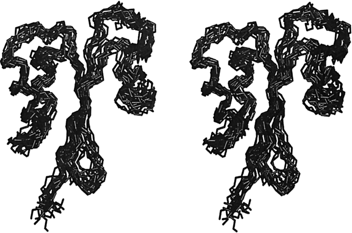
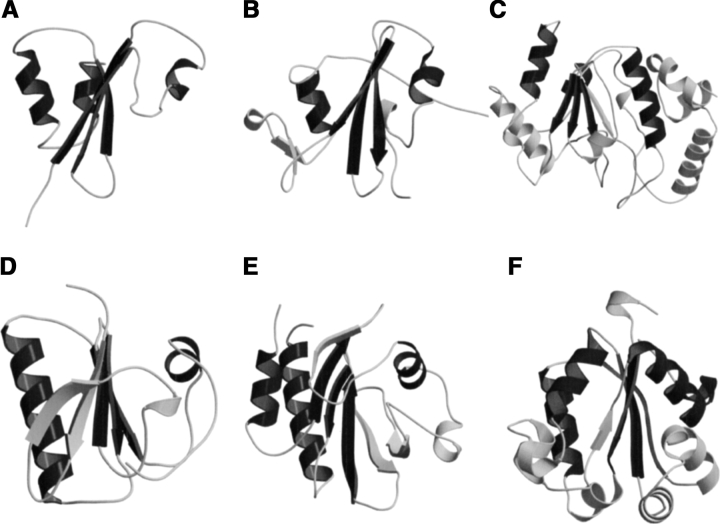
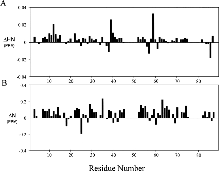
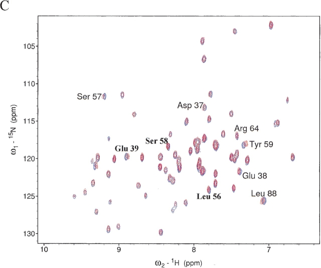
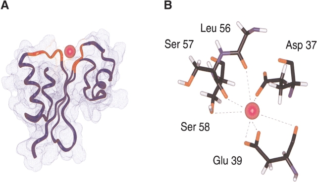
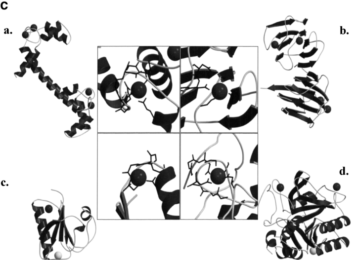
MTH1880 is a hypothetical protein from Methanobacterium thermoautotrophicum, a target organism of structural genomics. The solution structure determined by NMR spectroscopy demonstrates a typical alpha + beta-fold found in many proteins with different functions. The molecular surface of the protein reveals a small, highly acidic pocket comprising loop B (Asp36, Asp37, Asp38), the end of beta2 (Glu39), and loop D (Ser57, Ser58, Ser61), indicating that the protein would have a possible cation binding site. The NMR resonances of several amino acids within the acidic binding pocket in MTH1880, shifted upon addition of calcium ion. This calcium binding motif and overall topology of MTH1880 differ from those of other calcium binding proteins. MTH1880 did not show a calcium-induced conformational change typical of calcium sensor proteins. Therefore, we propose that the MTH1880 protein contains a novel motif for calcium-specific binding, and may function as a calcium buffering protein.


Figures

Figure 1.
Figure 1.
Solution structure of MTH1880. The backbone superposition of 20 final simulated annealing structures is displayed in stereo view.
Figure 2.
Figure 2.
The proteins with the α + β fold displayed in ribbon diagram together with MTH1880. All structures were based on the results of SCOP classification. (A) MTH1880 showing secondary structural topology, (B) SH2 domain of C-ABL (PDB: 1AB2), (C) uracil-DNA glycosylase (PDB: 1UDI), (D) severin domain 2 (PDB: 1SVY), (E) profilin (PDB: 1PNE), (F) nucleoside diphosphate kinase (PDB: 1EHW). The overall structures of SH2 and gelsolin are very similar to that of MTH1880; however, two proteins have different function. The structures are generated by MOLMOL (Koradi et al. 1996), MOLSCRIPT (Kraulis 1991), and Raster3D (Merritt and Bacon 1997) programs.
Figure 3.
Figure 3.
Chemical shift differences of (A) backbone amide proton and (B) nitrogen chemical shifts between free and calcium-bound form. The chemical shift difference (ppm) was calculated from the absolute value of the change in the 1H chemical shift and 15N chemical shift. (C) A superposition of the 2D 1H−15N HSQC spectra in the presence (red) and absence (blue) of calcium ions.
Figure 3.
Figure 3.
Chemical shift differences of (A) backbone amide proton and (B) nitrogen chemical shifts between free and calcium-bound form. The chemical shift difference (ppm) was calculated from the absolute value of the change in the 1H chemical shift and 15N chemical shift. (C) A superposition of the 2D 1H−15N HSQC spectra in the presence (red) and absence (blue) of calcium ions.
Figure 4.
Figure 4.
Calcium binding motif of MTH1880. (A) The structural model of MTH1880 bound to a calcium ion. Regions in red color represent the residues whose resonances changed upon calcium binding. (B) Proposed coordination of the calcium binding site based on NMR data. The residues involved in calcium binding were displayed with side-chain atoms. (C) A class of calcium binding motifs. (a) Calmodulin (PDB: 3CLN), (b) crystallin (PDB: 1HDF), (c) gelsolin (PDB: 4CPV), and (d) thermitase (PDB: 1THM). The calcium ion is shown as a black sphere and sodium ion as a white sphere.
Figure 4.
Figure 4.
Calcium binding motif of MTH1880. (A) The structural model of MTH1880 bound to a calcium ion. Regions in red color represent the residues whose resonances changed upon calcium binding. (B) Proposed coordination of the calcium binding site based on NMR data. The residues involved in calcium binding were displayed with side-chain atoms. (C) A class of calcium binding motifs. (a) Calmodulin (PDB: 3CLN), (b) crystallin (PDB: 1HDF), (c) gelsolin (PDB: 4CPV), and (d) thermitase (PDB: 1THM). The calcium ion is shown as a black sphere and sodium ion as a white sphere.

References

    1. Brunger, A.T., Adams, P.D., Clore, G.M., DeLano, W.L., Gros, P., Grosse-Kun-stleve, R.W., Jiang, J.S., Kuszewski, J., Nilges, M., Pannu, N.S., et al. 1998. Crystallography and NMR system: A new software suite for macromolecular structure determination. Acta Crystallogr. D. Biol. Crystallogr. 54 905–921. - PubMed
    1. Christendat, D., Yee, A., Dharamsi, A., Kluger, Y., Savchenko, A., Cort, J.R., Booth, V., Mackereth, C.D., Saridakis, V., Ekiel, I., et al. 2000. Structural proteomics of an archaeon. Nat. Struct. Biol. 7 903–909. - PubMed
    1. Delaglio, F., Grzesiek, S., Vuister, G.W., Zhu, G., Pfeifer, J., and Bax, A. 1995. NMRPipe: A multidimensional spectral processing system based on UNIX pipes. J. Biomol. NMR 6 277–293. - PubMed
    1. Goddard, T.D and Kneller, D.G. 2003. SPARKY 3. University of California, San Francisco, CA.
    1. Holm, L. and Sander, C. 1995. Dali: A network tool for protein structure comparison. Trends Biochem. Sci. 20 478–480. - PubMed

Publication types

MeSH terms

LinkOut - more resources