. 2004 Apr 7;23(7):1576-86.
doi: 10.1038/sj.emboj.7600173. Epub 2004 Mar 25.

GADD45beta/GADD45gamma and MEKK4 comprise a genetic pathway mediating STAT4-independent IFNgamma production in T cells

Affiliations

GADD45beta/GADD45gamma and MEKK4 comprise a genetic pathway mediating STAT4-independent IFNgamma production in T cells

Hongbo Chi et al. EMBO J. .

Abstract

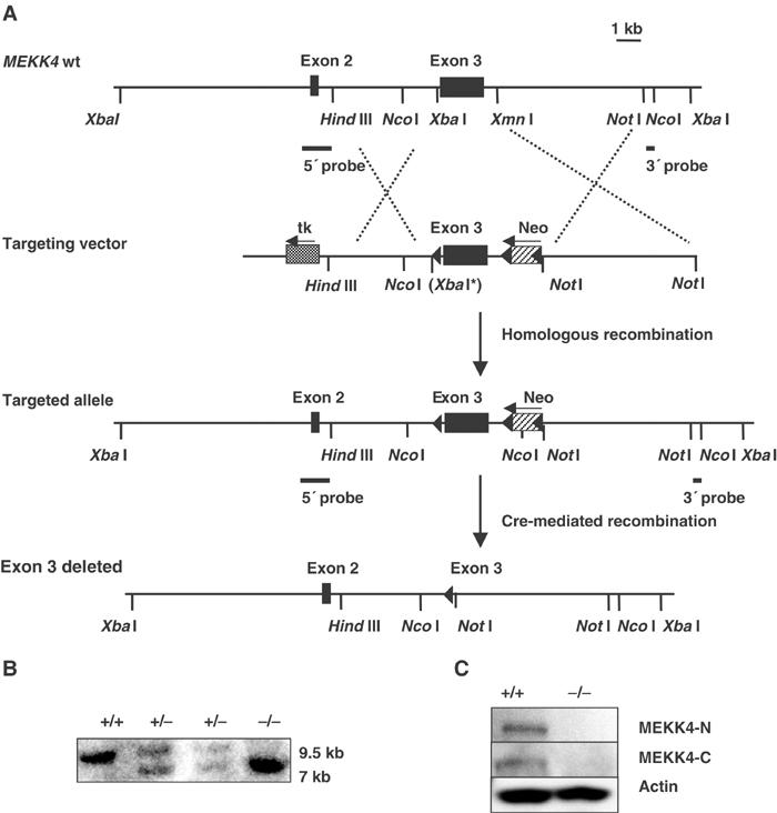
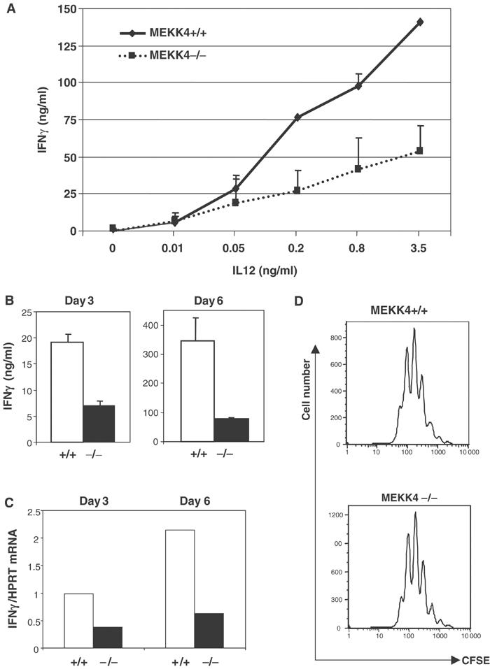
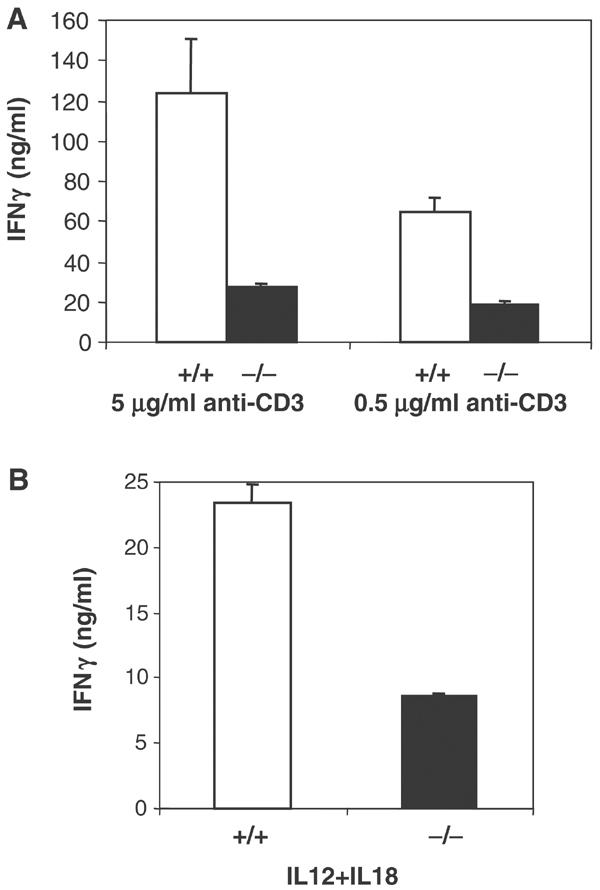
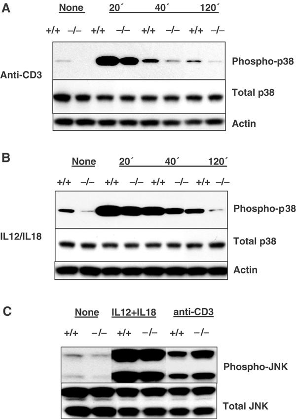
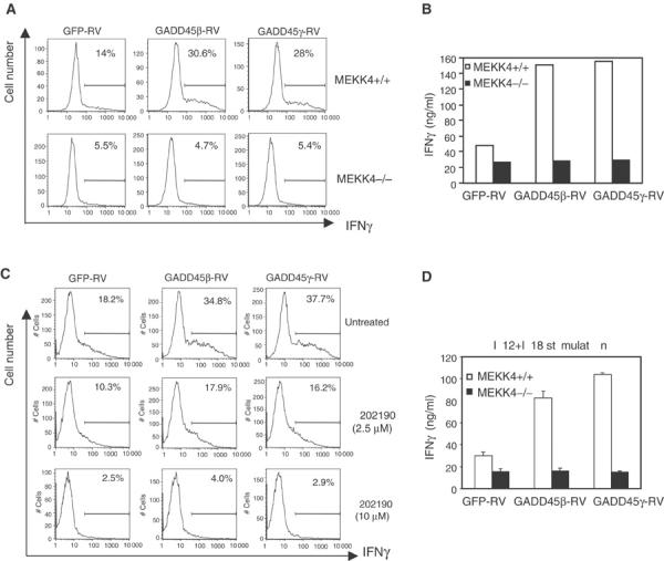
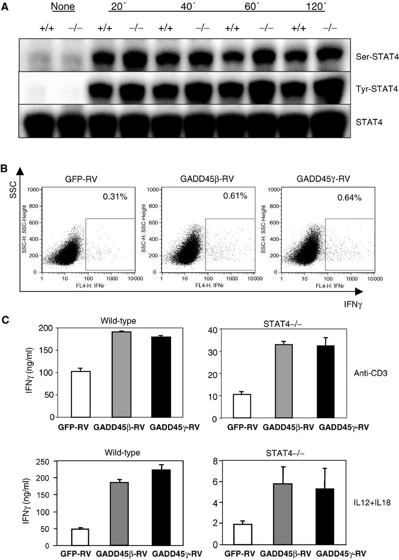
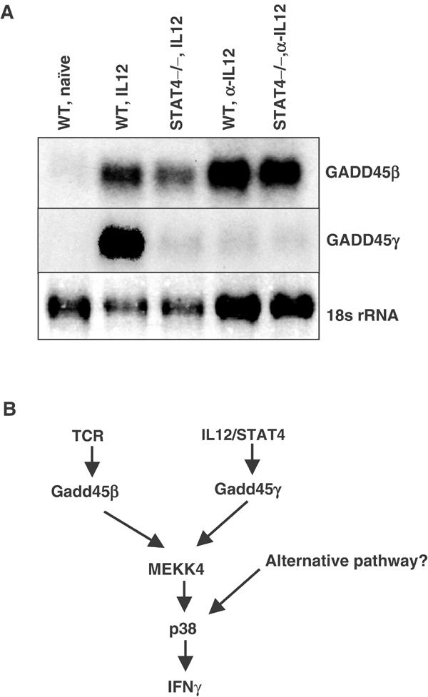
The stress-inducible molecules GADD45beta and GADD45gamma have been implicated in regulating IFNgamma production in CD4 T cells. However, how GADD45 proteins function has been controversial. MEKK4 is a MAP kinase kinase kinase that interacts with GADD45 in vitro. Here we generated MEKK4-deficient mice to define the function and regulation of this pathway. CD4 T cells from MEKK4-/- mice have reduced p38 activity and defective IFNgamma synthesis. Expression of GADD45beta or GADD45gamma promotes IFNgamma production in MEKK4+/+ T cells, but not in MEKK4-/- cells or in cells treated with a p38 inhibitor. Thus, MEKK4 mediates the action of GADD45beta and GADD45gamma on p38 activation and IFNgamma production. During Th1 differentiation, the GADD45beta/GADD45gamma/MEKK4 pathway appears to integrate upstream signals transduced by both T cell receptor and IL12/STAT4, leading to augmented IFNgamma production in a process independent of STAT4.


Figures

Figure 1
Figure 1
Generation of MEKK4 mutant mice. (A) Targeting strategy. The targeting vector to replace the endogenous MEKK4 exon 3 sequence contains a lox site (indicated by the filled arrow) inserted upstream of MEKK4 exon 3, and a neomycin cassette (neo) flanked by two lox sites downstream of exon 3. Cre-mediated recombination in vivo resulted in the complete deletion of MEKK4 exon 3. XbaI* indicates that the restriction site was destroyed. (B) Southern blot analysis of genomic DNA from MEKK4+/+, +/− and −/− mice. DNA was digested with NcoI and hybridized to the 3′ probe. The wild-type allele is 9.5 kb in length, and the mutant allele (with exon 3 deleted) is 7 kb. (C) Western blot analysis of activated CD4 T cells from MEKK4 +/+ and −/− mice. Two polyclonal antibodies that recognize the N-terminus (upper panel) and C-terminus (middle panel) of MTK1/MEKK4 coding sequence were used.
Figure 2
Figure 2
IFNγ synthesis is reduced in differentiating Th1 cells from MEKK4−/− mice. (A) CD4 T cells were activated by anti-CD3, anti-CD28, anti-IL4, and different doses of IL12 for 4 days, and IFNγ levels were measured by ELISA. (B) CD4 T cells were activated as in (A) in the presence of 2 ng/ml IL12, and IFNγ levels were measured by ELISA after 3 and 6 days of stimulation. (C) Real-time PCR analysis of IFNγ mRNA was performed using RNA isolated from cells after 3 and 6 days of Th1 differentiation. The amount of IFNγ mRNA was normalized to that of HPRT mRNA. (D) CFSE-labeled CD4 T cells were activated under Th1 conditions for 3 days, followed by FACS analysis.
Figure 3
Figure 3
IFNγ production is decreased in MEKK4−/− Th1 effector cells. CD4 T cells were activated under Th1 conditions for 6 days. Live cells were purified by Ficoll centrifugation and restimulated with 5 μg/ml anti-CD3, 0.5 μg/ml anti-CD3 (A) or 2 ng/ml IL12 and 20 ng/ml IL18 (B). IFNγ production was measured by ELISA.
Figure 4
Figure 4
MEKK4−/− Th1 cells exhibit defective p38 activation. CD4 T cells were activated under Th1 conditions for 6 days. Live cells were left untreated, or stimulated with 5 μg/ml anti-CD3 or 2 ng/ml IL12 and 20 ng/ml IL18. Protein extracts were isolated and analyzed by Western blot using antibodies recognizing phospho-p38, total p38, actin, phospho-JNK, and total JNK. (A) p38 activities after anti-CD3 stimulation. (B) p38 activities after IL12/IL18 stimulation. (C) JNK activities after 40 min stimulation by anti-CD3 or IL12/IL18.
Figure 5
Figure 5
MEKK4 and p38 are required for GADD45β and GADD45γ-induced IFNγ production. (A) IFNγ levels were analyzed by intracellular staining 2 days after retroviral transduction of MEKK4+/+ and −/− T cells. (B) IFNγ levels were measured by ELISA 4 days after retroviral transduction. (C) CD4 T cells were treated with SB202190 at the time of viral transduction. After 2 days, IFNγ levels were analyzed by intracellular staining. (D) At 4 days after viral transduction, GFP-positive cells were purified by FACS and restimulated with 2 ng/ml IL12 and 20 ng/ml IL18. IFNγ was measured by ELISA.
Figure 6
Figure 6
GADD45/MEKK4 induces STAT4-independent IFNγ production. (A) CD4 T cells were activated by anti-CD3 and anti-CD28 for 3 days. After washing and resting for 4 h, they were stimulated with 10 ng/ml IL12, and protein extracts were isolated at the times indicated. Western blot analysis was performed using antibodies against serine phosphorylated, tyrosine phosphorylated, and total STAT4. (B) STAT4−/− CD4 T cells were transduced with retroviruses, and analyzed by intracellular staining for IFNγ levels after 2 days. (C) STAT4+/+ and −/− CD4 T cells were transduced with retroviruses, and GFP-positive cells purified after 4 days were stimulated with 5 μg/ml anti-CD3 or 2 ng/ml IL12 and 20 ng/ml IL18. IFNγ was measured by ELISA.
Figure 7
Figure 7
Expression of GADD45β and GADD45γ is differentially regulated by STAT4. (A) CD4 T cells from wild-type and STAT4−/− were activated by anti-CD3 and anti-CD28 under Th1 conditions (2 ng/ml IL12 and 10 μg/ml anti-IL4) or neutralized conditions (anti-IL12 and anti-IL4) for 60 h. Lane 1: WT naïve T cells. Lane 2: WT Th1 cells. Lane 3: STAT4−/− Th1 cells. Lane 4: WT cells activated under neutralized conditions. Lane 5: STAT4−/− cells activated under neutralized conditions. Note the loading difference between Th1 and neutralized conditions. (B) A diagram for the regulation of the GADD45/MEKK4 pathway in CD4 T cells.

References

    1. Afkarian M, Sedy JR, Yang J, Jacobson NG, Cereb N, Yang SY, Murphy TL, Murphy KM (2002) T-bet is a STAT1-induced regulator of IL-12R expression in naive CD4+ T cells. Nat Immunol 3: 549–557 - PubMed
    1. Avni O, Lee D, Macian F, Szabo SJ, Glimcher LH, Rao A (2002) T(H) cell differentiation is accompanied by dynamic changes in histone acetylation of cytokine genes. Nat Immunol 3: 643–651 - PubMed
    1. Avni O, Rao A (2000) T cell differentiation: a mechanistic view. Curr Opin Immunol 12: 654–659 - PubMed
    1. Bulavin DV, Kovalsky O, Hollander MC, Fornace AJ Jr (2003) Loss of oncogenic H-ras-induced cell cycle arrest and p38 mitogen-activated protein kinase activation by disruption of Gadd45a. Mol Cell Biol 23: 3859–3871 - PMC - PubMed
    1. Camps M, Nichols A, Arkinstall S (2000) Dual specificity phosphatases: a gene family for control of MAP kinase function. FASEB J 14: 6–16 - PubMed

Publication types

MeSH terms