. 2004 Apr;2(4):E86.
doi: 10.1371/journal.pbio.0020086. Epub 2004 Mar 23.

Dissection and design of yeast prions

Affiliations

Dissection and design of yeast prions

Lev Z Osherovich et al. PLoS Biol. 2004 Apr.

Abstract

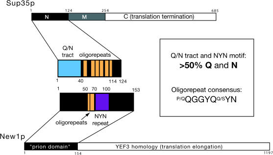
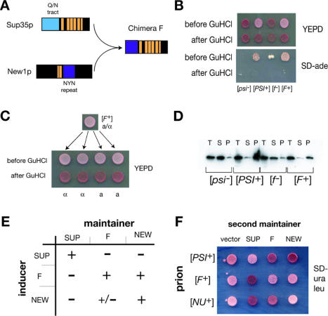
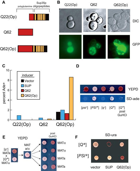
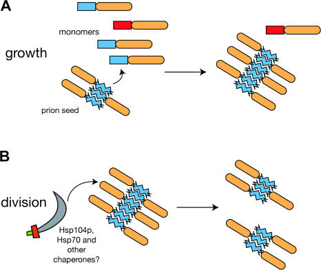
Many proteins can misfold into beta-sheet-rich, self-seeding polymers (amyloids). Prions are exceptional among such aggregates in that they are also infectious. In fungi, prions are not pathogenic but rather act as epigenetic regulators of cell physiology, providing a powerful model for studying the mechanism of prion replication. We used prion-forming domains from two budding yeast proteins (Sup35p and New1p) to examine the requirements for prion formation and inheritance. In both proteins, a glutamine/asparagine-rich (Q/N-rich) tract mediates sequence-specific aggregation, while an adjacent motif, the oligopeptide repeat, is required for the replication and stable inheritance of these aggregates. Our findings help to explain why although Q/N-rich proteins are relatively common, few form heritable aggregates: prion inheritance requires both an aggregation sequence responsible for self-seeded growth and an element that permits chaperone-dependent replication of the aggregate. Using this knowledge, we have designed novel artificial prions by fusing the replication element of Sup35p to aggregation-prone sequences from other proteins, including pathogenically expanded polyglutamine.


Conflict of interest statement

The authors have declared that no conflicts of interest exist.

Figures

Figure 1
Figure 1. Schematic Diagram of Sup35p and New1p
Prion domains of both proteins are enlarged in the center, highlighting the Q/N-rich tract of Sup35p (blue), the NYN tripeptide repeat of New1p (purple), and the oligopeptide repeat sequences (orange) found in both proteins. The sequence of the NEW1 oligopetide repeat (residues 50–70) is QQQRNWKQGGNYQQGGYQSYN, while that of the adjacent tripeptide repeat region (residues 71–100) is SNYNNYNNYNNYNNYNNYNNYNKYNGQGYQ.
Figure 2
Figure 2. Dissection of the New1p Prion Domain Reveals Distinct Regions Responsible for Aggregation and Prion Inheritance
(A) Indicated fragments of New1p (left) were expressed as GFP fusions (inducers) in a [nu] [pin] strain, examined by microscopy for GFP aggregation, then plated on SD-ade medium to assess induction of [NU+]. The symbol “+” indicates induction frequencies of at least 5%; the symbol “–” indicates no induction. Maintenance was assessed by the ability of an episomal maintainer version of the indicated fragment to support an Ade+ state after overexpression of New11–153-GFP (see Materials and Methods). The aggregation of New1-GFP fusions (second column) has been previously reported (Osherovich and Weissman 2001). (B) The NYN repeat of New1p induces [NU+] and [NU+]mini. New170–100-GFP was overexpressed in [nu] and [nu]mini strains ([pin] and [PIN+] derivatives of each), along with vector only or New11–153-GFP controls. Averages of three independent trials, representing 600–2000 colonies, are shown for most induction experiments; inductions using New170–100-GFP were conducted twice. Error bars show minimal and maximal observed induction efficiencies. (C) Reversibility of [NU+]mini. The [pin] Ade+ convertants obtained in (B) were colony purified on SD-ade medium and confirmed to have lost the inducer plasmid. A stable [NU+]mini isolate is shown before and after induction, as well as after GuHCl treatment, along with [nu] and [NU+] reference strains.
Figure 3
Figure 3. Dissection of the Sup35p Prion Domain
At top are schematic diagrams of these experiments; positive outcomes are shown below the arrows. In some cases, similar experiments have been reported by Parham et al. (2001) (indicated by “a”) and are repeated here as controls. Aggregation: Plasmid-borne M-GFP fusions of the indicated Sup35p N domain fragments (green) were overexpressed in a [psi] [PIN+] strain and examined for fluorescent focus formation. The symbol “+” indicates that 10% or more of cells displayed aggregates. Sup351–57-M-GFP displayed a lower frequency of aggregation (approximately 1%). Induction: Strains from the aggregation experiment were plated onto SD-ade medium and scored for growth to test whether aggregates of truncated protein (green) convert chromosomally encoded protein (blue) to [PSI+]. The symbol “+” indicates approximately 5–10% conversion frequency. Consistent with the aggregation experiment, Sup351–57-M-GFP displayed a lower frequency of [PSI+] induction (approximately 1%). Decoration: Indicated proteins were expressed as –M-GFP fusions in [PSI+] [PIN+] cells, which were examined to determine whether GFP-labeled truncations (green) decorate preexisting aggregates of full-length Sup35p (blue). Curiously, Sup351–49-M-GFP in [PSI+] cells formed abnormally large “ribbon” aggregates of the kind typically observed during de novo [PSI+] induction; furthermore, approximately 10% of the cells reverted to [psi] (indicated by “*”). Thus, this truncation was a potent dominant PNM mutant. Maintenance: A SUP35-deleted [PSI+] [PIN+] bearing wild-type SUP35 maintainer (blue) was transformed with maintainer plasmids containing the indicated truncation (purple). The wild-type maintainer was lost by counterselection, and the resulting strain was tested for [PSI+] by color and growth on SD-ade medium. The Sup351–93 mutant displayed an intermediate pink color and grew poorly on SD-ade medium, as previously reported (Parham et al. 2001). Note: King (2001) reports that Sup351–61-GFP fusion could decorate [PSI+] aggregates in certain strains and could induce [PSI+] de novo when overexpressed.
Figure 4
Figure 4. PNM2–1 (G58D) Prevents Inheritance But Not Aggregation of Sup35p Prions
(A) PNM2-1 protein can seed [PSI+]. A Sup35p inducer containing the PNM2-1 (G58D) mutation was overexpressed in [psi] [PIN+] cells; shown are cells (inset) with representative fluorescent foci, which were the same in frequency and appearance as cells with a wild-type inducer. Cells overexpressing inducer versions of wild-type Sup35p (SUP), an aggregation-defective N-terminal truncation (Δ1–38), and PNM2-1 were plated and scored for Ade+. Approximately 1000 colonies were counted. (B) PNM2-1 protein polymerization is similar to that of wild-type protein. (C) Preformed PNM2-1 polymers seed wild-type and PNM2-1 monomers with comparable efficiency. Endpoint PNM2-1 polymers were used to seed fresh reactions. (D) PNM2-1 displays a partially dominant, incompletely penetrant defect in [PSI+] maintenance. [psi] (1) and [PSI+] (2) SUP35::TRP1 pSUP35 controls are shown. [PSI+] [PIN+] SUP35::TRP1 pSUP35 was transformed with a second maintainer expressing PNM2-1 (3). The wild-type maintainer (pSUP35) was then lost through counterselection (4). Red sectors from (4) were isolated, retransformed with the wild-type maintainer, and allowed to lose the PNM2-1 maintainer (5). (E) Mitotic instability of [PSI+] in the PNM2-1 strain. A pink (Ade+) [PSI+] [PIN+] PNM2-1 isolate was grown to log phase in SD-ade liquid then shifted into nonselective (YEPD) medium. At indicated time points, aliquots were plated onto SD-ade and YEPD media to determine the fraction of [PSI+] cells (minimum of 200 colonies counted per time point). Whereas a wild-type control remained [PSI+] through the experiment, the PNM2-1 strain rapidly lost [PSI+] during logarithmic growth; during stationary phase (18 h and beyond), the percentage of [PSI+] PNM2-1 strains remained unchanged (approximately 5%). (F) Propagon count of PNM2-1 vs. wild-type [PSI+] strains. The majority of PNM2-1 cells had no [PSI+] propagons (i.e., were [psi]). In both strains, a small number of “jackpot” cells contained over 200 propagons; see Cox et al. (2003).
Figure 5
Figure 5. F, A New1p–Sup35p Chimera, Shows Prion Characteristics of New1p
(A) Schematic diagram illustrating the construction of chimera F. (B) Chimera F forms a prion, [F+]. The SUP35 gene in a [psi] [pin] strain was replaced with the F-M-C fusion; after transient overexpression of F-M-GFP, approximately 10% of these cells converted from an Ade- ([f ]) to an Ade+ ([F+]) state. Shown are examples of[f ] and [F+] strains, before and after GuHCl treatment, along with [psi] and [PSI+] controls. (C) Non-Mendelian inheritance of [F+]. A diploid made by mating a [F+] MATa strain against an [f ] MATα displayed a [F+] phenotype and, when sporulated, produced four [F+] meiotic progeny. All 11 tetrads examined showed this 4:0 pattern of inheritance. (D) Sedimentation analysis of F-M-C. Extracts of [f ] and [F+] strains, along with [psi] and [PSI+] controls, were subjected to 50K × g ultracentrifugation for 15 min. Total, supernatant, and pellet fractions were separated by SDS-PAGE, transferred to nitrocellulose, and probed with anti-SUP35NM serum. As with Sup35p, the prion form of F-M-C sediments primarily to the pellet but remains in the supernatant in [f ]. (E) F-M-GFP overexpression induces [NU+] but not [PSI+]. Indicated inducers and maintainers were used in an induction experiment. The symbol “+” indicates approximately 5–10% conversion to Ade+. F induced [NU+] at a comparable efficiency to New11–153; although New11–153 overexpression promoted the appearance of Ade+ colonies in the F-M-C strain, these were fewer in number (less than 5%) and reverted to Ade- after restreaking. (F) [F+] and [NU+] prion proteins interact with each other but not with [PSI+]. Episomal “second maintainers” were introduced into the indicated strains, along with an empty vector control. Antisuppression (red) indicates that the second maintainer is soluble, while white/pink indicates coaggregation of the endogenous and episomal maintainers.
Figure 6
Figure 6. [Q+], a Prion Form of Pathogenically Expanded Polyglutamine
(A) Schematic illustrating the construction of polyglutamine-derived prion domains. (Op) indicates the presence of the Sup35p oligopeptide repeats (residues 40–124). (B) Fluorescence micrographs of [psi] [PIN+] strains expressing indicated polyglutamine inducers. (C) Polyglutamine-based prion inheritance. Strains with indicated inducers and maintainers were plated onto SD-ade and YEPD media to determine the fraction of Ade+ after a standard induction experiment. Interestingly, Q62 inducer forms aggregates but does not promote Ade+ in the Q62(Op) maintainer strain. Note that Q62(Op) shows a high rate of spontaneous appearance of Ade+. (D) GuHCl sensitivity of the [Q+] state. An Ade+ convertant obtained in (C) was restreaked to lose the inducer plasmid, then grown on GuHCl. Shown are plates before and after GuHCl treatment, along with [psi] and [PSI+] controls. (E) Dominance and non-Mendelian inheritance of [Q+]. See Figure 5C. (F) [Q+] does not interact with Sup35p and vice versa. [Q+] and [PSI+] strains were transformed with indicated maintainers; mismatches between the maintainer and the chromosomally encoded allele result in antisuppression (red).
Figure 7
Figure 7. Model for Prion Growth and Division
(A) During prion growth, polymers seed the incorporation of monomers through interactions between Q/N-rich aggregation sequences (blue). Proteins with noncognate aggregation sequences (red) are excluded. (B) The division phase of prion replication requires the oligopeptide repeats (orange), which may facilitate the action of chaperones such as Hsp104p (scimitar) in breaking the polymer into smaller, heritable units.

Comment in

References

    1. Balbirnie M, Grothe R, Eisenberg DS. An amyloid-forming peptide from the yeast prion Sup35 reveals a dehydrated beta-sheet structure for amyloid. Proc Natl Acad Sci U S A. 2001;98:2375–2380. - PMC - PubMed
    1. Borchsenius AS, Wegrzyn RD, Newnam GP, Inge-Vechtomov SG, Chernoff YO. Yeast prion protein derivative defective in aggregate shearing and production of new “seeds.”. EMBO J. 2001;20:6683–6691. - PMC - PubMed
    1. Chernoff YO, Lindquist SL, Ono B, Inge-Vechtomov SG, Liebman SW. Role of the chaperone protein Hsp104 in propagation of the yeast prion-like factor [psi+] Science. 1995;268:880–884. - PubMed
    1. Chernoff YO, Galkin AP, Lewitin E, Chernova TA, Newnam GP, et al. Evolutionary conservation of prion-forming abilities of the yeast Sup35 protein. Mol Microbiol. 2000;35:865–876. - PubMed
    1. Chien P, Weissman JS. Conformational diversity in a yeast prion dictates its seeding specificity. Nature. 2001;410:223–227. - PubMed

Publication types

Associated data