. 2004 Apr;48(4):1168-74.
doi: 10.1128/AAC.48.4.1168-1174.2004.

Dynamics and spatial distribution of beta-lactamase expression in Pseudomonas aeruginosa biofilms

Affiliations

Dynamics and spatial distribution of beta-lactamase expression in Pseudomonas aeruginosa biofilms

Niels Bagge et al. Antimicrob Agents Chemother. 2004 Apr.

Abstract

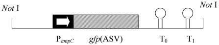
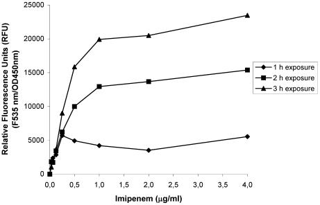
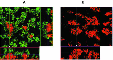
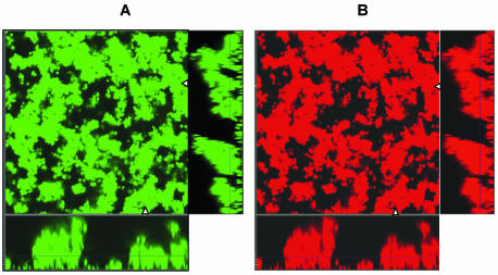
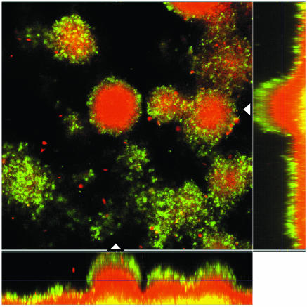
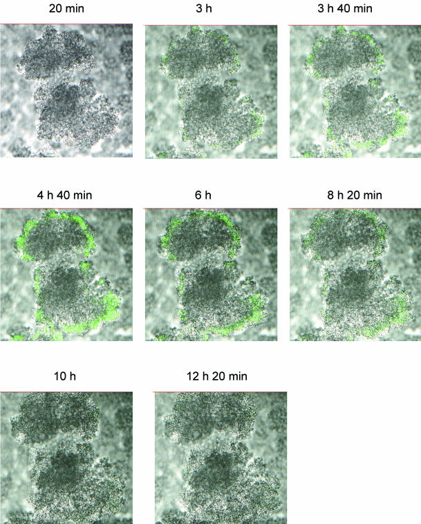
The development of resistance to beta-lactam antibiotics is a problem in the treatment of chronic Pseudomonas aeruginosa infection in the lungs of patients with cystic fibrosis. The main resistance mechanism is high-level expression of the chromosomally encoded AmpC beta-lactamase of P. aeruginosa cells growing in biofilms. Several genes have been shown to influence the level of ampC expression, but little is known about the regulation of ampC expression in P. aeruginosa biofilms. To study the expression of ampC in P. aeruginosa biofilms, we constructed a reporter that consisted of the fusion of the ampC promoter to gfp(ASV) encoding an unstable version of the green fluorescent protein. In vitro biofilms of P. aeruginosa were exposed to the beta-lactam antibiotics imipenem and ceftazidime. Sub-MICs of imipenem significantly induced the monitor system of the biofilm bacteria in the peripheries of the microcolonies, but the centers of the microcolonies remained uninduced. However, the centers of the microcolonies were physiologically active, as shown by experiments with another monitor construction consisting of an arabinose-inducible promoter fused to gfp(ASV). The whole biofilm was induced in the presence of increased imipenem concentrations. Ceftazidime induced the monitor system of the biofilm bacteria as well, but only bacteria in the peripheries of the microcolonies were induced in the presence of even very high concentrations. The experiments illustrate for the first time the dynamic and spatial distributions of beta-lactamase induction in P. aeruginosa cells growing in biofilms. Thus, our experiments show that P. aeruginosa cells growing in biofilms constitute a heterogeneous population unit which may create different antibiotic-selective environments for the bacteria in the biofilm.


Figures

FIG. 1.
FIG. 1.
Schematic drawing of the translational fusion PampC -gfp(ASV). The NotI fragment indicated was cloned into shuttle vector pNB100, a modified plasmid pUCP22Not. The first 12 codons of ampC were maintained and fused to the gfp(ASV) open reading frame. The 5′ region of the PampC fragment included the whole intergenic region of ampC-ampR. The translational fusion of PampC -gfp(ASV) was followed by translational stop codons in all three reading frames, in addition to the two strong transcriptional terminators T0 (the transcriptional terminator from phage lambda) and T1 (the transcriptional terminator from the rrnB operon of E. coli (1).
FIG. 2.
FIG. 2.
Expression of PampC-gfp(ASV) in planktonic cell cultures of P. aeruginosa exposed to imipenem. The relative fluorescence units in planktonic PAO1 cells growing in response to imipenem were measured after 1, 2, and 3 h. The cells were induced during exponential growth. A relative fluorescence unit of 0 corresponded to completely repressed Gfp expression. Fully induced single cells showed very distinct, visible Gfp expression. OD450nm, optical density at 450 nm.
FIG. 3.
FIG. 3.
Epifluorescence and scanning confocal laser micrographs of PAO1-J32 biofilms on day 6 showing the activity of the ampC promoter in response to imipenem exposure (0.5 μg/ml for 4 h) (A) and the activity of the ampC promoter in a noninduced biofilm (B). Each micrograph consists of a horizontal section and two vertical sections through the biofilms collected at the positions indicated by the white triangles in the horizontal section. The position of the horizontal image is indicated by the crossing lines in the vertical sections. The biofilms were stained red with SYTO 62, demonstrating the presence of biomass in the flow cell. The expression of Gfp indicates the expression of the AmpC β-lactamase in the biofilm.
FIG. 4.
FIG. 4.
Epifluorescence and scanning confocal laser photomicrographs of PAO1-J32 biofilms on day 6 showing the activity of the ampC promoter in response to imipenem exposure (10 μg/ml for 4 h) (A) and the same biofilm stained red with SYTO 62 to demonstrate the total biomass of the biofilm (B). Each micrograph consists of a horizontal section and two vertical sections through the biofilms collected at the positions indicated by the white triangles in the horizontal section. The position of the horizontal image is indicated by the crossing lines in the vertical sections. The expression of Gfp indicates the expressions of the AmpC β-lactamase in the biofilm.
FIG. 5.
FIG. 5.
Epifluorescence and scanning confocal laser photomicrographs of PAO1-J32 biofilms on day 6 showing the activity of the ampC promoter in response to ceftazidime exposure (100 μg/ml for 4 h). The micrograph consists of a horizontal section and two vertical sections through the biofilms collected at the positions indicated by the white triangles in the horizontal section. The position of the horizontal image is indicated by the crossing lines in the vertical sections. The biofilms were stained red with SYTO 62, demonstrating the presence of biomass in the flow cell. The expression of Gfp indicates the expression of the AmpC β-lactamase in the biofilm.
FIG. 6.
FIG. 6.
Epifluorescence and scanning confocal laser photomicrographs showing a time series of images of a horizontal section of a 6-day-old PAO1-J32 biofilm during induction with imipenem. Imipenem was administered from time zero (0 h), and the image of one microcolony was scanned every 10 min for 14 h. The result was increasing levels of AmpC β-lactamase expression in the first 5 h, as demonstrated by Gfp expression, followed by decreased levels of expression that faded out completely after 10 h.

References

    1. Andersen, J. B., C. Sternberg, L. K. Poulsen, S. P. Bjørn, M. Givskov, and S. Molin. 1998. New unstable variants of green fluorescent protein for studies of transient gene expression in bacteria. Appl. Environ. Microbiol. 64:2240-2246. - PMC - PubMed
    1. Anwar, H., J. L. Strap, and J. W. Costerton. 1992. Establishment of aging biofilms: possible mechanism of bacterial resistance to antimicrobial therapy. Antimicrob. Agents Chemother. 36:1347-1351. - PMC - PubMed
    1. Bagge, N., O. Ciofu, M. Hentzer, J. I. A. Campbell, M. Givskov, and N. Høiby. 2002. Constitutive high expression of chromosomal beta-lactamase in Pseudomonas aeruginosa caused by a new insertion sequence (IS1669) located in ampD. Antimicrob. Agents Chemother. 46:3406-3411. - PMC - PubMed
    1. Baquero, F., M.-C. Negri, M.-I. Morosini, and J. Blázquez. 1998. Antibiotic-selective environments. Clin. Infect. Dis. 27:S5-S11. - PubMed
    1. Christensen, B. B., C. Sternberg, J. B. Andersen, R. J. Palmer, A. T. Nielsen, M. Givskov, and S. Molin. 1999. Molecular tools in physiological studies of microbial biofilms. Methods Enzymol. 310:20-42. - PubMed

Publication types