. 2004 Apr;48(4):1281-8.
doi: 10.1128/AAC.48.4.1281-1288.2004.

Mycobacterium tuberculosis DNA gyrase: interaction with quinolones and correlation with antimycobacterial drug activity

Affiliations

Mycobacterium tuberculosis DNA gyrase: interaction with quinolones and correlation with antimycobacterial drug activity

Alexandra Aubry et al. Antimicrob Agents Chemother. 2004 Apr.

Abstract

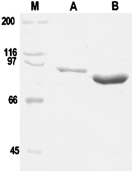
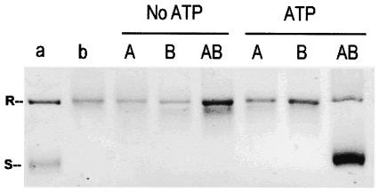
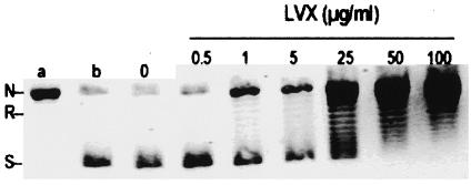
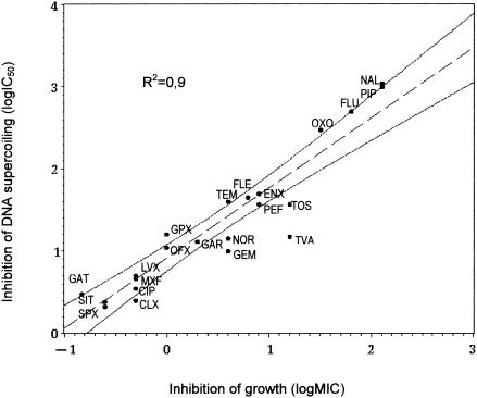
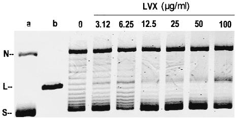
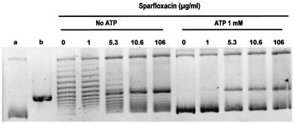
Genome studies suggest that DNA gyrase is the sole type II topoisomerase and likely the unique target of quinolones in Mycobacterium tuberculosis. Despite the emerging importance of quinolones in the treatment of mycobacterial disease, the slow growth and high pathogenicity of M. tuberculosis have precluded direct purification of its gyrase and detailed analysis of quinolone action. To address these issues, we separately overexpressed the M. tuberculosis DNA gyrase GyrA and GyrB subunits as His-tagged proteins in Escherichia coli from pET plasmids carrying gyrA and gyrB genes. The soluble 97-kDa GyrA and 72-kDa GyrB subunits were purified by nickel chelate chromatography and shown to reconstitute an ATP-dependent DNA supercoiling activity. The drug concentration that inhibited DNA supercoiling by 50% (IC(50)) was measured for 22 different quinolones, and values ranged from 2 to 3 microg/ml (sparfloxacin, sitafloxacin, clinafloxacin, and gatifloxacin) to >1,000 microg/ml (pipemidic acid and nalidixic acid). By comparison, MICs measured against M. tuberculosis ranged from 0.12 microg/ml (for gatifloxacin) to 128 microg/ml (both pipemidic acid and nalidixic acid) and correlated well with the gyrase IC(50)s (R(2) = 0.9). Quinolones promoted gyrase-mediated cleavage of plasmid pBR322 DNA due to stabilization of the cleavage complex, which is thought to be the lethal lesion. Surprisingly, the measured concentrations of drug inducing 50% plasmid linearization correlated less well with the MICs (R(2) = 0.7). These findings suggest that the DNA supercoiling inhibition assay may be a useful screening test in identifying quinolones with promising activity against M. tuberculosis. The quinolone structure-activity relationship demonstrated here shows that C-8, the C-7 ring, the C-6 fluorine, and the N-1 cyclopropyl substituents are desirable structural features in targeting M. tuberculosis gyrase.


Figures

FIG. 1.
FIG. 1.
SDS-PAGE analysis of purified M. tuberculosis GyrA and GyrB proteins. The His-tagged proteins were overexpressed in E. coli and purified by nickel resin chromatography, and approximately 16 μl of each protein sample was loaded on an SDS-9% polyacrylamide gel. Following electrophoresis, proteins were revealed by staining with Coomassie blue. Lane M, size markers (sizes are indicated to the left in kilodaltons).
FIG. 2.
FIG. 2.
M. tuberculosis GyrA and GyrB proteins generate an ATP-dependent DNA supercoiling activity. Relaxed pBR322 (0.4 μg) was incubated with DNA gyrase reconstituted from GyrA (1 U) and GyrB (1 U) in the absence and presence of 1 mM ATP. The reactions were stopped, and the DNA products were separated by electrophoresis in a 1% agarose gel. DNA was stained with ethidium bromide and photographed under UV illumination. Lanes: a, supercoiled pBR322 DNA; b, relaxed pBR322 DNA; A, relaxed pBR322 DNA and GyrA (1 U) protein; B, relaxed pBR322 DNA and GyrB (1 U) protein; AB, relaxed pBR322 DNA and both GyrA (1 U) and GyrB (1 U). R and S, relaxed and supercoiled DNA, respectively.
FIG. 3.
FIG. 3.
DNA supercoiling activity of wild-type M. tuberculosis DNA gyrase is sensitive to inhibition by levofloxacin (LVX). Relaxed pBR322 (0.4 μg) was incubated with DNA gyrase reconstituted from GyrA (2 U) and GyrB (2 U) in the absence and the presence of levofloxacin. The reactions were stopped, and the DNA products were analyzed by electrophoresis in a 1% agarose gel. Lanes a and b, relaxed and supercoiled pBR322 DNA, respectively. N, R, and S, nicked, relaxed, and supercoiled DNA, respectively.
FIG. 4.
FIG. 4.
Correlation between quinolone inhibition of M. tuberculosis gyrase (IC50 for DNA supercoiling) and quinolone MICs for M. tuberculosis. Dotted lines represent the confidence interval for 95% of the regression. R2 is the correlation coefficient. CIP, ciprofloxacin; CLX, clinafloxacin; ENX, enoxacin; FLE, fleroxacin; FLU, flumequine; GAR, garenoxacin; GAT, gatifloxacin; GEM, gemifloxacin, GRX, grepafloxacin; LVX, levofloxacin; MXF, moxifloxacin; NAL, nalidixic acid; NOR, norfloxacin, OFX, ofloxacin; OXO, oxolinic acid; PEF, pefloxacin; PIP, pipemidic acid; SIT, sitafloxacin; SPX, sparfloxacin; TEM, temafloxacin; TOS, tosufloxacin; TVA, trovafloxacin.
FIG. 5.
FIG. 5.
Levofloxacin-mediated DNA cleavage by M. tuberculosis DNA gyrase. Supercoiled pBR322 DNA (0.4 μg) was incubated with M. tuberculosis GyrA (2 U) and GyrB (2 U) proteins in the absence of ATP and in the presence of levofloxacin (LVX) at the concentrations indicated on the figure. After addition of SDS and proteinase K, DNA samples were analyzed by electrophoresis in 1% agarose. Lanes a and b, supercoiled pBR322 DNA and BamHI-linearized pBR322. N, L, and S, nicked, linear, and supercoiled DNA, respectively.
FIG. 6.
FIG. 6.
Effect of ATP on the cleavage assay for the DNA gyrase of M. tuberculosis in the presence of quinolones. This figure shows the assay in the presence of sparfloxacin. Supercoiled pBR322 DNA (0.4 μg) was incubated with the M. tuberculosis GyrA (1 U) and GyrB (1 U) proteins in the absence of ATP and in the presence ATP (1 mM) plus increasing concentrations of sparfloxacin at the concentrations indicated in the figure. After addition of SDS and proteinase K, DNA samples were analyzed by electrophoresis in 1% agarose. Lanes a and b, supercoiled pBR322 DNA and BamHI-linearized pBR322, respectively. N, L, and S, nicked, linear, and supercoiled DNA, respectively.

References

    1. Alovero, F. L., X. S. Pan, J. E. Morris, R. H. Manzo, and L. M. Fisher. 2000. Engineering the specificity of antibacterial fluoroquinolones: benzenesulfonamide modifications at C-7 of ciprofloxacin change its primary target in Streptococcus pneumoniae from topoisomerase IV to gyrase. Antimicrob. Agents Chemother. 44:320-325. - PMC - PubMed
    1. Banerjee, S. K., K. Bhatt, S. Rana, P. Misra, and P. K. Chakraborti. 1996. Involvement of an efflux system in mediating high level of fluoroquinolone resistance in Mycobacterium smegmatis. Biochem. Biophys. Res. Commun. 226:362-368. - PubMed
    1. Barnard, F. M., and A. Maxwell. 2001. Interaction between DNA gyrase and quinolones: effects of alanine mutations at GyrA subunit residues Ser83 and Asp87. Antimicrob. Agents Chemother. 45:1994-2000. - PMC - PubMed
    1. Barrett, J. F., J. I. Bernstein, H. M. Krause, J. J. Hilliard, and K. A. Ohemeng. 1993. Testing potential gyrase inhibitors of bacterial DNA gyrase: a comparison of the supercoiling inhibition assay and “cleavable complex” assay. Anal. Biochem. 214:313-317. - PubMed
    1. Blanche, F., B. Cameron, F. X. Bernard, L. Maton, B. Manse, L. Ferrero, N. Ratet, C. Lecoq, A. Goniot, D. Bisch, and J. Crouzet. 1996. Differential behaviors of Staphylococcus aureus and Escherichia coli type II DNA topoisomerases. Antimicrob. Agents Chemother. 40:2714-2720. - PMC - PubMed

Publication types

MeSH terms

LinkOut - more resources