

Distribution of constitutive (COX-1) and inducible (COX-2) cyclooxygenase in postviral human liver cirrhosis: a possible role for COX-2 in the pathogenesis of liver cirrhosis
- PMID: 15047734
- PMCID: PMC1770276
- DOI: 10.1136/jcp.2003.012120
Distribution of constitutive (COX-1) and inducible (COX-2) cyclooxygenase in postviral human liver cirrhosis: a possible role for COX-2 in the pathogenesis of liver cirrhosis
Erratum in
- J Clin Pathol. 2004 Oct;57(10):1119
- J Clin Pathol. 2004 Sep;57(9):1008. El-Aleem, SA [corrected to Abd El-Aleem, SA]
Abstract
Aims: Prostaglandins produced by the action of cyclooxygenases (COX) are important mediators of systemic vasodilatation and inflammation in liver cirrhosis. The aim of this study was to investigate the distribution of COX-1 and COX-2 in postviral cirrhosis.
Methods: The immunohistochemical expression of the constitutive (COX-1) and the inducible (COX-2) isoenzymes was investigated in 15 patients with cirrhosis after hepatitis B and C infection; three normal control livers were also analysed.
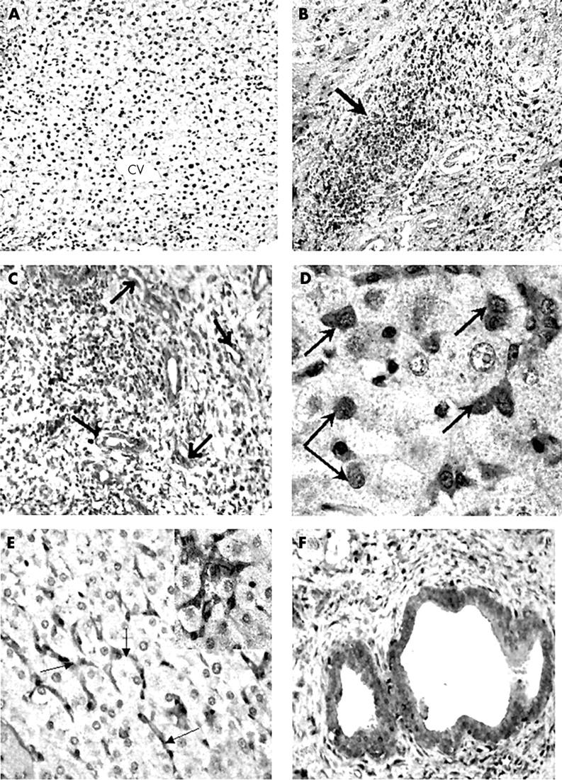
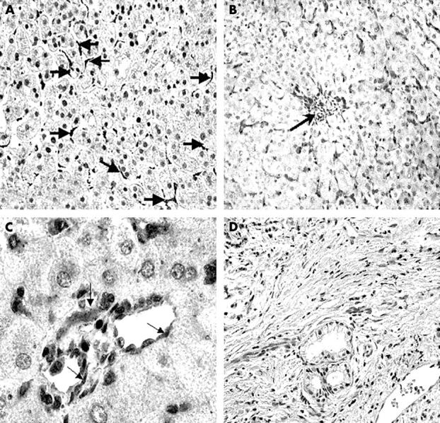
Results: COX-2 was absent from normal liver but was highly expressed in cirrhosis, mainly in the inflammatory, sinusoidal, vascular endothelial, and biliary epithelial cells. Low amounts of COX-1 were expressed in both normal and cirrhotic livers, exclusively in sinusoidal and vascular endothelial cells, with no differences seen between normal and cirrhotic livers.
Conclusions: COX-2 is overexpressed in liver cirrhosis, and possibly contributes to prostaglandin overproduction, which may be a major component of the inflammation and hyperdynamic circulation associated with cirrhosis. Because COX-2 is thought to contribute to tumour development, high COX-2 production could be a contributor to hepatocellular carcinoma development in cirrhosis. The finding of COX-2 and not COX-1 upregulation in cirrhosis could provide a possible new role for selective COX-2 inhibitors in reducing inflammation and minimising the occurrence of hepatocellular carcinoma in patients with cirrhosis.
Figures
References
-
- Appleton I, Tomlinson A, Willoughby D A. Induction of cyclooxygenase and nitric oxide synthase in inflammation. Adv Pharmacol 1996;35:27–78. - PubMed
-
- Willoughby DA, Tomlinson A. Inducible enzymes in inflammatory response. In: Willoughby DA, Tomlinson A, eds. Progress in inflammation research. Basel, Boston, Berlin: Birkhäuser, 1999:1–30.
-
- Jones DA, Carlton DP, Mcintyre TM, et al. Molecular cloning of human prostaglandin endoperoxide synthase type II and demonstration of expression in response to cytokines. J Biol Chem 1993;268:9049–54. - PubMed
-
- Hamasaki Y, Kitzler J, Hardman R, et al. Phorbol ester and epidermal growth factor enhance the expression of two inducible prostaglandin H synthase genes in rat tracheal epithelial cells. Arch Biochem Biophys 1993;304:226–34. - PubMed
Publication types
MeSH terms
Substances
LinkOut - more resources
Full Text Sources
Medical
Research Materials