. 2004 Apr;78(8):3953-64.
doi: 10.1128/jvi.78.8.3953-3964.2004.

NFI-Ski interactions mediate transforming growth factor beta modulation of human papillomavirus type 16 early gene expression

Affiliations

NFI-Ski interactions mediate transforming growth factor beta modulation of human papillomavirus type 16 early gene expression

Amy Baldwin et al. J Virol. 2004 Apr.

Abstract

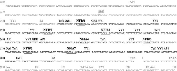
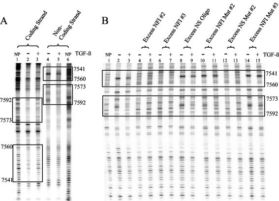
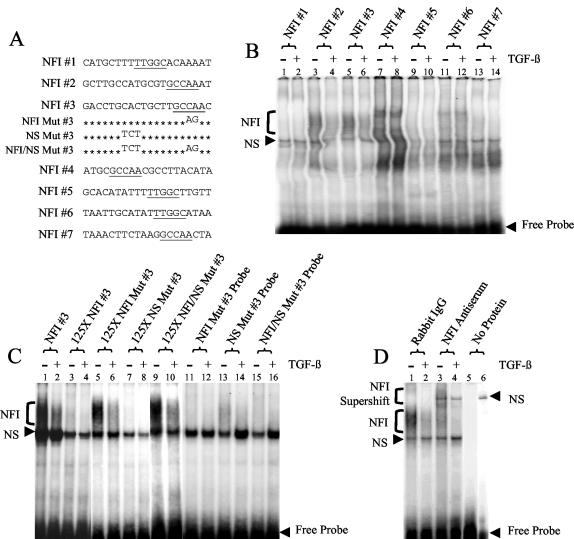
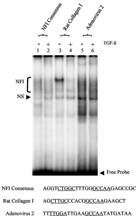
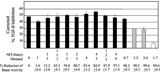
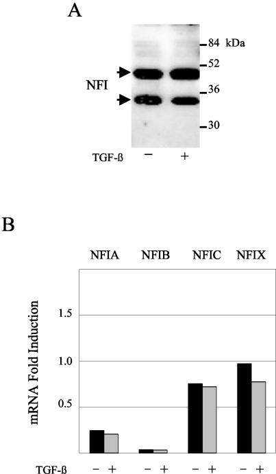
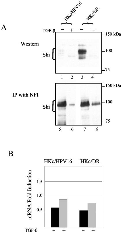
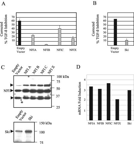
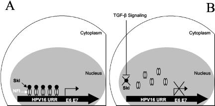
Human papillomaviruses (HPVs) are present in virtually all cervical cancers. An important step in the development of malignant disease, including cervical cancer, involves a loss of sensitivity to transforming growth factor beta (TGF-beta). HPV type 16 (HPV16) early gene expression, including that of the E6 and E7 oncoprotein genes, is under the control of the upstream regulatory region (URR), and E6 and E7 expression in HPV16-immortalized human epithelial cells is inhibited at the transcriptional level by TGF-beta. While the URR contains a myriad of transcription factor binding sites, including seven binding sites for nuclear factor I (NFI), the specific sequences within the URR or the transcription factors responsible for TGF-beta modulation of the URR remain unknown. To identify potential transcription factors and binding sites involved in TGF-beta modulation of the URR, we performed DNase I footprint analysis on the HPV16 URR using nuclear extracts from TGF-beta-sensitive HPV16-immortalized human keratinocytes (HKc/HPV16) treated with and without TGF-beta. Differentially protected regions were found to be located around NFI binding sites. Electrophoretic mobility shift assays, using the NFI binding sites as probes, showed decreased binding upon TGF-beta treatment. This decrease in binding was not due to reduced NFI protein or NFI mRNA levels. Mutational analysis of individual and multiple NFI binding sites in the URR defined their role in TGF-beta sensitivity of the promoter. Overexpression of the NFI family members in HKc/HPV16 decreased the ability of TGF-beta to inhibit the URR. Since the oncoprotein Ski has been shown to interact with and increase the transcriptional activity of NFI and since cellular Ski levels are decreased by TGF-beta treatment, we explored the possibility that Ski may provide a link between TGF-beta signaling and NFI activity. Anti-NFI antibodies coimmunoprecipitated endogenous Ski in nuclear extracts from HKc/HPV16, confirming that NFI and Ski interact in these cells. Ski levels dramatically decreased upon TGF-beta treatment of HKc/HPV16, and overexpression of Ski eliminated the ability of TGF-beta to inhibit the URR. Based on these studies, we propose that TGF-beta inhibition of HPV16 early gene expression is mediated by a decrease in Ski levels, which in turn dramatically reduces NFI activity.


Figures

FIG. 1.
FIG. 1.
Nucleotide sequence of the entire HPV16 URR (nucleotides 7232 to 119). Putative transcription factor binding sites are noted. The KDE is shown in black, and 3′ and 5′ segments of the URR are shown in gray. The seven potential NFI binding sites are numbered in sequential order, and their nucleotide sequences are underlined.
FIG. 2.
FIG. 2.
DNase I footprint analysis of the HPV16 URR. (A) A double-stranded segment of the KDE (nucleotides 7502 to 7677) was end labeled with 32P on either the coding (lanes 1 to 3) or the noncoding (lanes 4 to 6) strand. Nuclear extract from TGF-β-sensitive HKc/HPV16 treated for 48 h with (lanes 3 and 5) and without (lanes 2 and 4) 40 pM TGF-β was incubated with the labeled probes. Each probe was also incubated without protein (NP, lanes 1 and 6) to form a DNase I ladder. After DNase I digestion, unprotected fragments were resolved on a 6% denaturing polyacrylamide gel. Maxam and Gilbert A + G sequencing was performed on each probe for nucleotide identification (data not shown). Two areas of differential protection are located around NFI binding sites and are shown in boxes on each strand. The URR nucleotide number is given on the left and right sides of the footprint. NFI 2 is located between nucleotides 7541 and 7560, while NFI 3 is located between nucleotides 7573 and 7592. (B) Specificity of the differential binding was confirmed using the labeled noncoding strand probe described for panel A. Excess unlabeled oligonucleotides containing either intact NFI binding sites (lanes 4 to 7, 12, and 13) or without intact NFI binding sites (lanes 8 to 11, 14, and 15) were added to the binding reaction mixtures (described above). See Fig. 3A for the complete nucleotide sequence of the unlabeled competitor oligonucleotides, NFI 2, NFI 3, and NFI mutant (Mut) 3. The nucleotide sequence of the NS oligonucleotide (Oligo) was GCT TGT ACG GCG TGC AGA AT, the sequence of the NFI mutant (Mut) 2 was GCT TGC CAT GCG TGC AGA AT, and the sequence of NS mutant (Mut) 2 was GCT TGT ACG GCG TGC CAA AT. Competition and destruction of the differential binding (boxed areas, lanes 2 and 3) can be observed only in lanes containing oligonucleotides with intact NFI binding sites (lanes 4 to 7, 12, and 13).
FIG. 3.
FIG. 3.
Binding to multiple NFI sites in the HPV16 URR decreases upon treatment of HKc/HPV16 with TGF-β. (A) The nucleotide sequence is provided for each of the oligonucleotides representing the seven NFI half-sites in the HPV16 URR. Mutations made to the NFI 3 oligonucleotides are also shown. Putative NFI binding sites are underlined. (B) EMSAs were performed using each NFI site of the HPV16 URR as a probe. Nuclear extract from TGF-β-sensitive HKc/HPV16 treated with (even-numbered lanes) and without (odd-numbered lanes) 40 pM TGF-β for 48 h was incubated with each probe. Protein-probe complexes were separated from the free probe on a 5% nondenaturing polyacrylamide gel. Specific NFI binding (bracket) as well as NS binding is noted. (C) EMSAs were performed using probes made from the differentially protected area found around NFI site 3 of the HPV16 URR. Nuclear extract from TGF-β-sensitive HKc/HPV16 treated with (even-numbered lanes) and without (odd-numbered lanes) 40 pM TGF-β for 48 h was incubated with either a radiolabeled NFI 3 probe (lanes 1 to 10) or a radiolabeled mutant NFI 3 probe (lanes 11 to 16). Excess unlabeled oligonucleotides (125-fold) containing the intact NFI site (lanes 3, 4, 7, and 8) or the mutated NFI site (lanes 5, 6, 9, and 10) were added to the binding reaction mixture to demonstrate specificity. (D) NFI was verified as the transcription factor responsible for the differential binding by performing supershift analysis using NFI 3 oligonucleotide as the probe. Rabbit IgG (lanes 1 and 2) or anti-NFI antiserum (lanes 3 and 4) was added to the binding reaction mixtures (described above). NFI (bracket) and NS binding, as well as the resulting NFI supershift (bracket), is shown. Lane 5 contains labeled probe only (no nuclear extract). Lane 6 contains labeled probe and anti-NFI antiserum (no nuclear extract).
FIG. 4.
FIG. 4.
TGF-β modulation of NFI binding to TGF-β-responsive promoters. EMSAs were performed using an NFI consensus binding site (lanes 1 and 2) and sequences containing NFI binding sites from the rat collagen I (lanes 3 and 4) and adenovirus 2 (lanes 5 and 6) promoters as probes. The sequence of each probe is listed, and the NFI binding sites are underlined. Nuclear extract from TGF-β-sensitive HKc/HPV16 treated with (even-numbered lanes) and without (odd-numbered lanes) 40 pM TGF-β for 48 h was incubated with each probe. Protein-probe complexes were separated from the free probe on a 5% nondenaturing polyacrylamide gel. Specific NFI binding (bracket) as well as NS binding is noted.
FIG. 5.
FIG. 5.
Effects of single and multiple NFI mutations on TGF-β modulation of the HPV16 URR. The entire HPV16 URR (Fig. 1) was cloned into the luciferase reporter vector pGL3 (Promega) (pGL3/URR) where various NFI sites were mutated from GCCAA to GCAGA, which is unable to bind NFI. These constructs were transfected into TGF-β-sensitive HKc/HPV16 and treated with and without 40 pM TGF-β for 42 h. Luciferase activity was determined 68 h posttransfection. Corrected percent TGF-β inhibition for each construct was determined by subtracting the percent TGF-β inhibition obtained by transfection of a promoterless pGL3 plasmid from the total TGF-β inhibition obtained for each reporter construct. The specific NFI site(s) that was mutated and the percent reduction of basal URR activity are shown for each mutant construct at the bottom of the figure.
FIG. 6.
FIG. 6.
NFI protein and mRNA levels do not change following TGF-β treatment of HKc/HPV16. (A) Total NFI protein levels were determined by Western analysis. Nuclear extract (40 μg of protein) from TGF-β-sensitive HKc/HPV16 treated with and without 40 pM TGF-β for 48 h was separated on an SDS-12% polyacrylamide gel, transferred to nitrocellulose, and probed with an anti-NFI antibody (Santa Cruz). Molecular mass markers are shown on the right; arrows pointing to NFI bands are on the left. (B) mRNA expression of each of the four NFI family members (NFIA, NFIB, NFIC, and NFIX) was determined by real-time PCR. RNA was collected using RNeasy columns (Qiagen) from TGF-β-sensitive HKc/HPV16 treated with (gray) and without (black) 40 pM TGF-β for 46 h. Expression was determined using primers specific for each NFI family member, compared with a control set of primers, and expressed as fold induction. The average of two experiments for each family member is shown.
FIG. 7.
FIG. 7.
TGF-β treatment of HKc/HPV16 and HKc/DR decreases nuclear Ski levels, and Ski interacts with NFI. (A) Ski levels were demonstrated by Western analysis (lanes 1 to 4). Nuclear extract (30 μg of protein) from TGF-β-sensitive HKc/HPV16 and TGF-β-resistant HKc/DR treated with and without 40 pM TGF-β for 48 h was separated on an SDS-10% polyacrylamide gel. Proteins were transferred to nitrocellulose and probed with an anti-Ski antibody (Cascade Bioscience), which detects Ski products (brackets) ranging from 95 to 115 kDa. Endogenous Ski coimmunoprecipitated with NFI (lanes 5 to 8). Nuclear extract (850 μg of protein) from TGF-β-sensitive HKc/HPV16 and TGF-β-resistant HKc/DR treated with and without 40 pM TGF-β for 24 h was incubated (2 h, 25°C) with 5 μg of anti-NFI antibody preincubated with protein G agarose. After washing, the immunoprecipitates were resolved by SDS-polyacrylamide gel electrophoresis and probed for Ski as described above. Molecular mass markers are noted on the right. (B) mRNA expression of Ski was determined by real-time PCR. RNA was collected using RNeasy columns (Qiagen) from TGF-β-sensitive HKc/HPV16 treated with (gray) and without (black) 40 pM TGF-β for 46 h. Expression was determined using primers specific for Ski, compared with a control set of primers, and expressed as fold induction. The average of two experiments for each family member is shown.
FIG. 8.
FIG. 8.
TGF-β inhibition of the HPV16 URR is overcome by overexpression of either NFI or Ski. (A) Effects of NFI family member overexpression on TGF-β modulation of the HPV16 URR. pGL3/URR and pCMV vectors expressing each of the NFI family members were cotransfected into TGF-β-sensitive HKc/HPV16 using Transfast (Promega). Luciferase activity was determined after treatment with and without 40 pM TGF-β for 42 h, 68 h posttransfection. TGF-β inhibition resulting from transfection of an empty vector (black bar) is compared with the percent TGF-β inhibition upon overexpression of each NFI family member (gray bars). (B) Effects of Ski overexpression on TGF-β modulation of the HPV16 URR. pGL3/URR and a pcDNA3.1 vector expressing Ski were cotransfected and analyzed as described above (A). (C) NFI and Ski protein levels were determined by Western analysis. Lysates from TGF-β-sensitive HKc/HPV16 cotransfected with pGL3/URR and pCMV vectors expressing each NFI family member, empty pCMV vector, Ski, or an empty pcDNA3.1 vector were separated on an SDS-12% polyacrylamide gel, transferred to nitrocellulose, and probed with an anti-NFI or an anti-Ski antibody. Molecular mass markers are shown on the right with arrows pointing to three NFI bands (top panel) or Ski (bottom panel). (D) mRNA expression of each NFI family member and Ski was determined by real-time PCR analysis. RNA was collected using RNeasy columns (Qiagen) from TGF-β-sensitive HKc/HPV16 cotransfected with pGL3/URR and pCMV vectors expressing each NFI family member or a pcDNA vector expressing Ski. Expression was determined using primers specific for each NFI family member or Ski and is given as fold induction over empty vector.
FIG. 9.
FIG. 9.
Proposed model of TGF-β inhibition of HPV16 URR activity through NFI-Ski interactions in TGF-β-sensitive HKc/HPV16. (A) In the absence of TGF-β NFI-Ski complexes bind to and activate the HPV16 URR promoter. (B) In the presence of TGF-β signaling, Ski is degraded and no longer available to complex with NFI to induce promoter activity of the HPV16 URR.

References

    1. Apt, D., T. Chong, Y. Liu, and H. U. Bernard. 1993. Nuclear factor I and epithelial cell-specific transcription of human papillomavirus type 16. J. Virol. 67:4455-4463. - PMC - PubMed
    1. Armendariz-Borunda, J., C. P. Simkevich, N. Roy, R. Raghow, A. H. Kang, and J. M. Seyer. 1994. Activation of Ito cells involves regulation of AP-1 binding proteins and induction of type I collagen gene expression. Biochem. J. 304:817-824. - PMC - PubMed
    1. Berger, D. H., X. H. Feng, J. Yao, D. Saha, R. D. Beauchamp, and X. Lin. 2002. Resistance to transforming growth factor-beta occurs in the presence of normal Smad activation. Surgery 132:310-316. - PubMed
    1. Borger, D. R., Y. Mi, G. Geslani, L. L. Zyzak, A. Batova, T. S. Engin, L. Pirisi, and K. E. Creek. 2000. Retinoic acid resistance at late stages of human papillomavirus type 16-mediated transformation of human keratinocytes arises despite intact retinoid signaling and is due to a loss of sensitivity to transforming growth factor-beta. Virology 270:397-407. - PubMed
    1. Butz, K., and F. Hoppe-Seyler. 1993. Transcriptional control of human papillomavirus (HPV) oncogene expression: composition of the HPV type 18 upstream regulatory region. J. Virol. 67:6476-6486. - PMC - PubMed

Publication types

MeSH terms

LinkOut - more resources