. 2004 Apr;78(8):3965-76.
doi: 10.1128/jvi.78.8.3965-3976.2004.

Recombinant modified vaccinia virus Ankara expressing a soluble form of glycoprotein B causes durable immunity and neutralizing antibodies against multiple strains of human cytomegalovirus

Affiliations

Recombinant modified vaccinia virus Ankara expressing a soluble form of glycoprotein B causes durable immunity and neutralizing antibodies against multiple strains of human cytomegalovirus

Zhongde Wang et al. J Virol. 2004 Apr.

Abstract

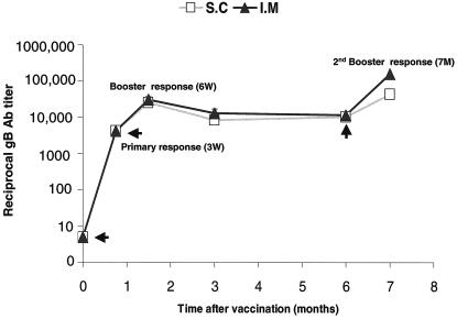
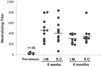
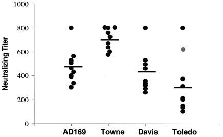
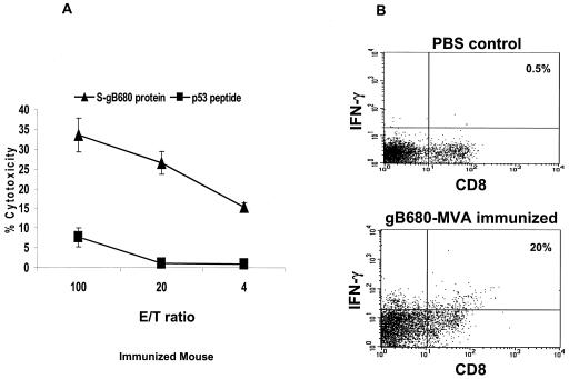
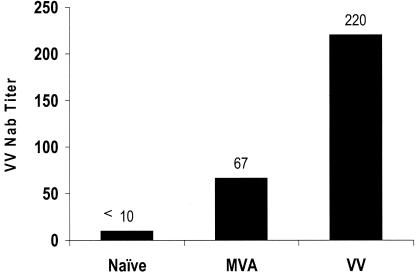
Human cytomegalovirus (CMV) is a viral pathogen that infects both genders, who remain asymptomatic unless they receive immunosuppressive drugs or acquire infections that cause reactivation of latent virus. CMV infection also causes serious birth defects following primary maternal infection during gestation. A safe and effective vaccine to limit disease in this population continues to be elusive. A well-studied antigen is glycoprotein B (gB), which is the principal target of neutralizing antibodies (NAb) towards CMV in humans and has been implicated as the viral partner in the receptor-mediated infection by CMV in a variety of cell types. Antibody-mediated virus neutralization has been proposed as a mechanism by which host immunity could modify primary infection. Towards this goal, an attenuated poxvirus, modified vaccinia virus Ankara (MVA), has been constructed to express soluble CMV gB (gB680-MVA) to induce CMV NAb. Very high levels of gB-specific CMV NAb were produced after two doses of the viral vaccine. NAb were durable within a twofold range for up to 6 months. Neutralization titers developed in immunized mice are equivalent to titers found clinically after natural infection. This viral vaccine, expressing gB derived from CMV strain AD169, induced antibodies that neutralized CMV strains of three different genotypes. Remarkably, preexisting MVA and vaccinia virus (poxvirus) immunity did not interfere with subsequent immunizations of gB680-MVA. The safety characteristics of MVA, combined with the robust immune response to CMV gB, suggest that this approach could be rapidly translated into the clinic.


Figures

FIG. 1.
FIG. 1.
Schematic map of the gB680-mpLW51 plasmid. pLW51 has four features: (i) flanking regions (FL1 and FL2); (ii) color screen marker gene, gus under control of VV promoter P11; (iii) identical direct repeats (DR); and (iv) two VV promoters, PsynII and mH5, for recombinant gene expression. The gB680 gene was cloned behind the mH5 promoter to create gB680-mpLW51. gB680-mpLW51 recombined into deletion III of wt MVA to create gB680-MVA by homologous recombination. More details can be found in Materials and Methods.
FIG. 2.
FIG. 2.
Characterization of gB680-MVA. (A) Western blot detection of intracellular gB680 protein from gB680-MVA-infected BHK-21 cells. Lane 1, cell lysate from gB680-MVA-infected BHK-21 cells; lane 2, cell lysate from gB-VV-infected BHK-21 cells; lane 3, cell lysate from HCMV (AD169)-infected MRC-5 cells. All lanes were loaded with the same amount of protein, as determined by the Bradford protein measurement method. (B) ELISA measurement of S-gB680 protein in culture medium collected from gB680-rMVA-infected BHK-21 cells. pp65-MVA served as a negative control (28). Concentration values were determined by using an internal reference standard of commercially bought soluble gB protein.
FIG. 3.
FIG. 3.
Purified S-gB680 protein does not cross-react with sera from MVA-immunized mice. (A) Coomassie blue-stained SDS-PAGE profile of affinity purified S-gB680 (see Materials and Methods). Lane 1, molecular mass markers; lane 2 is 2 μg of affinity-purified S-gB680, and lane 3 is 4 μg of affinity-purified S-gB680. The gel pattern shows possible dimers (200 kDa), monomer form (105 kDa), and proteolytically cleaved amino-terminal (59 kDa) and carboxyl-terminal (30 kDa) fragments, indicated by arrows (see the text for further description). (B) Sera from mice immunized with gB680-MVA and gus-MVA were used in ELISA as described in Materials and Methods. gB680-MVA-immunized sera were used as positive controls (▪). Sera from gus-MVA-immunized mice (• symbol) and unimmunized mice (▴ symbol) were still negative in ELISA even when serum dilution was less than 1:100 (data not shown in graph).
FIG. 4.
FIG. 4.
Time course of gB-specific antibody levels after immunization. Mice were immunized and boosted with gB680-MVA as indicated by arrows in the graph. Serum samples were collected at 3 weeks (primary response), 6 weeks (booster response), and 3, 6, and 7 months (second booster response) after initial immunization and analyzed by ELISA as described in Materials and Methods. The gB-specific antibody ELISA titer is expressed on a log scale as the reciprocal of the highest dilution of mouse serum that gives positive OD. The y axis represents the gB ELISA antibody titer on a log10 scale. The x axis indicates the time point (months) after initial immunization.
FIG. 5.
FIG. 5.
Titer of HCMV NAb 6 weeks and 6 months after initial immunization. Ten serum samples from the 6-week (short-term) and 6-month (long-term) immunizations were used in the CMV microneutralization assay. Sera from preimmunized mice were used as negative controls. The CMV NT is expressed as the reciprocal of the highest dilution of mouse sera that inhibits 50% of virus input compared to the control. Filled circles represent NT for each individual mouse. Horizontal bars in the graph represent geometric means of the NT. The i.m. and s.c. routes were examined.
FIG. 6.
FIG. 6.
Sera from gB680-MVA-immunized mice neutralized different CMV strains. The AD169, Towne, Davis, and Toledo CMV strains with different gB genotypes were used in CMV microneutralization assays. Filled circles represent NT for each mouse. Horizontal bars in the graph represent geometric means of the NT from 10 individual mice at the 6-week time point.
FIG. 7.
FIG. 7.
IgG subclass distribution of gB-specific antibodies after immunization. Serum samples from 6 weeks and 6 months after gB680-MVA immunization were used for an isotype-specific ELISA assay. Purified S-gB680 was used as the coating antigen. Biotinylated anti-mouse IgG1, IgG2a, IgG2b, and IgG3 were used as secondary antibodies. The percentage of gB-specific IgG subclasses was calculated as the ratio of the individual subclass OD divided by the total OD of all subclasses.
FIG. 8.
FIG. 8.
T-cell proliferation after gB680-MVA immunization. (A) Ten BALB/c mice were immunized with 5 × 10⁷ PFU of gB680-MVA via the s.c. route, followed with a booster immunization with the same dosage by the same route. Fresh spleen cells were harvested 6 weeks after the initial immunization and incubated with purified S-gB680 at the concentrations indicated. The SI is defined as a ratio of [3H]thymidine incorporated into cells in the presence of purified S-gB680 antigen over mock antigen (MRC-5 cell lysate). Each bar represents the average SI of 10 immunized mice. (B) ICC of IFN-γ expressed in CD4+ T-cells after IVS in the presence of S-gB680 protein in immunized mice. Representative plots are shown for each group. Numbers reflect the percentages of CD4+ T cells.
FIG. 9.
FIG. 9.
Induction of gB-specific CTL immune responses after immunization. Three BALB/c mice were immunized with gB680-MVA, and followed by booster immunization 3 weeks later. Spleens were removed 3 weeks after the booster. (a) An IVS was performed and was followed by a CRA as described in Materials and Methods. Purified S-gB680 protein (▴ symbol) and p53149-157 peptide (▪ symbol) sensitized splenocytes were used as targets. Means and SE were calculated at each effector-target (E/T) ratio for all evaluated mice. (b) ICC of IFN-γ expressed in CD8+ T-cells after IVS in the presence of S-gB680 protein in immunized or control PBS injected mice. One representative plot is shown for each group. Numbers reflect the percentages of CD8+ T cells.
FIG. 10.
FIG. 10.
Preexisting MVA and VV immunity. Mice were immunized twice with 5 × 10⁷ PFU of MVA or 1 × 10⁶ PFU of VV once by i.p. injection. Serum samples were collected 6 weeks after initial inoculation for measurement of MVA or VV immunity. MVA or VV immunity was measured by performing VV plaque reduction assays as described in Materials and Methods. VV NT is the highest dilution of serum producing at least 85% inhibition of the VV input. Bars represent the geometric mean of VV NT for eight individual mice.

Similar articles

Cited by

References

    1. Adler, S. P., S. H. Hempfling, S. E. Starr, S. A. Plotkin, and S. Riddell. 1998. Safety and immunogenicity of the Towne strain cytomegalovirus vaccine. Pediatr. Infect. Dis. J. 17:200-206. - PubMed
    1. Adler, S. P., S. A. Plotkin, E. Gonczol, M. Cadoz, C. Meric, J. B. Wang, P. Dellamonica, A. M. Best, J. Zahradnik, S. Pincus, K. Berencsi, W. I. Cox, and Z. Gyulai. 1999. A canarypox vector expressing cytomegalovirus (CMV) glycoprotein B primes for antibody responses to a live attenuated CMV vaccine (Towne). J. Infect. Dis. 180:843-846. - PubMed
    1. Amara, R. R., F. Villinger, J. D. Altman, S. L. Lydy, S. P. O'Neil, S. I. Staprans, D. C. Montefiori, Y. Xu, J. G. Herndon, et al. 2001. Control of a mucosal challenge and preventions of AIDS by a multiprotein DNA/MVA vaccine. Science 292:69-74. - PubMed
    1. Andreoni, M., M. Faircloth, L. Vugler, and W. J. Britt. 1989. A rapid microneutralization assay for the measurement of neutralizing antibody reactive with human cytomegalovirus. J. Virol. Methods 23:157-167. - PubMed
    1. Aquino, V. H., and L. T. Figueiredo. 2000. High prevalence of renal transplant recipients infected with more than one cytomegalovirus glycoprotein B genotype. J. Med. Virol. 61:138-142. - PubMed

Publication types

MeSH terms