. 2004 Apr;78(8):4003-10.
doi: 10.1128/jvi.78.8.4003-4010.2004.

tRNA-like structure regulates translation of Brome mosaic virus RNA

Affiliations

tRNA-like structure regulates translation of Brome mosaic virus RNA

Sharief Barends et al. J Virol. 2004 Apr.

Abstract

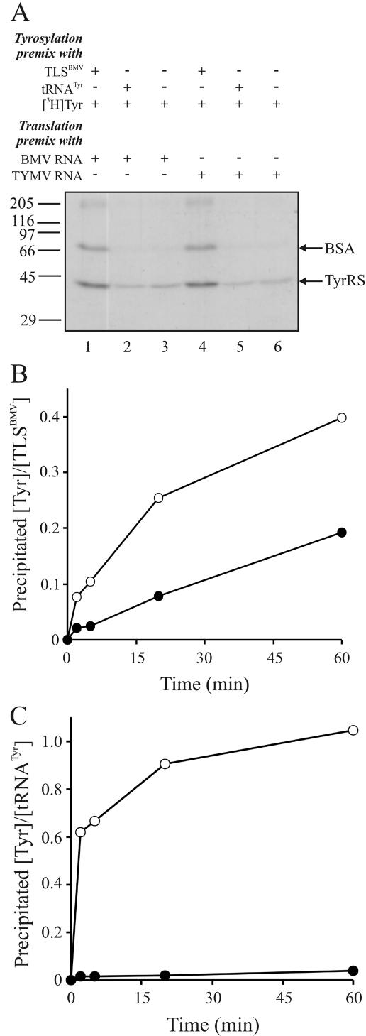
For various groups of plant viruses, the genomic RNAs end with a tRNA-like structure (TLS) instead of the 3' poly(A) tail of common mRNAs. The actual function of these TLSs has long been enigmatic. Recently, however, it became clear that for turnip yellow mosaic virus, a tymovirus, the valylated TLS(TYMV) of the single genomic RNA functions as a bait for host ribosomes and directs them to the internal initiation site of translation (with N-terminal valine) of the second open reading frame for the polyprotein. This discovery prompted us to investigate whether the much larger TLSs of a different genus of viruses have a comparable function in translation. Brome mosaic virus (BMV), a bromovirus, has a tripartite RNA genome with a subgenomic RNA4 for coat protein expression. All four RNAs carry a highly conserved and bulky 3' TLS(BMV) (about 200 nucleotides) with determinants for tyrosylation. We discovered TLS(BMV)-catalyzed self-tyrosylation of the tyrosyl-tRNA synthetase but could not clearly detect tyrosine incorporation into any virus-encoded protein. We established that BMV proteins do not need TLS(BMV) tyrosylation for their initiation. However, disruption of the TLSs strongly reduced the translation of genomic RNA1, RNA2, and less strongly, RNA3, whereas coat protein expression from RNA4 remained unaffected. This aberrant translation could be partially restored by providing the TLS(BMV) in trans. Intriguingly, a subdomain of the TLS(BMV) could even almost fully restore translation to the original pattern. We discuss here a model with a central and dominant role for the TLS(BMV) during the BMV infection cycle.


Figures

FIG. 1.
FIG. 1.
Genomic organization of BMV. (A) Schematic view of genomic RNAs 1 to 3 with ORFs for the following proteins (molecular masses are shown in parentheses in the figure): methyltransferase/helicase (Me/He), RdRp, MP, and CP, which is only translated from the sgRNA. All of the RNAs have the same 5′ cap (•) and 3′ TLSBMV. (B) Secondary structure of the TLSBMV with nucleotide numbering in the 3′→5′ direction. The nucleotides in the shaded boxes were hybridized to two DNA oligonucleotides for TLSBMV disruption by RNase H. The various stem-loop structures are indicated (A to E).
FIG. 2.
FIG. 2.
Effects of TLSBMV disruption on BMV RNA translation in a wheat germ system. The separate genomic RNAs 1, 2, and 3 (15 nM for each T7 transcript), their mixture (RNA1-3) (25 nM for each T7 transcript, i.e., 75 nM for the total population), and a native BMV RNA population mixture (8.6 nM for the total population) were subjected to TLSBMV disruption (−) and used for in vitro translation. The translation patterns of [35S]Met-labeled protein bands are compared to those for intact RNAs (+). The positions of molecular weight markers and the BMV proteins are indicated on the left and the right, respectively. The asterisk indicates an incomplete product of enhanced intensity after TLSBMV disruption. The bottom panels show the scanned relative intensities (%) of full-length protein bands together with their incomplete products, taking the intensities produced by the intact (+) RNAs as 100% (average values from at least two independent experiments).
FIG. 3.
FIG. 3.
[3H]Tyr-labeled proteins and RNAs in a coupled tyrosylation-translation system. (A) Autoradiogram of a protein gel after sodium dodecyl sulfate-PAGE of coupled tyrosylation-translation reaction mixtures. The premix compositions of the various samples are indicated above the gel, molecular weight markers are shown on the left, and reference positions of BSA and TyrRS are shown on the right. For further details, see the text. (B and C) Tyrosylation kinetics were monitored for the TLSBMV (B) and for yeast tRNATyr (C). During the 60-min incubation of the [3H]tyrosylation mixture, samples were either directly quenched in TCA (○) or incubated in the presence of a 150-fold molar excess of RNase A for 2 min at 37°C prior to TCA precipitation, filtration, and liquid scintillation counting (•). All data are representative of three independent experiments.
FIG. 4.
FIG. 4.
Translational complementation in trans by the free and complete TLSBMV. The defective translation of T7 transcripts of BMV RNAs 1 to 3 with a disrupted 3′ TLSBMV (−) was complemented with increasing concentrations of free TLSBMV (indicated in molar excess over the 3′-truncated RNAs) and was compared to the translation pattern of the intact RNAs (+). Optimal complementation was observed at about an eightfold molar excess of free TLSBMV. Relative band intensities were calculated from three independent experiments. See the legend for Fig. 2 for further information.
FIG. 5.
FIG. 5.
Translational complementation in trans with shortened TLSBMV variants and with yeast tRNATyr as a reference molecule. The effect of increasing concentrations of TLSBMV(-B2) and TLSBMV(-C) (left gel) and of TLSBMV(-A, -B1, -B3, -E) and yeast tRNATyr (right gel) on the translation of 3′-truncated BMV RNAs 1 to 3 was monitored. Above each gel, the schematic RNA structures of the TLSBMV variants are shown, with lacking parts indicated in gray. Relative band intensities were calculated from three independent experiments. See the legend for Fig. 2 for further information.

References

    1. Ahlquist, P., R. Dasgupta, and P. Kaesberg. 1981. Near identity of 3′ RNA secondary structure in bromoviruses and cucumber mosaic virus. Cell 23:183-189. - PubMed
    1. Ali, I. K., L. McKendrick, S. J. Morley, and R. J. Jackson. 2001. Truncated initiation factor eIF4G lacking an eIF4E binding site can support capped mRNA translation. EMBO J. 20:4233-4242. - PMC - PubMed
    1. Barends, S., H. H. J. Bink, S. H. E. van den Worm, C. W. A. Pleij, and B. Kraal. 2003. Entrapping ribosomes for viral translation: tRNA mimicry as a molecular Trojan horse. Cell 112:123-129. - PubMed
    1. Bink, H. H. J., K. Hellendoorn, J. van der Meulen, and C. W. A. Pleij. 2002. Protonation of non-Watson-Crick base pairs and encapsidation of turnip yellow mosaic virus RNA. Proc. Natl. Acad. Sci. USA 99:13465-13470. - PMC - PubMed
    1. Bol, J. F. 2003. Alfalfa mosaic virus: coat protein-dependent initiation of infection. Mol. Plant Pathol. 4:1-8. - PubMed

Publication types

MeSH terms

LinkOut - more resources