. 2004 Apr;78(8):4185-96.
doi: 10.1128/jvi.78.8.4185-4196.2004.

Herpes simplex virus 1 gene products occlude the interferon signaling pathway at multiple sites

Affiliations

Herpes simplex virus 1 gene products occlude the interferon signaling pathway at multiple sites

Ana Virginia Chee et al. J Virol. 2004 Apr.

Abstract

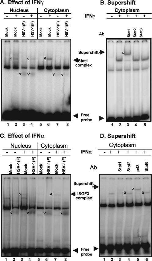
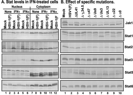
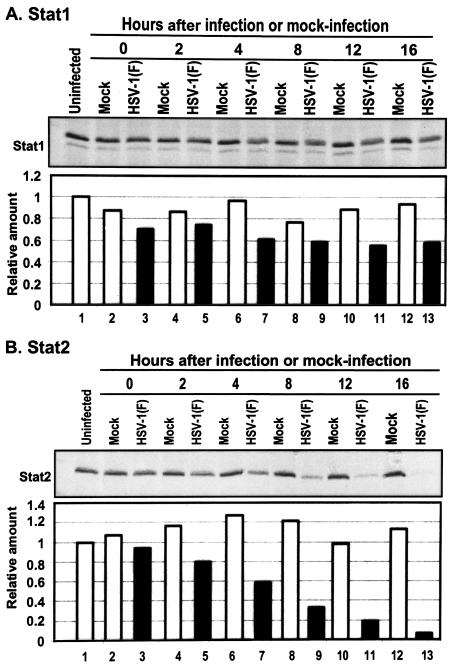
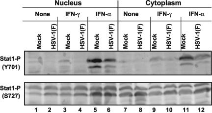
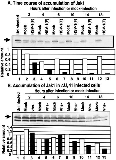
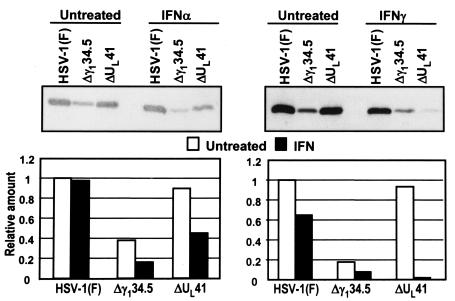
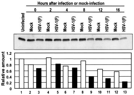
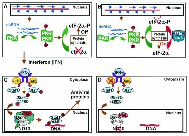
Earlier studies have shown that herpes simplex virus 1 (HSV-1) blocks the interferon response pathways, at least at two sites, by circumventing the effects of activation of protein kinase R by double-stranded RNA and interferon and through the degradation of promyelocytic leukemia protein (PML) since interferon has no antiviral effects in PML(-/-) cells. Here we report on two effects of viral genes on other sites of the interferon signaling pathway. (i) In infected cells, Jak1 kinase associated with interferon receptors and Stat2 associated with the interferon signaling pathway rapidly disappear from infected cells. The level of interferon alpha receptor is also reduced, albeit less drastically at times after 4 h postinfection. Other members of the Stat family of proteins were either decreased in amount or posttranslationally processed in a manner different from those of mock-infected cells. The decrease in the levels of Jak1 and Stat2 may account for the decrease in the formation of complexes consisting of Stat1 or ISGF3 and DNA sequences containing the interferon-stimulated response elements after exposure to interferon. (ii) The disappearance of Jak1 and Stat2 was related at least in part to the function of the virion host shutoff protein, the product of the viral U(L)41 gene. Consistent with this observation, a mutant lacking the U(L)41 gene and treated with interferon produced lesser amounts of a late protein (U(L)38) than the wild-type parent. We conclude that HSV-1 blocks the interferon signaling pathways at several sites.


Figures

FIG. 1.
FIG. 1.
HSV-1 blocks the formation of DNA protein complexes induced by IFN. Replicate HEp2 cells (A and B) or HeLa cells (C and D) were mock infected or exposed to10 PFU of HSV-1(F) per cell as described in Materials and Methods After 7 h of incubation, the cells were treated (+) or not treated (−) with 500 U of IFN-γ/ml for 1 h (A and B) or 5,000 U of IFN-α/ml for 15 min (C and D). Cells were harvested, fractionated, and processed as described in Materials and Methods. (A and C) EMSAs. Nuclear (lanes 1 to 4) or cytoplasmic fractions (40 μg/reaction) were reacted with either labeled M67 probe (A) or ISRE probe (C) in total volumes of 30 μl per lane. Complexes containing Stat1 (circles, A) or ISGF3 (C) are identified by open circles to the left of the bands. Bands containing virus-induced proteins are identified with a “v” placed to the left of the bands. Although not specifically identified as such here, HSV-1 induces proteins capable of binding DNA, and this could account for the virus-specific bands. (B and D) Supershift assays. To the reaction mixtures of untreated and IFN-treated cells described above were added 1 μl of Stat1 (lane 3), Stat2 (lane 4), or Stat3 (lane 5) antibody (A) or 1 μl of Stat1 (lane 3), Stat2 (lane 4), ISGF3 p48 (lane 5), or Stat6 (lane 6) antibody (D). The reaction mixtures were incubated for an additional 30 min and then subjected to electrophoresis in denaturing gels. The bands supershifted by antibodies are marked by an open triangle to the left of the band.
FIG. 2.
FIG. 2.
Effect of HSV-1 on members of the Stat family of proteins. (A) Replicate HeLa cells were mock infected or inoculated with 10 PFU of HSV-1(F) per cell as described in Materials and Methods. After 7 h of incubation, the cells were either not treated or treated with 500 U of IFN-γ/ml for 1 h or with 5,000 U of IFN-α/ml for 15 min. The cells were harvested and fractionated, and nuclear and cytoplasmic fractions (200 μg/lane) were electrophoretically separated on denaturing polyacrylamide gels, transferred to nitrocellulose membranes, and reacted with antibodies to Stat1, Stat2, Stat3, Stat5, or Stat6 (Transduction Laboratories) and then with secondary antibodies conjugated to AP. AP-conjugated antibodies were detected by using a colorimetric reaction (Bio-Rad). (B) Replicate HeLa cells were mock infected or exposed to 5 PFU of wild-type virus per cell or 5 PFU of mutant viruses lacking UL41, γ134.5, α47, US9, US3, UL13, α22, or α0 genes per cell. The cells were harvested at 18 h after infection and processed as described in Materials and Methods. Whole-cell lysates were subjected to electrophoresis in denaturing gels (200 μg/lane) and reacted with antibodies as described above. Bound peroxidase-conjugated antibodies were detected with the aid of enhanced chemiluminescence.
FIG. 3.
FIG. 3.
Effect of HSV-1 on the accumulation of Stat1 and Stat2 protein levels. Replicate HeLa cell cultures were either mock infected or exposed to 10 PFU of HSV-1(F) per cell and then harvested at 2, 4, 8, 12, or 16 h after infection, processed, subjected to electrophoresis in denaturing gels (200 μg of protein per lane), transferred to a nitrocellulose sheet, and reacted with anti-Stat1 (A) or Stat2 (B) antibodies and then with secondary antibodies conjugated to peroxidase. Bound antibodies were detected by using enhanced chemiluminescence and quantified with the aid of the Storm 860 PhosphorImager. The results were normalized to the Stat protein levels in the uninfected cell samples.
FIG. 4.
FIG. 4.
HSV-1 infection can inhibit tyrosine phosphorylation but not serine phosphorylation of Stat1 in HeLa cells. Replicate HeLa cell cultures were mock infected or exposed to 10 PFU of HSV-1(F) per cell. At 7 h after mock infection or infection the cells were mock treated or exposed to 500 U of IFN-γ/ml for 1 h or 5,000 U of IFN-α/ml for 15 min. At the conclusion of the treatment the cells were harvested and fractionated, and nuclear and cytoplasmic fractions were subjected to electrophoresis in denaturing gels (200 μg/lane). The electrophoretically separated proteins were transferred to a nitrocellulose sheet and reacted with primary antibodies to phospho-epitope-specific polyclonal antibodies tyrosine 701 or serine 727 of Stat1 (Upstate Biotechnology) and then with a secondary antibody conjugated to peroxidase. Bound peroxidase-conjugated antibodies were detected by using enhanced chemiluminescence.
FIG. 5.
FIG. 5.
Downregulation of Jak1 protein levels is delayed in HeLa cells infected with UL41 mutant virus. Replicate HeLa cell cultures were mock infected or were exposed to 10 PFU of the parent, HSV-1(F) virus (A) or the UL41 mutant virus (B) per cell and then harvested at 2, 4, 6, 10, 14, or 18 h postinfection. The harvested cells were solubilized, subjected to electrophoresis (200 μg/lane), transferred to a nitrocellulose sheet, and reacted first with primary antibodies to Jak1 and then with secondary conjugated antibodies. The bound antibodies were detected by enhanced luminescence and were quantified by using a Storm 860 PhosphorImager and then normalized with respect to the protein level detected in the uninfected samples.
FIG. 6.
FIG. 6.
IFN has a stronger inhibitory effect on the accumulation of late viral proteins in cells infected by UL41 mutant virus than in cells infected with the wild-type virus. Replicate HeLa cell cultures were exposed to either 1,000 U of IFN-γ or to 5,000 U of IFN-α per ml of medium for 24 h and then exposed to 10 PFU of parent HSV-1(F) virus to ΔUL41 or Δγ134.5 mutant viruses per cell. After 2 h, the inoculum was replaced with medium supplemented with 5% newborn calf serum with 1,000 U of IFN-γ per ml or with 5,000 U of IFN-α per ml. The cells were harvested at 12 h postinfection, solubilized, electrophoretically separated on denaturing 12% polyacrylamide gels, transferred to nitrocellulose membranes, and reacted first with polyclonal antibody to UL38 and then to anti-rabbit immunoglobulin G conjugated to peroxidase. Bound antibody was detected by using enhanced chemiluminescence, quantified with the aid of a Storm 860 PhosphorImager, and normalized with respect to the levels of UL38 protein levels detected in untreated HeLa cells infected by wild-type virus.
FIG. 7.
FIG. 7.
IFN-α receptor protein levels decrease late in HSV-1 infection. Replicate HeLa cell cultures were mock infected or exposed to 10 PFU of HSV-1(F) per cell and then at 0, 2, 4, 8, 12, or 16 h after infection. The harvested cells were processed as described in Materials and Methods, subjected to electrophoresis in denaturing polyacrylamide gels (200 μg/lane), transferred to a nitrocellulose sheet, and reacted with primary antibody to IFN-α receptor and a secondary antibody conjugated to peroxidase. Bound antibodies were detected by enhanced chemiluminescence, quantified with the aid of a Storm 860 PhosphorImager, and normalized to the protein level detected in mock-infected cells.
FIG. 8.
FIG. 8.
Schematic representation of the multiple mechanisms by which HSV-1 blocks cellular IFN-based defense systems. (A) Accumulation of complementary RNA capable of annealing induces the synthesis of IFN and activates PKR (25, 28). Activated PKR dimerizes, phosphorylates eIF-2α and, in the absence of viral anti-IFN gene function, shuts off protein synthesis. (B) In cells infected with wild-type virus the γ134.5 protein binds and redirects phosphatase 1α to dephosphorylate eIF-2α (22). Protein synthesis is not affected by activated PKR. (C) Secreted IFN binds IFN receptor to activate the signaling pathway. In the case of IFN-γ, the receptor-associated kinases Jak1 and Jak2 are activated and phosphorylate Stat1 (14). These and associated proteins are translocated into the nucleus, where they induce antiviral proteins. The activation of antiviral response is mediated by PML (9). (D) In wild-type-virus-infected cells, Jak1 disappears, PML is degraded, and the constituents of ND10 are dispersed (17). As a result, the infected cell does not respond to exogenous IFN to curtail viral replication.

References

    1. Abendroth, A., B. Slobedman, E. Lee, E. Mellins, M. Wallace, and A. M. Arvin. 2000. Modulation of major histocompatibility class II protein expression by varicella-zoster virus. J. Virol. 74:1900-1907. - PMC - PubMed
    1. Aubert, M., S. A. Rice, and J. A. Blaho. 2001. Accumulation of herpes simplex virus type 1 early and leaky-late proteins correlates with apoptosis prevention in infected human HEp-2 cells. J. Virol. 75:1013-1030. - PMC - PubMed
    1. Aurelian, L., and B. Roizman. 1965. Abortive infection of canine cells by herpes simplex virus. II. The alternative suppression of synthesis of interferon and viral constituents. J. Mol. Biol. 11:539-548. - PubMed
    1. Brandimarti, R., and B. Roizman. 1997. US9, a stable lysine-less herpes simplex virus 1 protein, is ubiquitinated before packaging into virions and associates with proteasomes. Proc. Natl. Acad. Sci. USA 94:13973-13978. - PMC - PubMed
    1. Caldenhoven, E., T. B. van Dijk, R. Solari, J. Armstrong, J. A. Raaijmakers, J. W. Lammers, L. Koenderman, and R. P. de Groot. 1996. STAT3beta, a splice variant of transcription factor STAT3, is a dominant negative regulator of transcription. J. Biol. Chem. 271:13221-13227. - PubMed

Publication types

MeSH terms

Substances