. 2004 Apr;78(8):4314-22.
doi: 10.1128/jvi.78.8.4314-4322.2004.

Rubella virus capsid protein modulates viral genome replication and virus infectivity

Affiliations

Rubella virus capsid protein modulates viral genome replication and virus infectivity

Min-Hsin Chen et al. J Virol. 2004 Apr.

Abstract

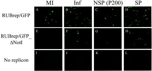
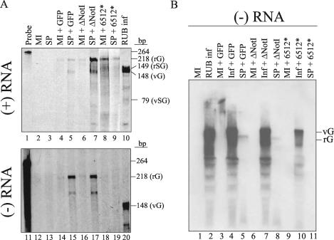
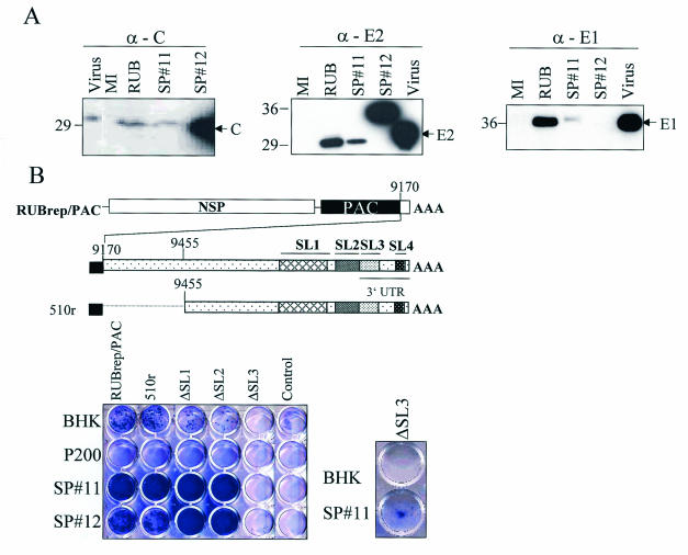
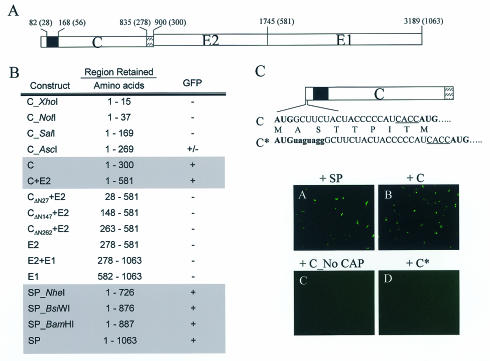
The structural proteins (SP) of the Togaviridae can be deleted in defective interfering RNAs. The dispensability of viral SP has allowed construction of noninfectious viral expression vectors and replicons from viruses of the Alphavirus and Rubivirus genera. Nevertheless, in this study, we found that the SP of rubella virus (RUB) could enhance expression of reporter genes from RUB replicons in trans. SP enhancement required capsid protein (CP) expression and was not due to RNA-RNA recombination. Accumulation of minus- and plus-strand RNAs from replicons was observed in the presence of SP, suggesting that SP specifically affects RNA synthesis. By using replicons containing an antibiotic resistance gene, we found 2- to 50-fold increases in the number of cells surviving selection in the presence of SP. The increases depended significantly on the amount of transfected RNA. Small amounts of RNA or templates that replicated inefficiently showed more enhancement. The infectivity of infectious RNA was increased by at least 10-fold in cells expressing CP. Moreover, virus infectivity was greatly enhanced in such cells. In other cells that expressed higher levels of CP, RNA replication of replicons was inhibited. Thus, depending on conditions, CP can markedly enhance or inhibit RUB RNA replication.


Figures

FIG. 1.
FIG. 1.
Enhanced GFP expression by RUB SP in trans; GFP expression in cotransfected cells. Vero cells were transfected with RUBrep/GFP (A to D) or RUBrep/GFP_ΔNotI (E to H) in the absence (MI) (A, E, and I) or presence (Inf) (B, F, and J) of helper virus or cotransfected with P200 RNA (C, G, and K) or SP RNA (D, H, and L). Infections were done with RUB F-therien at a multiplicity of infection of 1, and transfection was done at 24 h postinfection. GFP expression was examined 48 h after transfection.
FIG. 2.
FIG. 2.
Accumulation of plus- and minus-strand genomic RNAs by RUB SP. The synthesis of replicon genome in the absence or presence of RUB SP was investigated by use of an RNase protection assay (A) and Northern blot analysis (B). (A) Intracellular RNA from transfected Vero cells with replicons with or without SP RNA cotransfection was harvested 18 h (for detecting minus-strand RNA; upper panel) or 72 h (for detecting plus-strand RNA; lower panel) after transfection and subjected to an RNase protection assay using 35S-labeled RNA probes. The RNase-protected fragments corresponding to the rG, rSG, vG, and vSG RNAs are indicated. (B) Intracellular RNA prepared from infected (RUB inf) or uninfected (MI) cells transfected with specific replicons with or without RUB SP gene was prepared at 18 h after transfection. RNA was denatured by dimethyl sulfoxide and glyoxal and electrophoresed on an 0.85% agarose gel followed by hybridization using a DIG-labeled pGEM-GFP400 probe of positive polarity. The RNA species corresponding to rG and vG RNA are indicated at the right.
FIG. 3.
FIG. 3.
Replication of RUBrep/PAC and the 3′ mutants of RUBrep/PAC in BHK cells maintaining RUB NSP (P200) and SP. (A) Western blot showing the expression of RUB SP in SP#11 and SP#12. BHK clonal cell lines expressing RUB SP were obtained by antibiotic selection after transfecting a plasmid DNA containing RUB SP gene in pCI-Neo vector. Single colonies were isolated and maintained in media containing Geneticin. Cells grown in 10-cm2 plates were lysed in radioimmunoprecipitation buffer and 1/100 volume of the lysate were loaded on an sodium dodecyl sulfate-10% polyacrylamide gel. The proteins were transferred to a nitrocellulose membrane and blotted with a specific monoclonal antibody. Intracellular proteins from BHK cells infected with RUB F-therien (RUB) were harvested at 5 days postinfection. Virion proteins of RUB M33 (Virus) were purchased from Viral Antigens Inc. (B) Replication of 3′ mutants in cells expressing SP. BHK or clonal cell lines containing the indicated RUB genes were transfected with RUBrep/PAC or mutants with mutations at the 3′ end of RUBrep/PAC (unmodified RUBrepPAC is indicated). RNA transcripts (∼5 μg) were used for transfection to BHK or SP-expressing cells (∼106 cells). Cells were subjected to puromycin selection (5 μg/ml) 24 h after transfection and stained with crystal violet after 10 to 14 days of antibiotic selection. The control wells are cells without transfected RNA (Control). In order to visualize the small number of SP#11 cells containing the ΔSL3 construct surviving antibiotic selection, this culture and BHK control were passaged one more time.
FIG. 4.
FIG. 4.
Mapping the minimal region of the SP coding region required for enhancing GFP expression from RUBrep/GFP_ΔNotI. (A) Schematic representation of the gene organization of the RUB SP gene. The numbers on the top left represent the distances from the start codon AUG while the numbers in parentheses indicate amino acid position. In the C gene, the solid box indicates the major RNA binding site (19), and the box with slashes indicates the E2 signal peptide. (B) GFP expression in Vero cells cotransfected with RUBrep/GFP_ΔNotI and various SP RNAs. The GFP expression was examined 48 h after transfection. The GFP expression results were evaluated as follows: +, enhanced number of cells with GFP expression (>10) was detected; −, no GFP expression was detected at 24 h after transfection; ±, only a few GFP-expressing cells (3 to 10 cells over the complete field) were detected. (C) Modifications in the C* construct are shown. The GFP expression following transfection with either uncapped C RNA or capped C* are shown.
FIG. 5.
FIG. 5.
Viral infectivity regulation and replicon genome replication depends on the amount of CP. (A) RUB CP expression in C#26 and C#29 BHK clonal cell lines was determined by indirect immunofluorescent assay (IFA), using CP monoclonal antibody. A construct expressing RUB CP (F-therien strain) was created by cloning the RUB C gene, using pCI-Neo vector (Promega). C#26 and C#29 were isolated after transfection and selection. (B) Comparison of viral infectivity in cells expressing different amounts of CP. BHK, C#26, and C#29 were infected with three RUB strains (F-therien, Brd II, and RA27/3) at 10−1 PFU/ml. Four days after infection, virus growth was determined by IFA, using combined E1 and E2 monoclonal antibodies. MI, uninfected. (C) Comparison of RUBrep/PAC replication in cells expressing different amounts of CP. RUBrep/PAC RNA (5 μg) was transfected into BHK cells, C#26, or C#29 (∼106 cells) followed by selection with puromycin. The replication of RUBrep/PAC in all three cell lines was examined by reverse plaque assay. (D) Comparison of replication of RUBrep/GFP_ΔNotI in BHK, C#26, or C#29 cells. RUBrep/GFP_ΔNotI RNA was transfected into all three cell lines, and the GFP expression from the replicon was examined 48 h after transfection.

References

    1. Beatch, M. D., and T. C. Hobman. 2000. Rubella virus capsid associates with host cell protein p32 and localizes to mitochondria. J. Virol. 74:5569-5576. - PMC - PubMed
    1. Bol, J. F. 1999. Alfalfa mosaic virus and ilarviruses: involvement of coat protein in multiple steps of the replication cycle. J. Gen. Virol. 80:1089-1102. - PubMed
    1. Chen, M. H., and T. K. Frey. 1999. Mutagenic analysis of the 3′ cis-acting elements of the rubella virus genome. J. Virol. 73:3386-3403. - PMC - PubMed
    1. Chen, M. H., I. Frolov, J. Icenogle, and T. K. Frey. 2004. Analysis of the 3′ cis-acting elements of rubella virus using replicons expressing a puromycin resistance gene. J. Virol. 78:2553-2561. - PMC - PubMed
    1. Derdeyn, C. A. 1994. The characterization of rubella virus defective-interfering RNAs generated during serial undiluted passage and persistent infection. Ph.D. thesis. Georgia State University, Atlanta.

Publication types

MeSH terms

LinkOut - more resources