. 2004 Mar 26;32(6):1886-93.
doi: 10.1093/nar/gkh343. Print 2004.

Structural and functional analysis of Mre11-3

Affiliations

Structural and functional analysis of Mre11-3

L Matthew Arthur et al. Nucleic Acids Res. .

Abstract

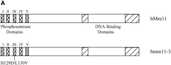
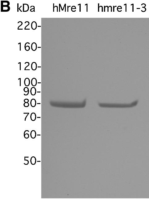
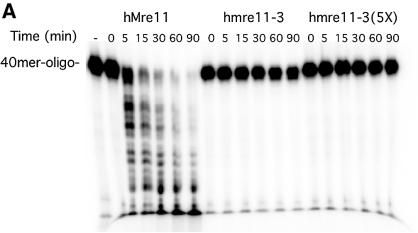
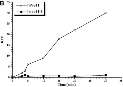
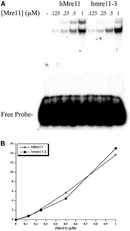
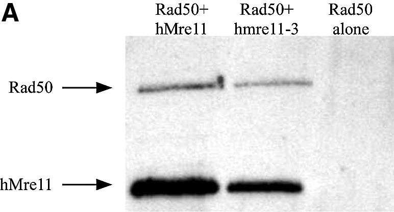
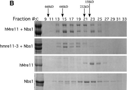
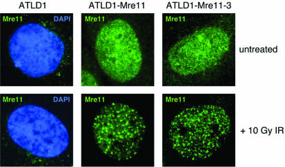
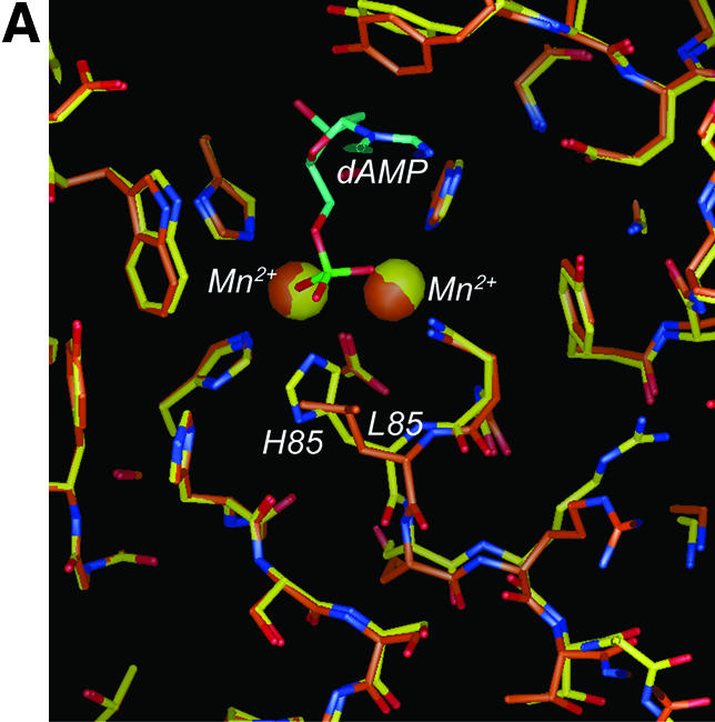
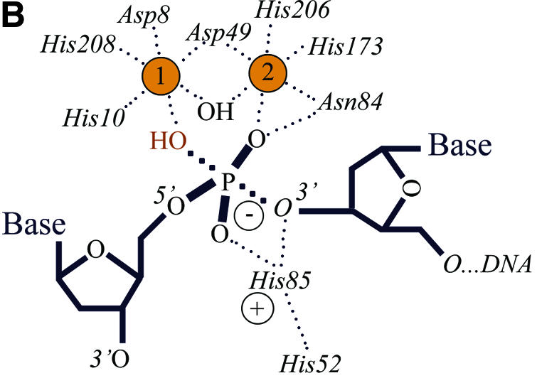
The Mre11, Rad50 and Nbs1 proteins make up the conserved multi-functional Mre11 (MRN) complex involved in multiple, critical DNA metabolic processes including double-strand break repair and telomere maintenance. The Mre11 protein is a nuclease with broad substrate recognition, but MRN-dependent processes requiring the nuclease activity are not clearly defined. Here, we report the functional and structural characterization of a nuclease-deficient Mre11 protein termed mre11-3. Importantly, the hmre11-3 protein has wild-type ability to bind DNA, Rad50 and Nbs1; however, nuclease activity was completely abrogated. When expressed in cell lines from patients with ataxia telangiectasia-like disorder (ATLD), hmre11-3 restored the formation of ionizing radiation-induced foci. Consistent with the biochemical results, the 2.3 A crystal structure of mre11-3 from Pyrococcus furiosus revealed an active site structure with a wild-type-like metal-binding environment. The structural analysis of the H85L mutation provides a detailed molecular basis for the ability of mre11-3 to bind but not hydrolyze DNA. Together, these results establish that the mre11-3 protein provides an excellent system for dissecting nuclease-dependent and independent functions of the Mre11 complex.


Figures

Figure 1
Figure 1
Schematic sequence and purity of wild-type and hmre11-3 mutant Mre11 proteins. (A) The N-terminus of Mre11 is evolutionarily conserved, specifically the five phosphoesterase domains (cross-hatch). The C-terminal domain contains two putative DNA-binding domains (diagonal hatch). The hmre11-3 protein has a mutation in the third phosphoesterase domain, specifically a histidine and aspartic acid at position 129 and 130 were converted to a leucine and valine, respectively. (B) Purified hMre11 (WT) and hmre11-3 analyzed by SDS–PAGE and Coomassie blue staining (left panel) and western blot (right panel).
Figure 1
Figure 1
Schematic sequence and purity of wild-type and hmre11-3 mutant Mre11 proteins. (A) The N-terminus of Mre11 is evolutionarily conserved, specifically the five phosphoesterase domains (cross-hatch). The C-terminal domain contains two putative DNA-binding domains (diagonal hatch). The hmre11-3 protein has a mutation in the third phosphoesterase domain, specifically a histidine and aspartic acid at position 129 and 130 were converted to a leucine and valine, respectively. (B) Purified hMre11 (WT) and hmre11-3 analyzed by SDS–PAGE and Coomassie blue staining (left panel) and western blot (right panel).
Figure 2
Figure 2
Exonuclease activities of the hmre11-3 mutant protein. (A) Proteins were tested for activity on a double-stranded 40mer oligonucleotide as described in Materials and Methods. Aliquots were removed from the reactions at the indicated time points, and at the conclusion of the reaction (90 min) the reaction products were separated on a denaturing acrylamide gel and visualized by phosphoimager. The mre11-3 protein showed no activity at five times the concentration of wild type. (B) Exonuclease assays were also performed using 25 nM protein with 2.5 mM of 10mer double-stranded oligo containing a single 2-amino purine that is six bases from the 3′ end. The release of the 2-amino purine over a time course was measured on a spectrophotometer.
Figure 2
Figure 2
Exonuclease activities of the hmre11-3 mutant protein. (A) Proteins were tested for activity on a double-stranded 40mer oligonucleotide as described in Materials and Methods. Aliquots were removed from the reactions at the indicated time points, and at the conclusion of the reaction (90 min) the reaction products were separated on a denaturing acrylamide gel and visualized by phosphoimager. The mre11-3 protein showed no activity at five times the concentration of wild type. (B) Exonuclease assays were also performed using 25 nM protein with 2.5 mM of 10mer double-stranded oligo containing a single 2-amino purine that is six bases from the 3′ end. The release of the 2-amino purine over a time course was measured on a spectrophotometer.
Figure 3
Figure 3
DNA binding tested by DNA gel shift assay. (A) DNA binding capabilities of hMre11 and hmre11-3 were assessed by gel shift assay using a single-stranded 40mer oligonucleotide as a probe. The probe was incubated with increasing protein concentrations (µM). Protein:DNA complexes were resolved on a native polyacrylamide gel and visualized by a phosphoimager. (B) The gel in (A) was quantitated and percent DNA shifted is plotted versus amount of protein added. No difference in binding was observed between hMre11 and hmre11-3.
Figure 4
Figure 4
Preserved interactions in the Mre11 protein complex proteins. (A) Crude samples from co-infections of Sf9 cells using His-tagged hMre11 (lane 1) or hmre11-3 (lane 2) virus with an untagged Rad50, and Rad50 alone (lane 3) were applied to Ni-NTA beads and eluted using imidazole. Elutions were run on a polyacrylamide gel and a western blot was performed using Mre11 and Rad50 antibodies. (B) Fractions from a gel filtration column run on a polyacrylamide gel. Co-infections of Sf9 cells using His-tagged hMre11 or hmre11-3 virus with a His-tagged Nbs1 virus were purified on a nickel column. The samples were subsequently fractionated on a Superdex 200 column. The hMre11 (top panel) and hmre11-3 (second from top panel) proteins, co-expressed with Nbs1, demonstrate a shift from a lower molecular weight to a higher molecular weight when compared with hMre11 (second from bottom panel) or Nbs1 (bottom panel) alone.
Figure 4
Figure 4
Preserved interactions in the Mre11 protein complex proteins. (A) Crude samples from co-infections of Sf9 cells using His-tagged hMre11 (lane 1) or hmre11-3 (lane 2) virus with an untagged Rad50, and Rad50 alone (lane 3) were applied to Ni-NTA beads and eluted using imidazole. Elutions were run on a polyacrylamide gel and a western blot was performed using Mre11 and Rad50 antibodies. (B) Fractions from a gel filtration column run on a polyacrylamide gel. Co-infections of Sf9 cells using His-tagged hMre11 or hmre11-3 virus with a His-tagged Nbs1 virus were purified on a nickel column. The samples were subsequently fractionated on a Superdex 200 column. The hMre11 (top panel) and hmre11-3 (second from top panel) proteins, co-expressed with Nbs1, demonstrate a shift from a lower molecular weight to a higher molecular weight when compared with hMre11 (second from bottom panel) or Nbs1 (bottom panel) alone.
Figure 5
Figure 5
Mre11 complex foci formation in cells complemented with wild-type and hmre11-3 proteins. ATLD1 cells were complemented by transduction with retrovirus vector expressing wild-type hMre11 and the hmre11-3 mutant. Cells were untreated or were exposed to 10 Gy IR and allowed to recover for 2 h prior to immunofluorescence with an Mre11 antibody. In ATLD1 cells, faint staining for the truncated Mre11 protein was observed mainly in the cytoplasm (DAPI staining marks the nuclei). In complemented cells, the hMre11 and hmre11-3 proteins appear in the nucleus and both form foci upon IR treatment. Focus formation was analyzed in pools of transduced cells. In both cases, foci were observed in the majority of cells expressing detectable levels of the Mre11 protein in the nucleus. The appearance of the foci for wild-type hMre11 and mutant hmre11-3 was similar.
Figure 6
Figure 6
Crystal structure of pfMre11-3, active site preservation and implicated transition state alterations. (A) Superposition of the refined crystal structure of pfMre11-3 (tube model with orange carbons, red oxygen and blue nitrogen atoms) with the previously determined structure of manganese-dAMP-bound wild-type pfMre11 (tube model with yellow carbons, red oxygen and blue nitrogen atoms). The two active site manganese ions are shown as orange (Mre11-3) or yellow (wild-type) spheres. The dAMP molecule (wild-type) is shown in cyan. With the exception of the backbone loop at His85, the active sites of wild-type and mutated Mre11 are structurally conserved, including the position and coordination of the manganese ions. This structural conservation suggests that metal and substrate binding by Mre11 is mostly undisturbed, whereas transition state stabilization is implied to be defective, due to disruption of the conserved His52–His85 charge relay system. (B) Proposed structure and mechanism of transition state stabilization by Mre11 as deduced from wild-type and mutant Mre11 structure and biochemistry. The DNA backbone at the scissile bond is bound to one of two metals (yellow spheres), whereas the other metal activates a hydroxide ion for nucleophilic attack. In the expected pentacoordinated phosphate transition state, His85 evidently stabilizes the developing negative charge by binding to the unliganded phosphate oxygen and possibly also the leaving group oxygen.
Figure 6
Figure 6
Crystal structure of pfMre11-3, active site preservation and implicated transition state alterations. (A) Superposition of the refined crystal structure of pfMre11-3 (tube model with orange carbons, red oxygen and blue nitrogen atoms) with the previously determined structure of manganese-dAMP-bound wild-type pfMre11 (tube model with yellow carbons, red oxygen and blue nitrogen atoms). The two active site manganese ions are shown as orange (Mre11-3) or yellow (wild-type) spheres. The dAMP molecule (wild-type) is shown in cyan. With the exception of the backbone loop at His85, the active sites of wild-type and mutated Mre11 are structurally conserved, including the position and coordination of the manganese ions. This structural conservation suggests that metal and substrate binding by Mre11 is mostly undisturbed, whereas transition state stabilization is implied to be defective, due to disruption of the conserved His52–His85 charge relay system. (B) Proposed structure and mechanism of transition state stabilization by Mre11 as deduced from wild-type and mutant Mre11 structure and biochemistry. The DNA backbone at the scissile bond is bound to one of two metals (yellow spheres), whereas the other metal activates a hydroxide ion for nucleophilic attack. In the expected pentacoordinated phosphate transition state, His85 evidently stabilizes the developing negative charge by binding to the unliganded phosphate oxygen and possibly also the leaving group oxygen.

References

    1. Critchlow S.E. and Jackson,S.P. (1998) DNA end-joining: from yeast to man. Trends Biochem. Sci., 23, 394–398. - PubMed
    1. Varon R., Vissinga,C., Platzer,M., Cerosaletti,K.M., Chrzanowska,K.H., Saar,K., Beckmann,G., Seemanova,E., Cooper,P.R., Nowak,N.J. et al. (1998) Nibrin, a novel DNA double-strand break repair protein, is mutated in Nijmegen breakage syndrome. Cell, 93, 467–476. - PubMed
    1. Carney J.P., Maser,R.S., Olivares,H., Davis,E.M., Le Beau,M., Yates,J.R., 3rd, Hays,L., Morgan,W.F. and Petrini,J.H. (1998) The hMre11/hRad50 protein complex and Nijmegen breakage syndrome: linkage of double-strand break repair to the cellular DNA damage response. Cell, 93, 477–486. - PubMed
    1. Haber J.E. (1998) The many interfaces of Mre11. Cell, 95, 583–586. - PubMed
    1. Stewart G.S., Maser,R.S., Stankovic,T., Bressan,D.A., Kaplan,M.I., Jaspers,N.G., Raams,A., Byrd,P.J., Petrini,J.H. and Taylor,A.M. (1999) The DNA double-strand break repair gene hMRE11 is mutated in individuals with an ataxia-telangiectasia-like disorder. Cell, 99, 577–587. - PubMed

Publication types

Associated data