. 2004;12(2):163-73.
doi: 10.1023/b:chro.0000013166.04629.97.

Non-random positioning of chromosomes in human sperm nuclei

Affiliations

Non-random positioning of chromosomes in human sperm nuclei

Irina A Zalenskaya et al. Chromosome Res. 2004.

Abstract

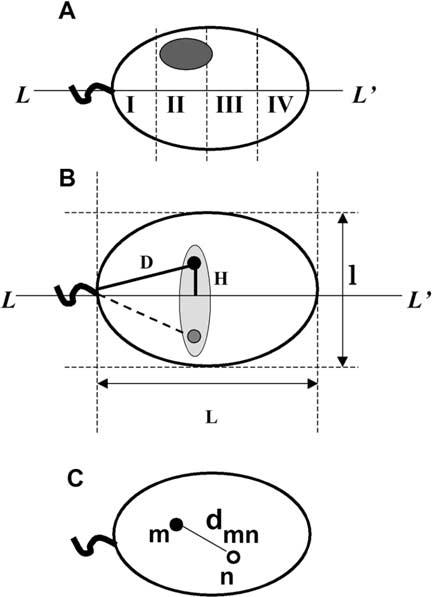
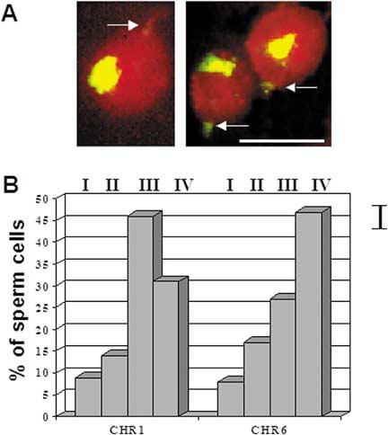
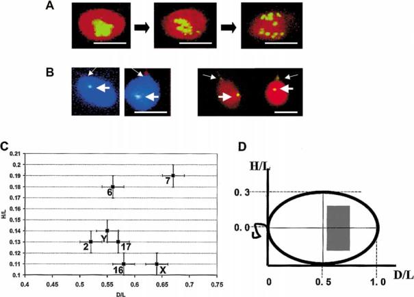
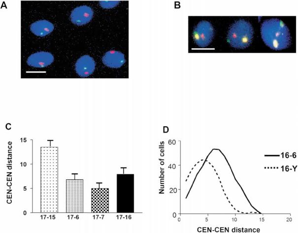
In human spermatozoa, the arrangement of chromosomes is non-random. Characteristic features are association of centromeres in the interior chromocenter and peripheral location of telomeres. In this paper, we have investigated the highest level of order in DNA packing in sperm--absolute and relative intranuclear chromosome positioning. Asymmetrical nuclear shape, existence of a defined spatial marker, and the haploid complement of chromosomes facilitated an experimental approach using in situ hybridization. Our results showed the tendency for non-random intranuclear location of individual chromosome territories. Moreover, centromeres demonstrated specific intranuclear position, and were located within a limited area of nuclear volume. Additionally, the relative positions of centromeres were non-random; some were found in close proximity, while other pairs showed significantly greater intercentromere distances. Therefore, a unique and specific adherence may exist between chromosomes in sperm. The observed chromosome order is discussed in relation to sperm nuclei decondensation, and reactivation during fertilization.


Figures

Figure 1.
Figure 1.
Geometrical parameters of human sperm cell used to determine chromosome positioning. A. Schematic representation of a sperm cell nuclei approximated by an ellipse with the axis of symmetry L-L′. Shaded oval shows an area of hybridization with a chromosome-painting probe. Sperm nucleus is divided into four zones I-IV starting from the basal side that is determined by tail attachment site. B. Parameters, which have been used to describe intranuclear localization of the chromosome-specific centromere hybridization signals. D - distance from the tail attachment point, H - elevation over the long axis. L and l are lengths of the long and short axes. Because of anticipated freedom of rotation along L-L′ we expected random distribution of signals within a schematically shown shaded area. Filled and shaded circles are experimentally observed mirror positions of a centromere. C. Scheme of the sperm nucleus image after multicolor hybridization with two centromere-specific probes. Distances between centers of the FISH signals (dmn) were used to determine relative positioning of centromeres within nuclei.
Figure 2.
Figure 2.
Determination of the intranuclear position of chromosomes in human sperm using FISH with whole chromosome painting probes. A. Representative hybridization pattern: chromosome 6 territory (yellow/green), total nuclear DNA counterstained with propidium iodide (red). Arrows show tail attachment points. Bar corresponds to 5 μm. B. Diagram of the distribution of chromosome FISH signals detected in nuclear sectors [I (basal) - IV (apical)] as defined in Figure 1A. Bar on the right shows confidence interval.
Figure 3.
Figure 3.
Determination of the intranuclear position of chromosomes using hybridization with centromere-specific probes. A. FISH localization of centromeres of all nonhomologous chromosomes in sperm nuclei using alphoid DNA probe. Typical patterns of FISH demonstrating dispersion of sperm chromocenter induced by cell swelling with Heparin in the presence of 10 mmol/L DTT. Panels from left to right correspond to 0 mg/ml, 0.01 mg/ml, and 0.1 mg/ml Heparin. B. Representative FISH patterns after hybridization with chromosome-specific centromere probes (thick arrows), left panels - CEN Y, total DNA counterstained with DAPI (blue); right panels - CEN 17, total DNA counterstained with PI (red). Thin arrows show tail attachment points. Bars in A and B correspond to 5 μm. C. Localization of individual centromeres within sperm nuclei. Each spot have the mean normalized coordinates experimentally determined (as explained in Methods, Figure 1B) for each chromosome-specific centromere. Bars show standard errors. D. Schematic view of the sperm nucleus showing the area within which all studied centromeres were localized (shaded rectangular). The same system of coordinates as in C is used.
Figure 4.
Figure 4.
Determination of relative position of chromosomes using FISH with chromosome-specific centromere probes. Typical patterns of centromere localization by multicolor FISH which were used to measure intercentromere distances dmn, total DNA was counterstained with DAPI (blue). A. CEN16 (green), CEN18 (red). B. CEN16 (green), CEN17 (yellow) CEN X (red). Bars in A and B correspond to 5 μm. C. Chart of intercentromere distances between chromosome 17 and four selected chromosomes. Bars indicate standard errors. D. Frequency distribution plot for intercentromere distances for the chromosome pairs 16-6 and 16-Y.

Similar articles

Cited by

References

    1. Allison DC, Nestor AL. Evidence for a relatively random array of human chromosomes on the mitotic ring. J Cell Biol. 1999;145:1–14. - PMC - PubMed
    1. Ashley T. Specific end-to-end attachment of chromosomes in Ornithogalum virens. J Cell Sci. 1979;38:357–367. - PubMed
    1. Balhorn R. A model for the structure of chromatin in mammalian sperm. J Cell Biol. 1982;93:298–305. - PMC - PubMed
    1. Bickmore WA, Chubb JR. Chromosome position: now, where was I? Curr Biol. 2003;13:357–359. - PubMed
    1. Bojanowski K, Maniotis AJ, Plisov S, Larsen AK, Ingber DE. DNA topoisomerase II can drive changes in higher order chromosome architecture without enzymatically modifying DNA. J Cell Biochem. 1998;69:127–142. - PubMed

Publication types