Comparative Study
. 2004 Mar 26;13(6):805-15.
doi: 10.1016/s1097-2765(04)00124-8.

Respiratory complex III is required to maintain complex I in mammalian mitochondria

Affiliations
Comparative Study

Respiratory complex III is required to maintain complex I in mammalian mitochondria

Rebeca Acín-Pérez et al. Mol Cell. .

Abstract

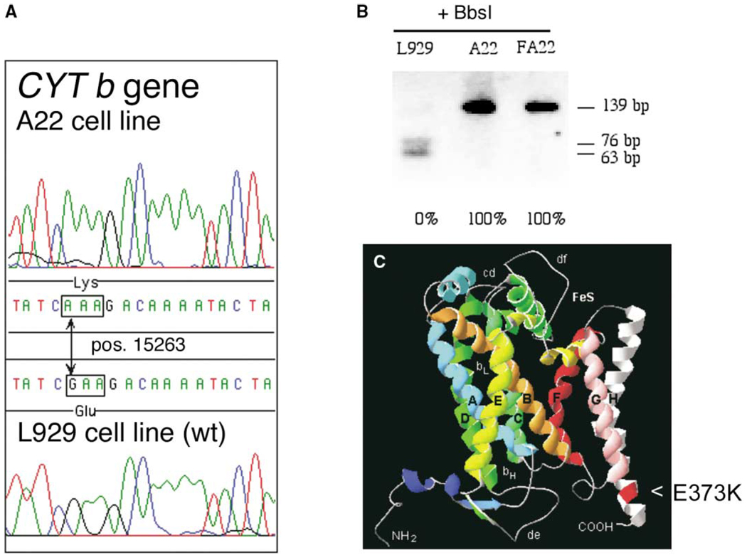
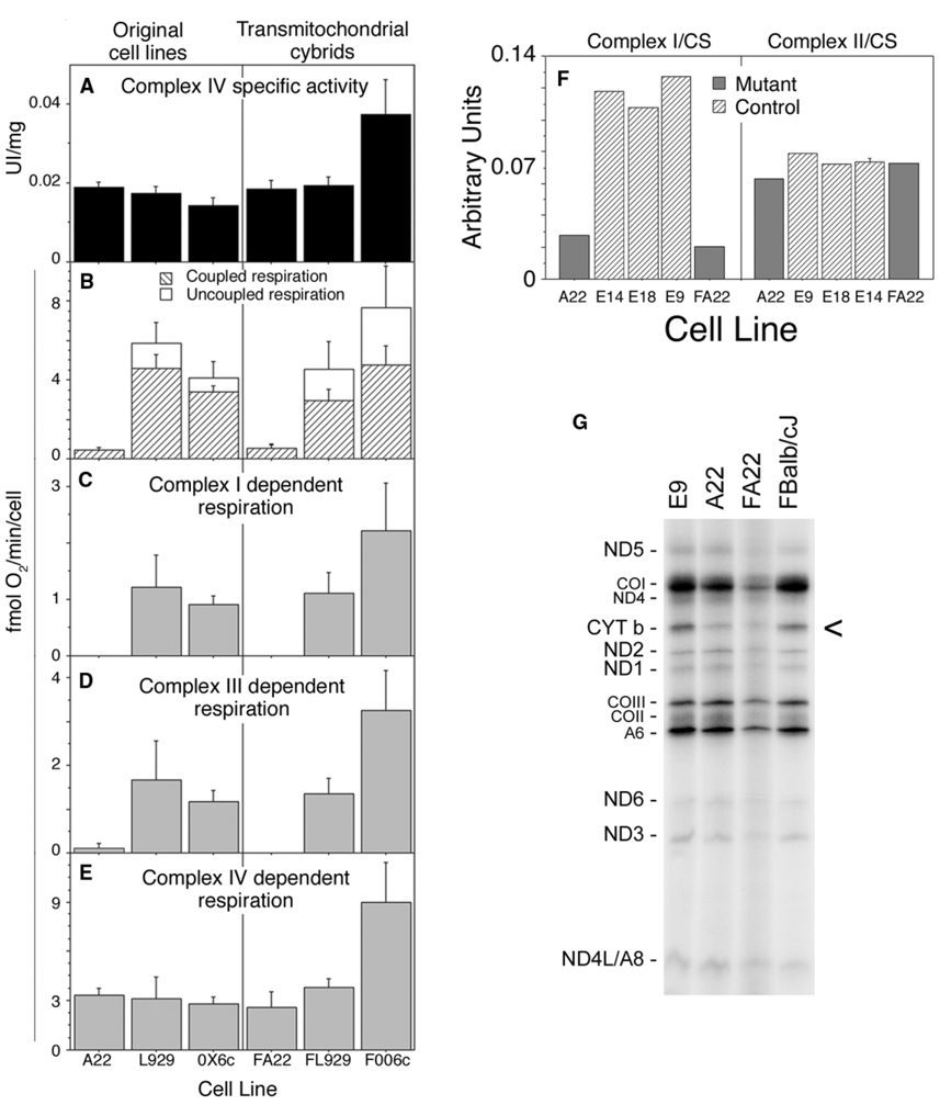
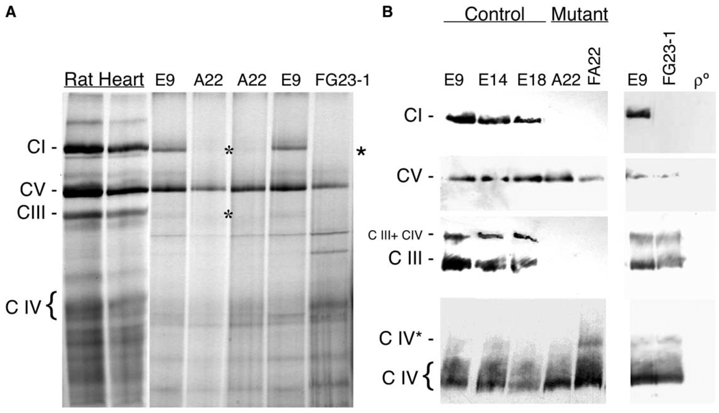
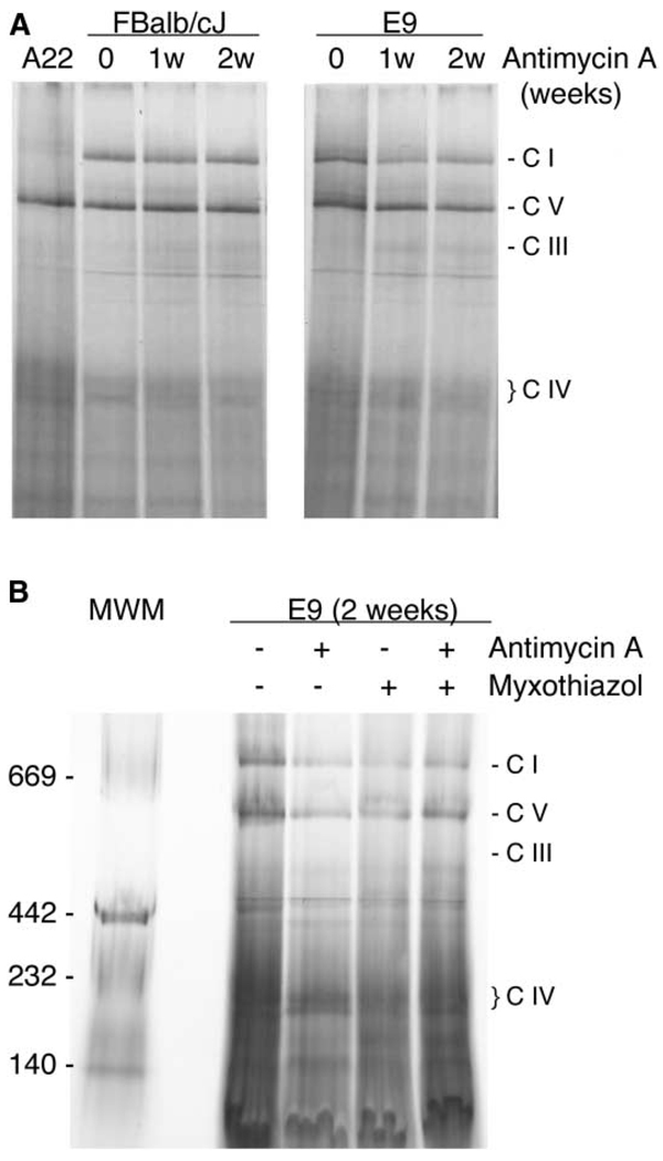
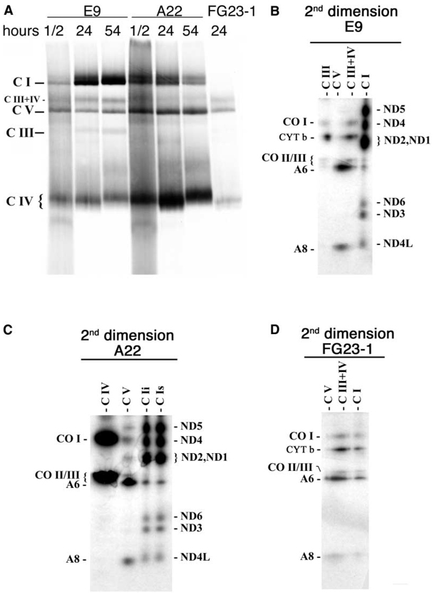
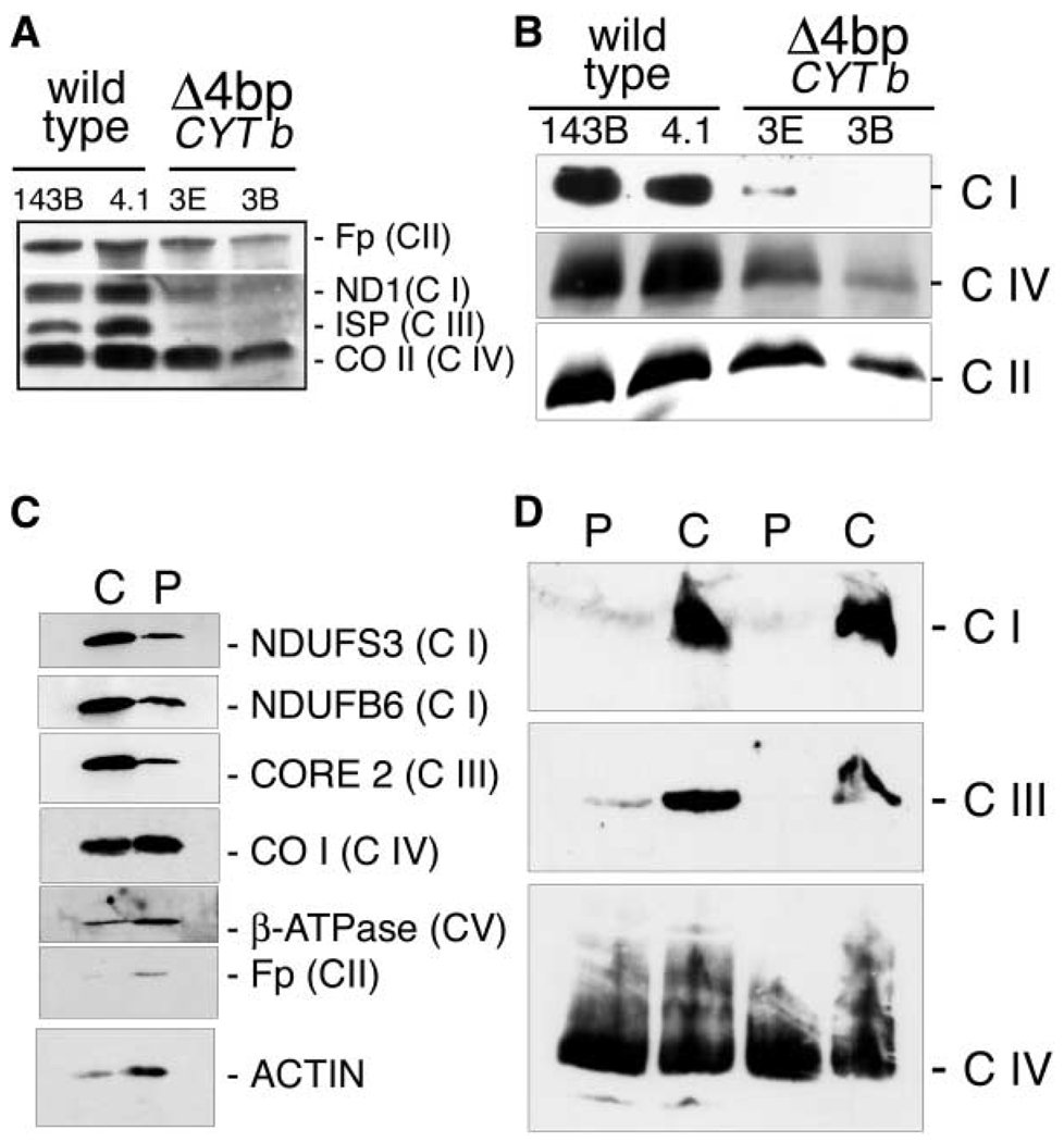
A puzzling observation in patients with oxidative phosphorylation (OXPHOS) deficiencies is the presence of combined enzyme complex defects associated with a genetic alteration in only one protein-coding gene. In particular, mutations in the mtDNA encoded cytochrome b gene are associated either with combined complex I+III deficiency or with only complex III deficiency. We have reproduced the combined complex I+III defect in mouse and human cultured cell models harboring cytochrome b mutations. In both, complex III assembly is impeded and causes a severe reduction in the amount of complex I, not observed when complex III activity was pharmacologically inhibited. Metabolic labeling in mouse cells revealed that complex I was assembled, although its stability was severely hampered. Conversely, complex III stability was not influenced by the absence of complex I. This structural dependence among complexes I and III was confirmed in a muscle biopsy of a patient harboring a nonsense cytochrome b mutation.


Figures

Figure 1
Figure 1. Characterization of the Cytochrome b Mutation
(A) Chromatogram showing the G15263A transition within the CYT b gene of A22 cells. (B) RFLP analysis of the presence and homoplasmic status for the G15263A transition that disrupts a restriction site for BbsI. (C) Structural model of the mouse CYT b protein derived by analogy with the bovine crystallographic data (Tsukihara et al., 1996). The E373K substitution is located at the helix H close to the carboxy terminus end of the protein.
Figure 2
Figure 2. Functional Analysis of OXPHOS Performance and Mitochondrial Protein Synthesis
Spectrophotometry measurements of complex IV activity in total cell extracts (A), or oxygraphic measurement of overall respiration (B), glutamate plus malate-dependent respiration (C), succinate plus glycerol-3-phosphate-dependent respiration (D), or TMPD (N,N,N′,N′-TetraMethyl-p-PhenyleneDiamine)-dependent respiration (E) of the original cell clones (left) and their transmitochondrial cell lines (right). OX6c: average values of cell lines E9, E14, and E18 isogenic with A22 but without the mutation in CYT b. FOO6c: average values of the transmitochondrial control cell lines FBalb/cJ, FC57BL/6J, and FCBA/J generated as described in Experimental Procedures. (F) Spectroscopic measurement of isolated complex I and II in mutant (A22, FA22) and control cells (E9, E14, E18) normalized by citrate synthase activity (values for Complex I-specific activities were in UI/g: E9 = 89.31 ± 11; E14 = 84.85 ± 16.9; E18 = 71.54 ± 3.9; A22 = 13.43 ± 2.7 and FA22 = 10.81 ± 2.4 and for Complex II: E9 = 91.5 ± 15; E14 = 83.5 ± 12; E18 = 89.8 ± 17; A22 = 82.8 ± 18 and FA22 = 79.2 ± 15.4). (G) Fluorogram, after electrophoresis through an SDS-polyacrylamide gradient gel, of the mitochondrial translation products of the indicated mutant and wild-type cells, labeled with [35S]-methionine for 1 hr in the presence of emetine. COI, COII, and COIII, subunits I, II, and III of cytochrome c oxidase; ND1 ND2, ND3, ND4, ND4L, ND5, and ND6, subunits 1, 2, 3, 4, 4L, 5, and 6 of the respiratory chain NADH dehydrogenase; A6 and A8, subunits 6 and 8 of H+-ATPase; CYT b, apocytochrome b.
Figure 3
Figure 3. Analysis of the Assembly Status of the Different OXPHOS Complexes
(A) Blue-Native gel electrophoresis (BNGE) of the mitocondrial OXPHOS complexes in E9 and A22 cells using rat heart mitochondria as a control and showing also the ND6-deficient cell line FG23-1. (B) Western blot of the different assembled complexes probed with anti NDUFS3 for complex I (C I), anti β F1-ATPase for complex V (C V), anti-Core 2 for complex III (C III), anti-CO I for complex IV (C IV), in the indicated control (E9, E14, E18), cytochrome b mutant (A22, FA22), ND6 mutant (FG23-1), and mtDNA-less cells (ρ°). CI, complex I, C III, complex III, C IV, complex IV, C III + IV, combined complex III + IV, CIV* probable complex IV dimer. Asterisks indicate the absence of CI or CIII in A22 and FG23-1 cells, respectively.
Figure 4
Figure 4. The Absence of Complex III Activity Does Not Influence the Assembly of Complex I
(A) Blue-Native gel electrophoresis showing the assembly status of the OXPHOS complexes in two different control cell lines after one or two weeks of treatment with antimycin A. (B) Blue-Native gel electrophoresis showing the assembly status of the OXPHOS complexes in control cells treated for 2 weeks with the indicated complex III inhibitors.
Figure 5
Figure 5. Metabolic Labeling of the Assembled OXPHOS Complexes
(A) Fluorogram, after Blue-Native electrophoresis of the mitochondrial translation products of mutant and wild-type cells, pulse-labeled with [35S]-methionine for 1 hr in the presence of cycloheximide and chased for the indicated number of hours. (B to D) Fluorograms of bidimensional electrophoresis (Blue-Native electrophoresis followed by SDS-polyacrylamide gel electrophoresis), of the mitochondrial translation products of mutant and wild-type cells shown in (A) (24 hr chase). Nomenclature over the lines indicates the putative complex that is expected to migrate in the first dimension in the area of the gel taken for the second dimension regardless if the complex is observed or not. The double band observed in the area of complex I in the first dimension from A22 cells was analyzed separately by second dimension in (C): faster migrating band (C Ii) and slower migrating band (C Is). The remaining symbols are as indicated in Figure 4.
Figure 6
Figure 6. Human Cells Lacking Cytochrome b Do Not Assemble Complex III and Show a Concomitant Defect in the Assembly of Complex I
(A and C) Quantification by SDS-PAGE and Western blot of the relative level of proteins belonging to the indicated OXPHOS complex in total protein extracts from (A) human cultured control cells (143B and 4.1) and mutant cells (3E and 3B) and (C) from control (C) and patient (P) muscle biopsies. (B and D) Blue-Native gel electrophoresis of mitochondrial proteins isolated from the indicated human cultured cells (B) and muscle biopsies (D).

References

    1. Acin-Perez R, Bayona-Bafaluy MP, Bueno M, Machicado C, Fernandez-Silva P, Perez-Martos A, Montoya J, Lopez-Perez MJ, Sancho J, Enriquez JA. An intragenic suppressor in the cytochrome c oxidase I gene of mouse mitochondrial DNA. Hum. Mol. Genet. 2003;12:329–339. - PubMed
    1. Andreu AL, Bruno C, Shanske S, Shtilbans A, Hirano M, Krishna S, Hayward L, Systrom DS, Brown RH, Jr, Di-Mauro S. Missense mutation in the mtDNA cytochrome b gene in a patient with myopathy. Neurology. 1998;51:1444–1447. - PubMed
    1. Andreu AL, Hanna MG, Reichmann H, Bruno C, Penn AS, Tanji K, Pallotti F, Iwata S, Bonilla E, Lach B, et al. Exercise intolerance due to mutations in the cytochrome b gene of mitochondrial DNA. N. Engl. J. Med. 1999;341:1037–1044. - PubMed
    1. Antonicka H, Ogilvie I, Taivassalo T, Anitori RP, Haller RG, Vissing J, Kennaway NG, Shoubridge EA. Identification and characterization of a common set of complex I assembly intermediates in mitochondria from patients with complex I deficiency. J. Biol. Chem. 2003;278:43081–43088. - PubMed
    1. Appleby RD, Porteous WK, Hughes G, James AM, Shannon D, Wei YH, Murphy MP. Quantitation and origin of the mitochondrial membrane potential in human cells lacking mitochondrial DNA. Eur. J. Biochem. 1999;262:108–116. - PubMed

Publication types