

Breast adenocarcinoma liver metastases, in contrast to colorectal cancer liver metastases, display a non-angiogenic growth pattern that preserves the stroma and lacks hypoxia
- PMID: 15054467
- PMCID: PMC2409675
- DOI: 10.1038/sj.bjc.6601727
Breast adenocarcinoma liver metastases, in contrast to colorectal cancer liver metastases, display a non-angiogenic growth pattern that preserves the stroma and lacks hypoxia
Abstract
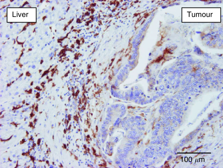
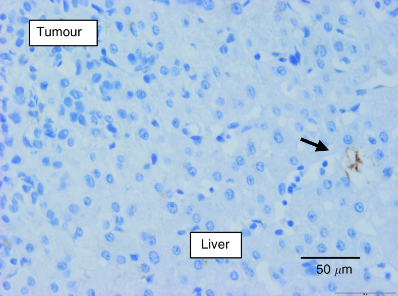
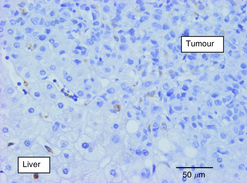
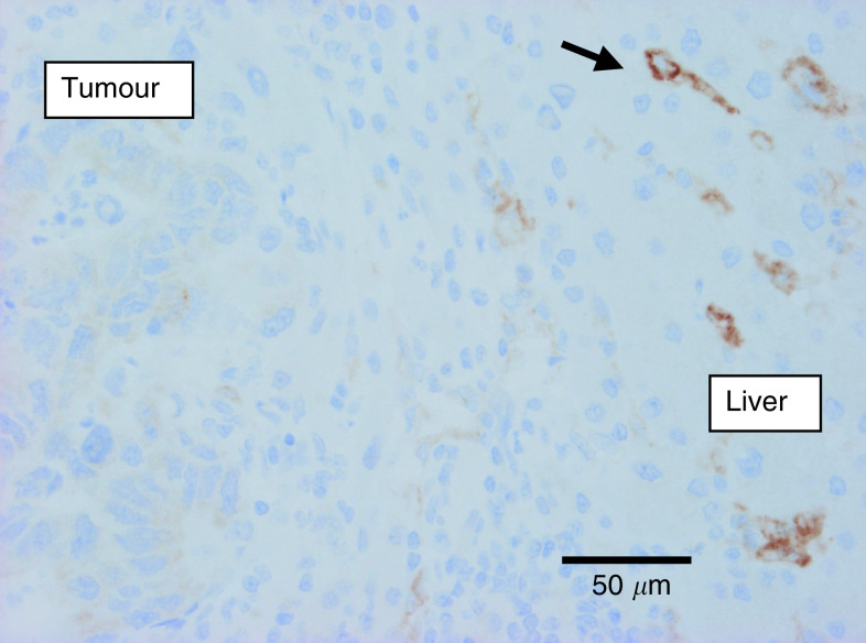
Although angiogenesis is a prerequisite for the growth of most human solid tumours, alternative mechanisms of vascularisation can be adopted. We have previously described a non-angiogenic growth pattern in liver metastases of colorectal adenocarcinomas (CRC) in which tumour cells replace hepatocytes at the tumour-liver interface, preserving the liver architecture and co-opting the sinusoidal blood vessels. The aim of this study was to determine whether this replacement pattern occurs during liver metastasis of breast adenocarcinomas (BC) and whether the lack of an angiogenic switch in such metastases is due to the absence of hypoxia and subsequent vascular fibrinogen leakage. The growth pattern of 45 BC liver metastases and 28 CRC liver metastases (73 consecutive patients) was assessed on haematoxylin- and eosin-stained tissue sections. The majority of the BC liver metastases had a replacement growth pattern (96%), in contrast to only 32% of the CRC metastases (P<0.0001). The median carbonic anhydrase 9 (CA9) expression (M75 antibody), as a marker of hypoxia, (intensity x % of stained tumour cells) was 0 in the BC metastases and 53 in the CRC metastases (P<0.0001). There was CA9 expression at the tumour-liver interface in only 16% of the BC liver metastases vs 54% of the CRC metastases (P=0.002). There was fibrin (T2G1 antibody) at the tumour-liver interface in only 21% of the BC metastases vs 56% of the CRC metastases (P=0.04). The median macrophage count (Chalkley morphometry; KP-1 anti-CD68 antibody) at the interface was 4.3 and 7.5, respectively (P<0.0001). Carbonic anhydrase 9 score and macrophage count were positively correlated (r=0.42; P=0.002) in all metastases. Glandular differentiation was less in the BC liver metastases: 80% had less than 10% gland formation vs only 7% of the CRC metastases (P<0.0001). The liver is a densely vascularised organ and can host metastases that exploit this environment by replacing the hepatocytes and co-opting the vasculature. Our findings confirm that a non-angiogenic pattern of liver metastasis indeed occurs in BC, that this pattern of replacement growth is even more prevalent than in CRC, and that the process induces neither hypoxia nor vascular leakage.
Figures


References
-
- Alpaugh ML, Tomlinson JS, Kasraeian S, Barsky SH (2002) Cooperative role of E-Cadherin and sialyl-Lewis X/A-deficient MUC1 in the passive dissimination of tumor emboli in inflammatory breast carcinoma. Oncogene 16: 3631–3643 - PubMed
-
- Beasley NJ, Prevo R, Banerji S, Leek RD, Moore J, van Trappen P, Cox G, Harris AL, Jackson DG (2002) Intratumoral lymphangiogenesis and lymph node metastasis in head and neck cancer. Cancer Res 62: 1315–1320 - PubMed
-
- Burke B, Tang N, Corke KP, Tazzyman D, Ameri K, Wells M, Lewis CE (2002) Expression of HIF-1alpha by human macrophages: implications for the use of macrophages in hypoxia-regulated cancer gene therapy. J Pathol 196: 204–212 - PubMed
-
- Chalkley HW (1943) Method for the quantitative morphologic analysis of tissues. J Natl Cancer Inst 4: 47–53
Publication types
MeSH terms
Substances
LinkOut - more resources
Full Text Sources
Other Literature Sources
Medical
Miscellaneous
