. 2004 Apr;111(4):400-6.
doi: 10.1111/j.1365-2567.2004.01824.x.

Alpha6-integrin is expressed on germinal centre B cells and modifies growth of a B-cell line

Affiliations

Alpha6-integrin is expressed on germinal centre B cells and modifies growth of a B-cell line

Helen E Ambrose et al. Immunology. 2004 Apr.

Abstract

The production of high-affinity antibodies requires diversification of the antibody repertoire by somatic hypermutation followed by selection of those B cells bearing the highest affinity antibodies. Whilst many surface molecules that mediate the cell-cell interactions required for germinal centre formation have been identified, little is known of the importance of interactions with components of the extracellular matrix, i.e. fibronectin, collagen and laminin. We demonstrate that the laminin-binding alpha6-integrin is expressed on germinal centre B cells and is induced during the in vitro activation of naïve splenic B cells. A laminin network is demonstrated within the germinal centre. Analysis of an alpha6-integrin-expressing mouse B-cell line, A20, demonstrates that this molecule is essential for binding to laminin, and that blocking by anti-alpha6-integrin immunoglobulin causes loss of adhesion associated with an increase in proliferation. There is no correlation with changes in BCL-6 or Blimp-1 expression, suggesting that alpha6-integrin does not play a role in differentiation.


Figures

Figure 1
Figure 1
Flow cytometry of mouse Peyer's patch and spleen cells. (a) Single-cell suspensions made from mouse Peyer's patches were incubated with an antibody directed against B220 and the lectin, peanut agglutinin (PNA), together with anti-α-integrin immunoglobulin. Histograms of the expression of integrin in B220+ PNA and B220+ PNA+ populations were obtained. Dotted lines represent isotype controls and solid lines integrin expression. (b) Histograms showing expression of selected β-integrins in B220+ PNA and B220+ PNA+ populations. (c) Histogram showing the expression of α6-integrin in B220+ PNA and B220+ PNA+ populations derived from mouse spleen.
Figure 2
Figure 2
Immunohistochemistry of mouse spleen. (a) Sections of spleen taken from immunized mice were stained with anti-α6-integrin immunoglobulin. Serial sections were stained with anti-mouse immunoglobulin D (IgD) in order to define the mantle zone and germinal centres (GC). Two separate primary follicles are represented. The anti-α6-integrin immunoglobulin also stains the endothelial cells of the marginal zone sinus (MZS), thus delineating the entire primary follicle. T, T-cell zone. Magnification ×40. (b) Anti-α6-integrin and anti-IgD staining of a secondary follicle (magnification ×40). (c) Anti-β1-integrin, anti-β4-integrin and anti-IgD staining of a secondary follicle. As positive controls for the anti-β-integrin immunoglobulin, images of arterial endothelium from the same sections have been included as insets (magnification ×100). (d) Serial sections showing a splenic follicle stained with anti-IgD and anti-laminin immunoglobulin. There is a coarse laminin network in the T-cell zones, with finer fibres present in the B-cell areas, including the GC. The laminin basement membrane of the MZS is demonstrated, as are basement membranes of the central arterioles (A). Magnification ×25. (e) High power (×100) view of a T-cell area and a B-cell area stained with anti-laminin immunoglobulin. The central arteriole in the T-cell area is indicated (A).
Figure 3
Figure 3
In vitro induction of α6-integrin expression on mouse splenocytes. (a) Mouse splenocytes were incubated with anti-CD40 (1 µg/ml) + interleukin-4 (IL-4) (20 ng/ml), with and without anti-µ (10 µg/ml), or with IL-4 or anti-µ alone, and the expression of α6-integrin was measured by flow cytometry after 12 hr and 36 hr. Dotted lines represent isotype controls and solid lines represent α6-integrin expression on cells exposed to the indicated antibodies or growth factors. (b) Adhesion assay. After 36 hr of stimulation with anti-CD40/IL-4 + anti-µ, anti-CD40/IL-4, or anti-µ alone, adhesion of mouse splenocytes to laminin was assayed. Black columns represent adhesion in the presence of blocking anti-α6-integrin immunoglobulin (GoH3) and white columns represent adhesion without blocking antibody. An increase of α6-integrin expression is associated with an increase in adhesion.
Figure 4
Figure 4
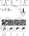
The A20 mouse B-cell lymphoma cell line expresses α6-integrin. (a) Flow cytometry of the EpH4 mouse mammary epithelial cell line and A20 cells, demonstrating α6-integrin expression and the J558 mouse plasma cell line lacking α6-integrin expression. (b) Western blot showing the predominant expression of α6A-integrin on A20 cells. (c) Adhesion to different extracellular membrane substrates. White columns again represent adhesion without blocking antibody and black columns adhesion in the presence of antibody. A20 cells bind strongly to fibronectin, but anti-α6-integrin immunoglobulin is not inhibitory. There is specific blocking of binding of A20 cells to laminin. (d) Effect of blocking laminin binding. A20 cells lose adherence and clump when cells are cultured on laminin-coated plates in the presence of anti-α6-integrin immunoglobulin. The cells remain adherent, despite anti-α6-integrin immunoglobulin, if other means of binding are permitted, e.g. culturing the cells in tissue culture plastic. (e) Loss of adherence is associated with an increase in the proportion of cells in the S phase of the cell cycle. Plots show anti-BrdU versus propidium iodide staining of A20 cells, and the figure panels show the proportion of cells in G1/G0, S and G2/M stages of the cell cycle. Cells growing in clumps are proliferating more than adherent cells. The results presented are representative of three experiments. BrdU, bromodeoxyuridine; PI, propidium iodide.
Figure 5
Figure 5
Reverse transcription–polymerase chain reaction (RT–PCR) of BCL-6 and Blimp-1 cDNA produced at various time-points after culture of A20 cells on laminin-coated plates with anti-α6-integrin immunoglobulin. Lane 1, time 0; lane 2, 1 hr; lane 3, 4 hr; lane 4, 24 hr; and lane 5, control PCR from cDNA extracted from a Blimp-1-expressing mouse plasma cell line (MPC11). RT–PCR was also carried out on A20 cells that had been kept rolling for specified lengths of time, in order to show that the effects were specific to blocking of binding to laminin and not simply to loss of adhesion. BCL-6 expression did not vary and Blimp-1 was not induced, suggesting that neither non-specific loss of adhesion nor that caused by anti-α6-integrin immunoglobulin resulted in changes indicative of terminal differentiation.

Comment in

References

    1. MacLennan I, Liu Y-J, Johnson G. Maturation and dispersal of B-cell clones during T cell-dependent antibody responses. Immunol Rev. 1992;126:143. - PubMed
    1. Jacob J, Kelsoe G, Rajewsky K, Weiss U. Intraclonal generation of antibody mutants in germinal centres. Nature. 1991;354:389. - PubMed
    1. Kepler T, Perelson A. Cyclic re-entry of germinal centre B cells and the efficiency of affinity maturation. Immunol Today. 1993;14:412. - PubMed
    1. Hynes R. Integrins: bidirectional allosteric signalling machines. Cell. 2002;110:673. - PubMed
    1. Lu T, Cyster J. Integrin mediated long-term B cell retention in the splenic marginal zone. Science. 2002;297:409. - PubMed

Publication types

LinkOut - more resources