. 2004 Apr;111(4):444-52.
doi: 10.1111/j.0019-2805.2004.01837.x.

The interaction of streptococcal inhibitor of complement (SIC) and its proteolytic fragments with the human beta defensins

Affiliations

The interaction of streptococcal inhibitor of complement (SIC) and its proteolytic fragments with the human beta defensins

Barbara A Fernie-King et al. Immunology. 2004 Apr.

Abstract

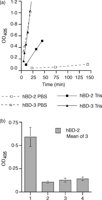
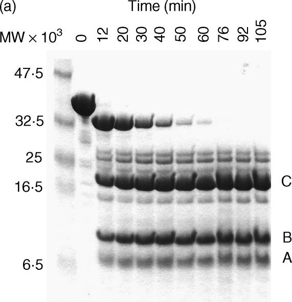
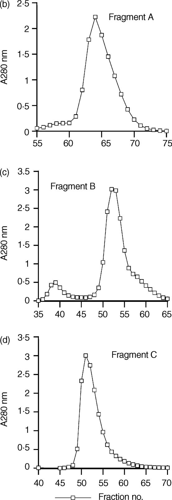
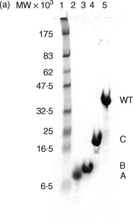
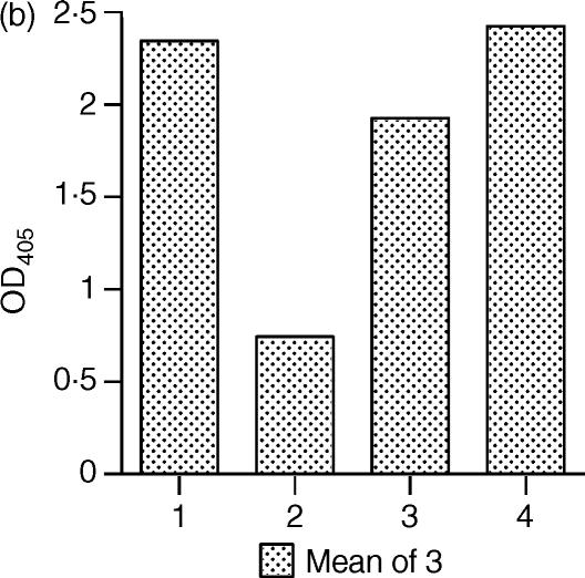
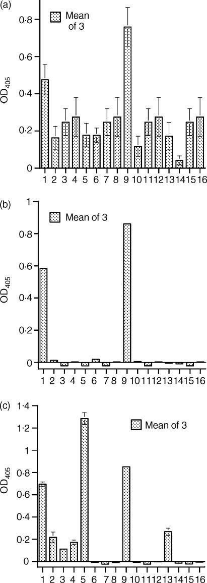
Streptococcal inhibitor of complement (SIC) is a 31 kDa extracellular protein produced by a few highly virulent strains of Streptococcus pyogenes (in particular the M1 strain). It has been shown additionally to inhibit four further components of the mucosal innate response-lysozyme, secretory leucocyte proteinase inhibitor, human alpha-defensin 1 and the cathelicidin LL-37 which are all bactericidal against Group A Streptococci (GAS). We now show that SIC also inhibits variably the antibacterial action of hBD-1, -2 and -3. By enzyme-linked immunosorbent assay (ELISA), SIC binds strongly to hBD-2 and hBD-3, but not at all to hBD-1. Investigation of the antimicrobial action of beta-defensins hBD-1, -2 and -3 against GAS in two different buffer systems shows that both the killing efficiencies of all three defensins, and the binding of SIC to them, occurs more efficiently in 10 mm Tris buffer than in 10 mm phosphate. The lower ionic strength of the Tris buffer may underlie this effect. hBD-1 kills the M1 strain of GAS only in 10 mm Tris, but is able to kill an M6 (SIC negative) strain in 10 mm phosphate. The inhibition of hBD-3 by SIC is clearly of physiological relevance, that of hBD-2 is likely to be so, but the inhibition of hBD-1 occurs only at lower ionic strength than is likely to be encountered in vivo. Elastase digestion of SIC yields three major fragments of MW 3.843 kDa comprising residues 1-33 (fragment A); 10.369 kDa comprising residues 34-126 (fragment B); and MW 16.487 kDa, comprising residues 127-273 (fragment C). By ELISA, only fragment B binds to hBD-2 and hBD-3 and this may indicate the inhibitory portion of the SIC molecule.


Figures

Figure 1
Figure 1
Binding of SIC to hBD-2 and hBD-3 in PBS or Tris buffer by ELISA. (a) Comparison of the binding efficiency between SIC and the β-defensins in different buffers, coating protein hBD-2 or hBD-3; ligand SIC; diluted in either PBS/Tween/0·1% gelatine or 10 mm Tris-HCl, pH 7·5/Tween/0·1% gelatine. Dotted lines and open symbols = PBS; solid lines and filled symbols = Tris. □ = hBD-2; ▵ = hBD-3. (b) Binding of hBD-2 to SIC in Tris buffer. Coating protein SIC; ligand hBD-2; dilution buffer 10 mm Tris-HCl, pH 7·5/Tween/0·5% hydrolysed casein. Bar 1 = SIC plus ligand plus first and second antibodies; bar 2 = SIC plus first and second antibodies; bar 3 = SIC plus second antibody; and bar 4 = SIC plus ligand plus second antibody. Results are mean ± SEM. In both panels, results are expressed as net OD405 after deduction of background values from matching uncoated wells.
Figure 2
Figure 2
SIC protects Group A Streptococci from killing by hBD-1, -2 and -3 in both (a) phosphate and (b) Tris buffer. Inhibition of the antibacterial action of the β-defensins by SIC. (a) Killing of M1 GAS by hBD-2 and hBD-3 in phosphate buffer is inhibited by SIC. Doubling dilutions of SIC from 6 µm were combined with 2 µm hBD-2 or SIC from 19·5 µm with 6·5 µm hBD-3, and incubated for 2 h at 37°. Bacteria at 2 × 10⁵/ml were added, incubated for a further 2 h at 37°, then diluted and plated. Final overall concentrations were either SIC from 1·5 µm plus 0·5 µm hBD-2 or SIC from 4·875 µm plus 1·625 µm hBD-3, and bacteria at 105/ml. (b) Killing of M1 GAS by hBD-1, hBD-2 and hBD-3 in Tris buffer is inhibited by SIC. Doubling dilutions of SIC from 48 µm were combined with 12 µm hBD-1, SIC from 2·4 µm with 0·6 µm hBD-2 or SIC from 9·6 µm with 2·4 µm hBD-3, and incubated for 2 h at 37°. Bacteria at 2 × 10⁵/ml were added, incubated for a further 2 h at 37°, then diluted and plated. Final overall concentrations were either SIC from 12 µm plus 3 µm hBD-1, SIC from 0·6 µm plus 0·15 µm hBD-2 or SIC from 2·4 µm plus 0·6 µm hBD-3, and bacteria at 105/ml. In both panels, results are expressed as percentage survival compared to bacteria incubated in the equivalent concentration of SIC alone ± SEM, against the number of molecules of defensin per molecule of SIC (hBD-1: three experiments; hBD-2: two experiments in each buffer; hBD-3: three experiments in phosphate, two experiments in Tris).
Figure 3
Figure 3
Purification of elastase-digested fragments of SIC. (a) Time course digestion of SIC analysed on a Tricine-SDS gel. (b) Purification of Fragment A by gel filtration. (c) Purification of Fragment B by gel filtration. (d) Purification of Fragment C by gel filtration. (a) A time–course experiment was performed in which SIC was digested with 0·1% w/w human neutrophil elastase. Samples taken at approximately 10-min intervals were analysed on a 16% Tricine SDS-PAGE gel and Coomassie stained. Lane 1, molecular weight markers; lane 2, undigested SIC; lanes 3–11, digested SIC. The 50-min digest was chosen for purification of the three major fragments designated. (b) (c) and (d) Purification of Fragments A, B and C by gel filtration. Pooled fractions containing predominantly either fragments A, B or C, separated initially by ion-exchange chromatography on a Source Q column, were purified further on a Superdex 75 column and resolved into sharp peaks.
Figure 3
Figure 3
Purification of elastase-digested fragments of SIC. (a) Time course digestion of SIC analysed on a Tricine-SDS gel. (b) Purification of Fragment A by gel filtration. (c) Purification of Fragment B by gel filtration. (d) Purification of Fragment C by gel filtration. (a) A time–course experiment was performed in which SIC was digested with 0·1% w/w human neutrophil elastase. Samples taken at approximately 10-min intervals were analysed on a 16% Tricine SDS-PAGE gel and Coomassie stained. Lane 1, molecular weight markers; lane 2, undigested SIC; lanes 3–11, digested SIC. The 50-min digest was chosen for purification of the three major fragments designated. (b) (c) and (d) Purification of Fragments A, B and C by gel filtration. Pooled fractions containing predominantly either fragments A, B or C, separated initially by ion-exchange chromatography on a Source Q column, were purified further on a Superdex 75 column and resolved into sharp peaks.
Figure 4
Figure 4
Purified SIC fragments. (a) Tricine-SDS gel of purified SIC and fragments. (b) ELISA—detection of SIC and fragments by rabbit anti-SIC. Analysis of purified elastase-digested fragments of SIC. (a) SIC and purified elastase-digested fragments of SIC were run on a 16% Tricine SDS-PAGE gel and Coomassie stained. Lane 1, molecular weight markers; lane 2, fragment A, amino acids 1–33; lane 3, fragment B, amino acids 34–126; lane 4, fragment C, amino acids 127–273; lane 5, whole SIC. (b) An ELISA plate was coated with SIC or purified elastase-digested fragments of SIC, and bound protein detected. Bar 1 = intact SIC, bar 2 = fragment A, bar 3 = fragment B, bar 4 = fragment C. Results are expressed as net OD405 after deduction of background values from matching uncoated wells, ± SEM. Where no error bars are shown, the SEM is too small to be seen. Controls of coated wells plus second antibody are not shown, but all values were < 0·07.
Figure 4
Figure 4
Purified SIC fragments. (a) Tricine-SDS gel of purified SIC and fragments. (b) ELISA—detection of SIC and fragments by rabbit anti-SIC. Analysis of purified elastase-digested fragments of SIC. (a) SIC and purified elastase-digested fragments of SIC were run on a 16% Tricine SDS-PAGE gel and Coomassie stained. Lane 1, molecular weight markers; lane 2, fragment A, amino acids 1–33; lane 3, fragment B, amino acids 34–126; lane 4, fragment C, amino acids 127–273; lane 5, whole SIC. (b) An ELISA plate was coated with SIC or purified elastase-digested fragments of SIC, and bound protein detected. Bar 1 = intact SIC, bar 2 = fragment A, bar 3 = fragment B, bar 4 = fragment C. Results are expressed as net OD405 after deduction of background values from matching uncoated wells, ± SEM. Where no error bars are shown, the SEM is too small to be seen. Controls of coated wells plus second antibody are not shown, but all values were < 0·07.
Figure 5
Figure 5
ELISAs. Binding of SIC and SIC fragments to (a) hBD-2 and (b) hBD-3; (c) binding of hBD-2 to SIC and SIC fragments. In each panel, bars 1–4 = intact SIC, bars 5–8 = fragment A, bars 9–12 = fragment B, bars 13–16 = fragment C. Within each group of 4 bars, bar 1 = coating protein + ligand + both antibodies; bar 2 = coating protein + both antibodies; bar 3 = coating protein + second antibody; bar 4 = coating protein + ligand + second antibody. (a) coating peptide hBD-2, ligands SIC or fragments. Buffer = 10 mm Tris-HCl/Tween/gelatine. (b) coating protein hBD-3, ligands SIC or fragments. Buffer = PBS/Tween/gelatine. (c) coating protein SIC or fragments; ligand hBD-2. Buffer = 10 mm Tris-HCl/Tween/casein. Results are expressed as net OD405 after deduction of background values from matching uncoated wells, ± SEM. Where no error bars are shown, the SEM is too small to be seen.

Similar articles

Cited by

References

    1. Åkesson P, Sjöholm AG, Björck L. Protein SIC, a novel extracellular protein of Streptococcus pyogenes interfering with complement function. J Biol Chem. 1996;271:1081–8. - PubMed
    1. Fernie-King BA, Seilly DJ, Willers C, Würzner R, Davies A, Lachmann PJ. Streptococcal inhibitor of complement (SIC) inhibits the membrane attack complex by preventing uptake of C567 onto cell membranes. Immunol. 2001;103:390–8. - PMC - PubMed
    1. Fernie-King BA, Seilly DJ, Davies A, Lachmann PJ. Streptococcal Inhibitor of Complement (SIC) inhibits two further components of the mucosal innate immune system: secretory leucokyte proteinase inhibitor (SLPI) and lysozyme. Infect Immun. 2002;70:4908–16. - PMC - PubMed
    1. Frick I-M, Åkesson P, Rasmussen M, Schmidtchen A, Björck L. SIC—a secreted protein of Streptococcus pyogenes that inactivates antibacterial peptides. J Biol Chem. 2003;278:16561–6. - PubMed
    1. Lukomski S, Hoe NP, Abdi I, et al. Nonpolar inactivation of the hypervariable Streptococcal Inhibitor of Complement gene (sic) in serotype M1 Streptococcus pyogenes significantly decreases mouse mucosal colonisation. Infect Immun. 2000;68:535–42. - PMC - PubMed

Publication types

MeSH terms

LinkOut - more resources