. 2004 Mar 31;24(13):3186-98.
doi: 10.1523/JNEUROSCI.5146-03.2004.

Connexin32-containing gap junctions in Schwann cells at the internodal zone of partial myelin compaction and in Schmidt-Lanterman incisures

Affiliations

Connexin32-containing gap junctions in Schwann cells at the internodal zone of partial myelin compaction and in Schmidt-Lanterman incisures

Carola Meier et al. J Neurosci. .

Abstract

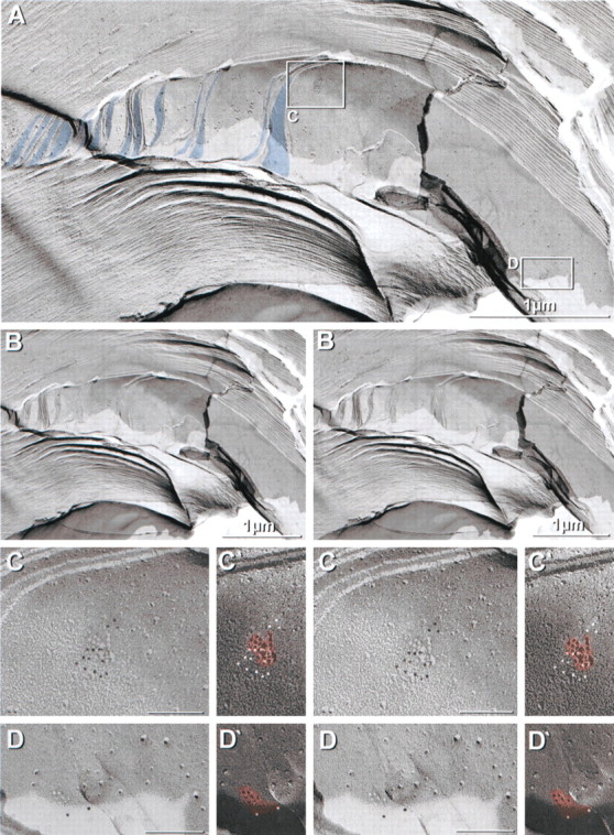
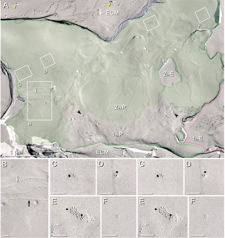
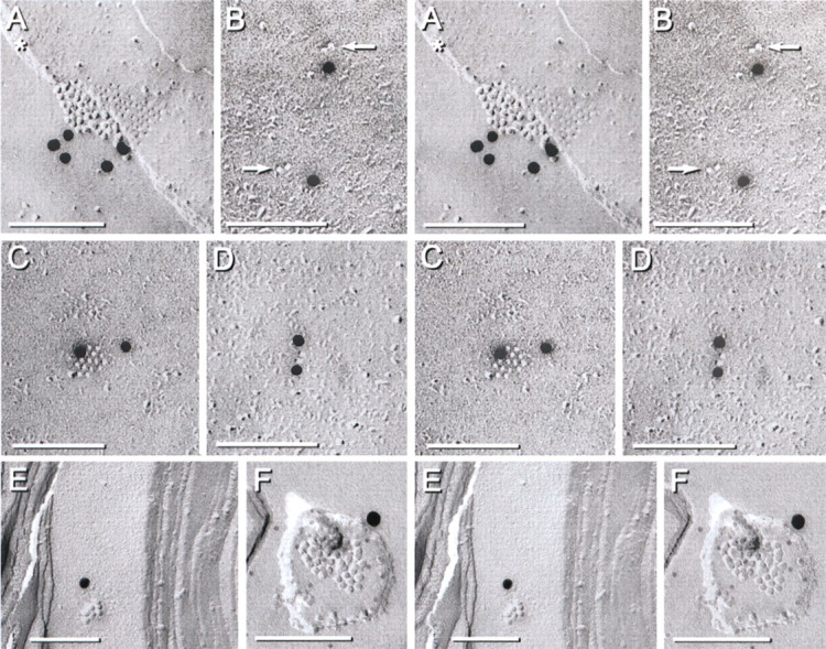
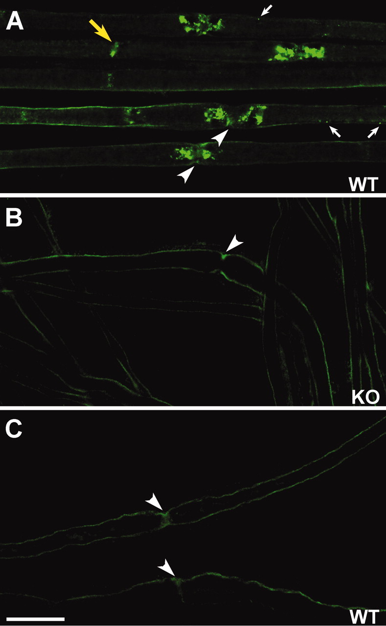
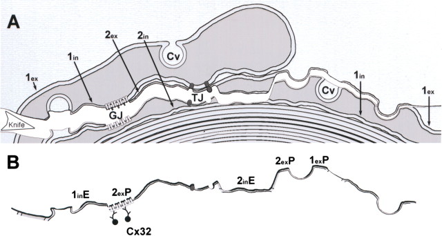
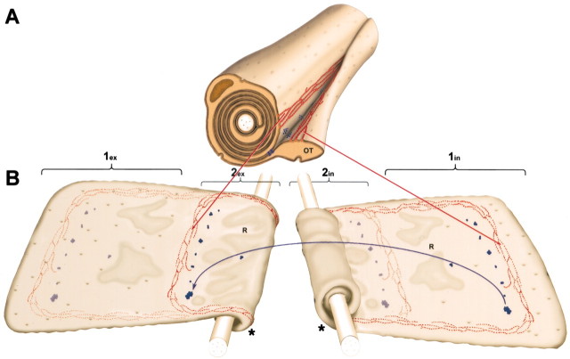
In vertebrate peripheral nerves, the insulating myelin sheath is formed by Schwann cells, which generate flattened membrane processes that spiral around axons and form compact myelin by extrusion of cytoplasm and adhesion of apposed intracellular and extracellular membrane surfaces. Cytoplasm remains within the innermost and outermost tongues, in the paranodal loops bordering nodes of Ranvier and in Schmidt-Lanterman incisures. By immunocytochemistry, connexin32 (Cx32) protein has been demonstrated at paranodal loops and Schmidt-Lanterman incisures, and it is widely assumed that gap junctions are present in these locations, thereby providing a direct radial route for transport of ions and metabolites between cytoplasmic myelin layers. This study used freeze-fracture replica immunogold labeling to detect Cx32 in ultrastructurally defined gap junctions in Schmidt-Lanterman incisures, as well as in a novel location, between the outer two layers of internodal myelin, approximately every micrometer along the entire length of myelin, at the zone between compact myelin and noncompact myelin. Thus, these gap junctions link the partially compacted second layer of myelin to the noncompact outer tongue. Although these gap junctions are unusually small (average, 11 connexon channels), their relative abundance and regular distribution along the zone that is structurally intermediate between compact and noncompact myelin demonstrates the existence of multiple sites for unidirectional or bidirectional transport of water, ions, and small molecules between these two distinct cytoplasmic compartments, possibly to regulate or facilitate myelin compaction or to maintain the transition zone between noncompact and compact myelin.


Figures

Figure 2.
Figure 2.
Cx32 immunogold labeling in freeze-fracture replicas of gap junctions in Schmidt–Lanterman incisures. A, Low-magnification view of an extensive stair-step arrangement of cytoplasmic expansions (blue shading) that are characteristic of Schmidt–Lanterman incisures. C, D, The inscribed areas are shown at higher magnification. B, Stereoscopic view of Schmidt–Lanterman incisures. C, D, High-magnification images of Cx32-labeled E-face gap junctions within the Schmidt–Lanterman incisure. Cx32 is labeled with 6 nm gold beads. C', D', High-magnification views of the same gap junctions, presented with black shadows (i.e., in reversed photographic contrast). At high magnification, white shadows (as seen in C and D) are unnatural and are difficult to interpret by most viewers (Steere et al., 1980). E-face pits of the gap junction are highlighted (red area). Immunogold beads are white in black shadow images. Gold beads smaller than 10 nm are difficult to detect or discriminate from shadowed IMPs without stereoscopic imaging. Scale bars (in electron micrographs), 0.1 μm; unless otherwise indicated.
Figure 4.
Figure 4.
FRIL image of a broad expanse of outer myelin membranes labeled for Cx32. A, The outermost layer of myelin (1exP) is characterized by abundant caveolae (black arrowheads). The outer tongue has been fractured away, revealing its cross-fractured cytoplasm (blue). Where the outer tongue has been removed by fracturing, the second layer of myelin (2exP; shaded green) is recognized by the absence or reduced density of caveolae (white arrowheads), lower density of IMPs, or areas devoid of IMPs and having smooth contour. The extracellular matrix (ECM) contains collagen fibers (yellow arrowheads). Tight junctions (B, white arrows) and gap junctions (C–F) are shown at higher magnification. B, Higher magnification view of tight junction strands, with rows of IMPs (black arrow) intermixed with grooves of pits (white arrow), which represent the sites where some of the tight junction particles–proteins were removed by fracturing. C–F, Stereoscopic P-face images of gap junctions, three of which are labeled for Cx32 (C–E; 12 nm gold beads) and one is unlabeled (F). 1exP, P-face of the outer membrane of the outermost (first) Schwann cell wrapping; 1inE, E-face of the inner membrane of the outermost Schwann cell wrapping; 2exP, P-face of the outer membrane of the second Schwann cell wrapping.
Figure 5.
Figure 5.
FRIL images of outer myelin layers after immunogold labeling for Cx32. A, Low-magnification image of myelin membrane 1exP, which is characterized by abundant caveolae. D, Layer 2exP, the outer membrane of the underlying Schwann cell wrapping, is characterized by the presence of rivulets containing residual cytoplasm, as documented at different tilt angle, 5D, adjacent. Two gap junctions are visible (inscribed areas B and C). B, A gap junction located where the fracture plane stepped from layer 2exP to 1inE consists of both P-face IMPs and E-face pits. The extracellular space (*) is narrowed to <3 nm within the area of the gap junction. C, Gap junction in membrane 1inE. The E-face pits are immunogold labeled for Cx32. D, At a high tilt angle, rivulets at the margin of cross-fractured myelin (M) are seen to contain cytoplasm (blue shading). E, Removal of the top rivulet membrane (1exP) reveals a view of the E-face of the underlying membrane 1inE. Gap junctions frequently were found on rivulet membranes (inscribed area F). F, Cx32 immunogold-labeled gap junction (yellow arrowhead) in particle-rich myelin membrane 2inE. ECM, Extracellular matrix; 1exP, P-face of the outer membrane of the outermost Schwann cell wrapping; 1inE, E-face of the inner membrane of the outermost (first) Schwann cell wrapping; 2exP, P-face of the outer membrane of the second Schwann cell wrapping.
Figure 6.
Figure 6.
High-magnification FRIL images of immunogold-labeled gap junctions. A, Cx32-labeled gap junction located at the step from P- to E-face. Note the narrowing of extracellular space (*) at the area of junctional contact. B, Two gap junctions, each composed of two connexons (arrows), with each gap junction labeled by one immunogold bead. C, Gap junction composed of 15 E-face pits, labeled by two immunogold beads. D, Gap junction composed of two connexons, with 12 nm immunogold beads indicating the presence of Cx32. E, Rare orphaned gap junction located on smooth myelin membrane deep within the stack of compact myelin. F, Gap junction at an indeterminate location in myelin. Gap junction P-face IMPs are double-labeled for Cx32 by one 18-nm and 13 6-nm gold beads.
Figure 1.
Figure 1.
Confocal immunofluorescence images of teased sciatic nerves (composite images of individually teased fibers) after immunolabeling for Cx32. A, In the sciatic nerve of wild-type mice, immunofluorescence was present at paranodal loops (white arrowheads pointing to nodes of Ranvier) and Schmidt–Lanterman incisures (yellow arrow). Additional fluorescent puncta (white arrows) may also represent discrete immunolabeling along outer layers of myelin, as found by FRIL (Figs. 4, 5, 6). B, In the sciatic nerve of Cx32 null-mutant mice, Cx32 was not detected (white arrowhead at nodes of Ranvier). C, Background immunofluorescence in wild-type mice after omission of monoclonal primary antibody to Cx32 (white arrowheads at nodes of Ranvier). Scale bar, 20 μm.
Figure 3.
Figure 3.
Diagrammatic representation of FRIL images of sciatic nerve, according to the low-magnification image in Figure 4. A, Beginning at the outside, myelin membranes are sequentially designated 1ex and 1in (outer and inner membranes of the outer wrapping of myelin), 2ex and 2in (outer and inner layers of second wrapping), and so on. The fracture plane through a myelinated fiber cross-fractured the cytoplasm (gray) of the outer tongue and then sequentially exposed several membrane faces. Left, The fracture plane first exposed the E-face of the inner plasma membrane of the outer tongue (1inE) and then the P-face IMPs of a gap junction (GJ) and IMPs and pits of a tight junction (TJ) linking the inner plasma membrane of the outer tongue to the P-face of the outer plasma membrane of the second wrapping of myelin (2exP). In the center, the fracture plane exposed the E-face of the inner plasma membrane of the second wrapping of myelin (2inE) and then returned to the P-face of the outer plasma membrane of the first or outermost layer of myelin, which at that point is exposed beyond the tip of the Schwann cell outer tongue. Caveolae (Cv) are cross-fractured and surface-fractured (right margin). B, After SDS washing and immunogold labeling for Cx32, immunogold beads are found almost exclusively at gap junctions, which are identified as hexagonally packed clusters of 9 nm P-face IMPs (Fig. 4) or E-face pits (Fig. 5C).
Figure 7.
Figure 7.
Diagrams of cross-sectional view versus view of unrolled outer layers of myelin in an internodal segment of peripheral nerve. A, Perspective view of a partially unrolled outer myelin layer, showing the outer tongue (OT) of myelin and the relative locations of tight junctions (red) and gap junctions (blue). Tight junctions and gap junctions link the inner membrane of the outer tongue to the outer membrane of the second wrapping of myelin. B, View of outer membrane of the first two unrolled wrappings of myelin (1ex and 2ex). Tight junctions are located away from the tip of the myelin tongue a distance equal to or slightly greater than the circumference of the outermost layer of myelin (π × D). The pattern of tight junction strands and gap junction hemichannels in 2ex is identical to the distribution of their pairing partners in the inner surface of the first turn of the outer wrapping (1in), as seen in the right panel of B. Inverted view of the same Schwann cell, revealing the inner membrane of the first (1in) and second (2in) Schwann cell wrappings. Few or no caveolae are present in the zone of partial compaction inside the borders of tight junctions in layer 1in. Rivulets (R) of cytoplasm are present in the second wrapping but extend into the first wrapping, past the gap junctions, almost to the tight junctions. Additional myelin layers are not visible in this partially unrolled segment of myelin, because each successive myelin layer is wider than the previous layer, and each successive paranodal loop covers the previous loop (*).

Similar articles

Cited by

References

    1. Altevogt BM, Kleopa KA, Postma FR, Scherer SS, Paul DL (2002) Connexin29 is uniquely distributed within myelinating glial cells of the central and peripheral nervous systems. J Neurosci 22: 6458–6470. - PMC - PubMed
    1. Anzini P, Neuberg DH, Schachner M, Nelles E, Willecke K, Zielasek J, Toyka KV, Suter U, Martini R (1997) Structural abnormalities and deficient maintenance of peripheral nerve myelin in mice lacking the gap junction protein connexin 32. J Neurosci 17: 4545–4551. - PMC - PubMed
    1. Balice-Gordon RJ, Bone LJ, Scherer SS (1998) Functional gap junctions in the Schwann cell myelin sheath. J Cell Biol 142: 1095–1104. - PMC - PubMed
    1. Bergoffen J, Scherer SS, Wang S, Oronzi-Scott M, Bone L, Paul DL, Chen K, Lensch MW, Chance P, Fischbeck K (1993) Connexin mutations in X-linked Charcot-Marie-Tooth disease. Science 262: 2039–2042. - PubMed
    1. Branton D, Bullivant S, Gilula NB, Karnovsky MJ, Moor H, Muhlethaler K, Northcote DH, Packer L, Satir B, Satir P, Speth V, Staehlin LA, Steere RL, Weinstein RS (1975) Freeze-etching nomenclature. Science 190: 54–56. - PubMed

Publication types

MeSH terms

LinkOut - more resources