. 2004 Mar 31;24(13):3199-209.
doi: 10.1523/JNEUROSCI.4419-03.2004.

Common sensory inputs and differential excitability of segmentally homologous reticulospinal neurons in the hindbrain

Affiliations

Common sensory inputs and differential excitability of segmentally homologous reticulospinal neurons in the hindbrain

Hisako Nakayama et al. J Neurosci. .

Abstract

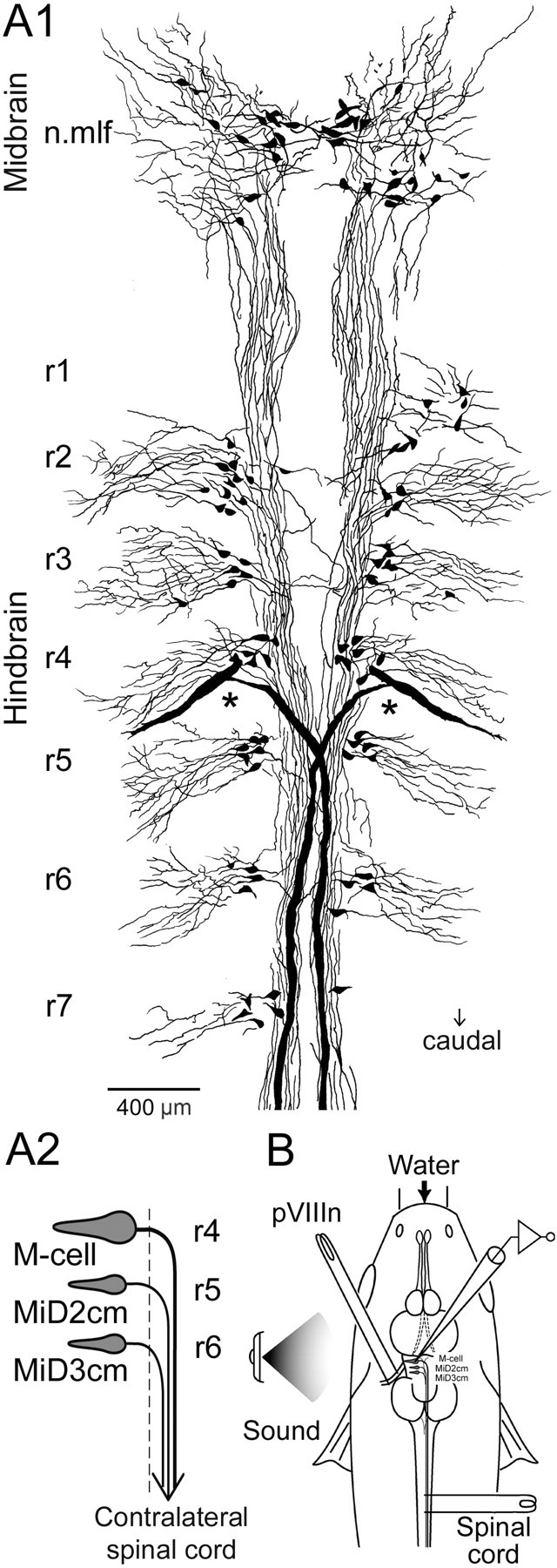
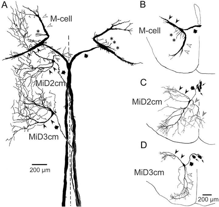
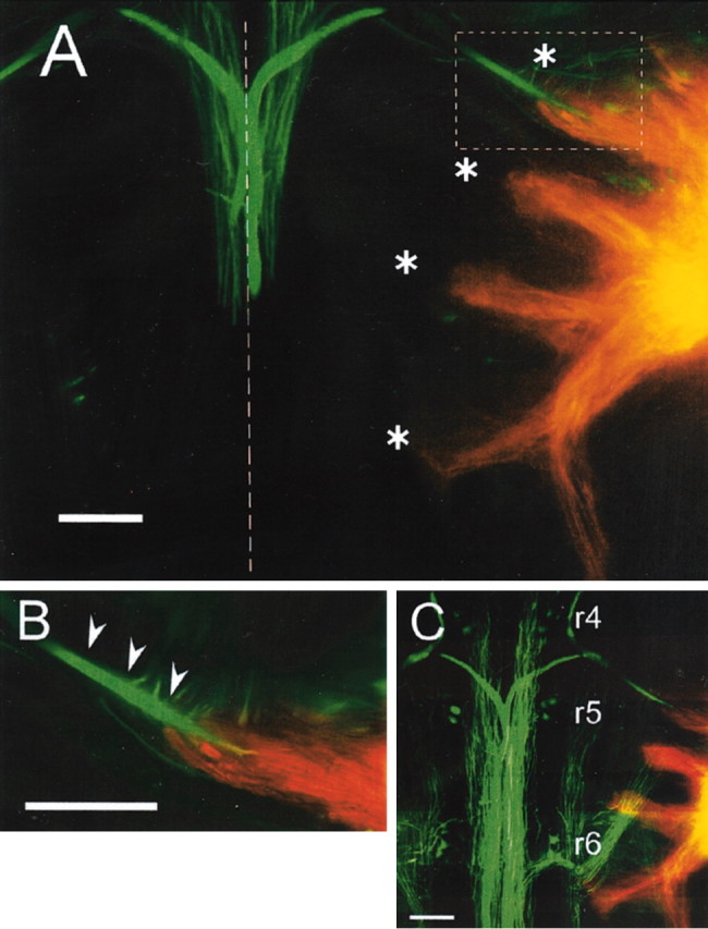
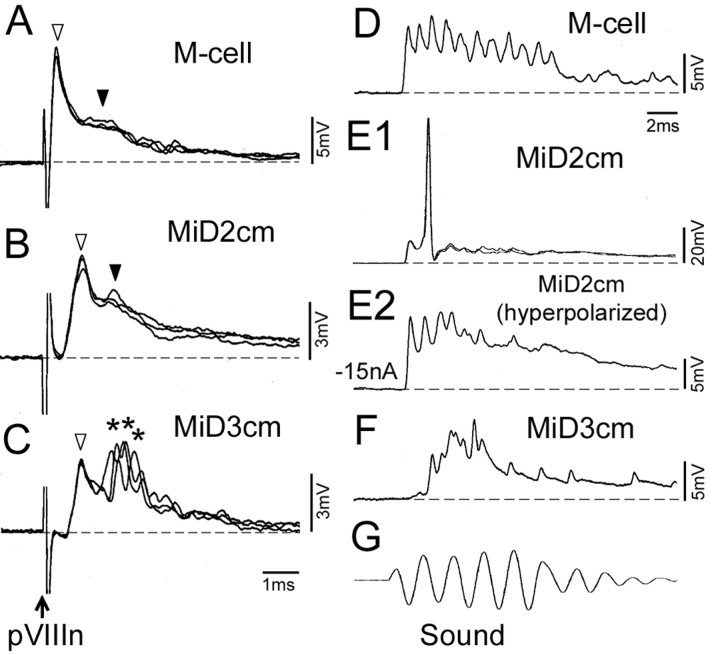
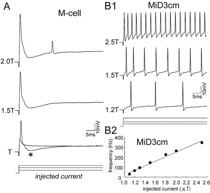
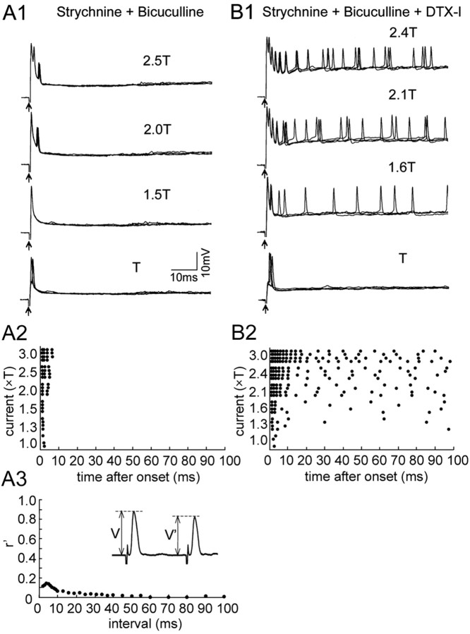
In the hindbrain of zebrafish and goldfish, reticulospinal (RS) neurons are arranged in seven segments, with segmental homologs in adjacent segments. The Mauthner cell (M-cell) in the fourth segment (r4) is known to trigger fast escape behavior. Its serial homologs, MiD2cm in r5 and MiD3cm in r6, are predicted to contribute to this behavior, which can be evoked by head-tap stimuli. However, little is known about their input-output properties. Therefore, we studied afferent projections from the auditory posterior eighth nerve (pVIIIn) and firing properties of MiD2cm and MiD3cm for comparison with the M-cell in adult goldfish. Labeling of RS neurons and the pVIIIn afferents with fluorescent tracers showed that the pVIIIn projected to r4-r6. Tone burst and electrical stimulation of the pVIIIn evoked EPSPs in the M-cell, MiD2cm, and MiD3cm. Stepwise depolarization typically elicited a single spike at the onset in the M-cell but repetitive spiking in MiD2cm and MiD3cm. This atypical property of the M-cell was mediated by dendrotoxin-I (DTX-I)-sensitive voltage-gated potassium channels together with recurrent inhibition, because combined application of DTX-I, strychnine, and bicuculline led to continuous repetitive firing in M-cells. The M-cell but not MiD2cm or MiD3cm expressed Kv1.2, a DTX-I-sensitive potassium channel subunit. Thus, the M-cell and its segmental homologs may sense common auditory information but send different outputs to the spinal circuits to control adaptive escape behavior.


Figures

Figure 1.
Figure 1.
A, Segmental arrangement of hindbrain RS neurons in an adult goldfish. A1, RS neurons labeled retrogradely with biocytin were reconstructed from 15 serial horizontal sections, each 50μm in thickness. RS neurons were divided into seven clusters along the neuraxis, r1–r7, rostral to caudal. The largest RS neurons, M-cells, were bilaterally located in r4 (asterisks). Neurons in the nucleus of mlf (n.mlf) are located in the midbrain. The difference in labeled RS neurons between the right and the left side is a result of incomplete transport of biocytin from the injection site. A2, Schematic representation of the M-series neurons: M-cell, MiD2cm, and MiD3cm. Somata of the paired M-series neurons are bilaterally located dorsally in r4–r6 (the first to third of middle segments of hindbrain), respectively, and their axons descend into the contralateral spinal cord via the mlfD after decussating in the hindbrain. The dashed line indicates the midline. B, The schematic diagram indicates the experimental set-up. Intracellular recordings were performed from M-series neurons with glass micropipettes filled with 4 m potassium acetate containing neurobiotin to label the recorded neurons. Bipolar stimulating electrodes were placed on the vertebral column for antidromic activation of the RS axons and on the pVIIIn for orthodromic activation of the synaptic inputs. Sound stimulation was applied from a loudspeaker positioned on the left side of the fish.
Figure 4.
Figure 4.
Antidromically evoked action potentials of M-series neurons. A, B, AD spikes recorded intracellularly (Intra.) in an M-cell and a MiD2cm, respectively, in response to spinal cord stimulation at threshold intensities (top traces). Corresponding extracellular (Extra.) field potentials are shown below. Failures were represented by gray traces. Three responses were overlaid for each. The onset latency of AD spikes was 0.26 msec (A) and 0.46 msec (B), respectively. Erest was –76 mV in the M-cell and –77 mV in the MiD2cm. Note that the large negative field potentials were observed in the axon cap of the M-cell (A, bottom). C, Frequency histogram of onset latencies of AD spikes from spinal stimulation. The latencies of M-cells (30 cells) were shorter than those of MiD2cm (17 cells) and MiD3cm (17 cells).
Figure 2.
Figure 2.
Intracellularly labeled M-series neurons. Horizontal sections include bilateral M-cells, left MiD2cm, and left MiD3cm from one fish. A1–A4 depict dorsal to ventral sections, as do B and C. A1, Distal lateral dendrite (filled arrowhead) of the left M-cell and thick crossing axons of bilateral M-cells (filled arrows) were labeled. A2, Somata (asterisks) and descending axons (filled arrows) of bilateral M-cells and thin axon of left MiD2cm (open arrows). A3, A4, Horizontal sections of M-cell ventral dendrite extending rostroventrally (open arrowheads). B1, Axons of bilateral M-cells (filled arrows) and crossing axon of the left MiD2cm (open arrows). B2–B4, Lateral dendrite (B2, B3, filled arrowheads), soma (B3, asterisk), and ventral dendrites with small branches (B4, open arrowheads) of the left MiD2cm were labeled. C1, Lateral dendrites (filled arrowheads) and crossing axon (gray arrow) of the left MiD3cm were clearly labeled. Filled and open arrows indicate axons of the bilateral M-cell and the left MiD2cm, respectively. Another fiber (thin arrow) was an axon of right MiD3cm. C2–C4, Soma (C2, asterisk), proximal (C3, open arrowhead), and distal ventral dendrites (C4, open arrowheads) of the left MiD3cm. Dashed lines indicate the midline. Calibration: A1, A2, C1, 200 μm; A3, A4, B1–B4, C2–C4, 100 μm. These labeled neurons were reconstructed by camera lucida drawing, as shown in Figure 3A.
Figure 3.
Figure 3.
Camera lucida reconstruction of the M-series from serial horizontal (A) and coronal (B–D) sections. A, Intracellularly labeled bilateral M-cells, left MiD2cm, and left MiD3cm in a fish, which were shown in Figure 2. The axons, lateral dendrites, and ventral dendrites are indicated by thick arrows, filled arrowheads, and open arrowheads, respectively.B–D, Coronal views of the M-series neurons in different fish. Axons of the M-series neurons (thick arrows) crossed the midline and descended along the contralateral mlfD. Lateral dendrites (filled arrowheads) of the M-series neurons extended caudolaterally, and ventral dendrites (open arrowheads) extended rostroventrally. The M-cell had additional small dendrites extending from the soma (asterisks; also in A). The MiD2cm had a medial dendrite extending ventromedially, the peripheral part of which entered into the contralateral brain (gray arrowheads; also in A). The thin arrows in C and D indicate axons of bilateral M-cells. The calibration in D is also applied to B and C. A–D are a composite respectively of 24, 6,7, and 15 serial sections, each 50 μm in thickness. The dashed line indicates the midline. Up is rostral in A and dorsal in B–D.
Figure 5.
Figure 5.
Labeling of pVIIIn and RS neurons. RS neurons (green) and pVIIIn (orange) were labeled with Oregon Green 488 and DiI, respectively. A, Two adjacent 80 μm sections were superimposed where axons and lateral dendrites of bilateral M-cells (green) and right pVIIIn (orange) were labeled. Four fascicles of pVIIIn (asterisks) were observed to project medially to where lateral dendrites of the M-series neurons likely exist (compare Fig. 3). The first bundle projected to the vicinity of the distal region of the M-cell lateral dendrite. The second and fourth bundles appeared to project into r5 and r6, respectively (also see C). Another bundle descending along the lateral edge of the brain was labeled. The dashed line indicates the midline. B, Higher-magnification image of the inset in A. The pVIIIn afferents appeared to contact the distal part of the M-cell lateral dendrite (arrowheads). C, Eight serial sections were superimposed, including two dorsal sections shown in A. Somata of dorsal RS neurons in r4–r6 were labeled. Rostral is up. Scale bars, 200 μm.
Figure 6.
Figure 6.
Synaptic responses evoked in M-series neurons by auditory stimulation. A–C, Postsynaptic potentials evoked in an M-cell, a MiD2cm, and a MiD3cm, respectively, by electrical stimulation of pVIIIn. A, Depolarizing responses elicited in the M-cell had two components, indicated by open and filled arrowheads, that have been established to be mediated by electrical and glutamatergic synapses, respectively. Erest, –82 mV. B, Depolarizing potentials of the MiD2cm consisted of two components: fast potentials (open arrowhead) with a constant onset latency followed by slow potentials (filled arrowhead). Erest, –73 mV. C, Depolarizing potentials induced in the MiD3cm consisted of fast potentials (open arrowhead) with delayed but locked-in onset followed by fluctuating fast potentials (asterisks). Erest, –78 mV. Dashed lines represent the baseline. D–F, Postsynaptic potentials induced by sound (500 Hz, 95–100 dB) (G) in M-series neurons different from A–C. D, Sharp depolarizing potentials were evoked in the M-cell. Erest, –78 mV. E1, An action potential from an Erest of –77 mV was elicited in the MiD2cm. E2, Depolarizing potentials with sharp peaks were observed when the membrane was hyperpolarized by passing cathodal current (–15 nA). F, Delayed responses were observed in the MiD3cm. Erest, –75 mV. The time scales in C and D apply to A–C and D–G, respectively.
Figure 7.
Figure 7.
Different patterns of firing elicited by stepwise depolarization. A, Step-depolarizing currents were injected into an M-cell soma through one of the two channels of θ microelectrode, and voltage responses were recorded with the other channel. A single action potential was elicited in response to threshold current intensity (T, 120 nA in this cell) and 1.5T. The recurrent IPSPs (asterisk) were visible as hyperpolarizing potentials after the initial spike. An additional spike was elicited after the IPSPs at 2T. The amplitude of the first spike in each trace included capacitive transient potentials caused by an incomplete compensation of a coupling capacitance between two channels of a θ micropipette. B1, Repetitive firing was elicited in a MiD3cm by injection of much smaller currents than the M-cell (T, 10 nA). The MiD3cm spiked with almost regular frequencies proportional to the injected currents. Note that there was no sign of the recurrent IPSPs after the spikes. B2, Relationship between injected current and mean firing frequency of the MiD3cm shown in B1. The firing frequency increased almost linearly with the amplitude of the injected current; the slope was 23.1 Hz/nA.
Figure 8.
Figure 8.
Effects of DTX-I on the firing of M-cell. Depolarization-induced firing of an M-cell before (A) and during (B) the application of DTX-I (1 μm) to the brain surface. A1, An action potential followed by recurrent IPSPs was elicited by stepwise depolarization with intensities of T (80 nA) and 1.6T stimulus. The onset of step currents was denoted by arrows (also in Fig. 9). An additional action potential was elicited after the recurrent IPSPs in response to a stronger stimulus (2.1T). Four responses were superimposed for each (also in B1). A2, Raster plots showing the peak time of each action potential from the onset of current pulse. One or two action potentials were evoked at the onset of stepwise depolarization. Additional spikes were observed, but after ∼15 msec intervals from the initial firing in response to >2T depolarization. A3, Synaptic conductance underlying the recurrent IPSPs (GIPSP) was estimated from its shunting effect on the test AD spikes, which were applied after the control AD spike at various inter-spike intervals (inset). Relative synaptic conductance (r′= GIPSP /Gm, where Gm is the resting conductance) was calculated from amplitude of the control (V) and shunted (V′) AD spikes as r′= (V/V′–1). B1, In the presence of DTX-I, the M-cell fired repetitively (1.5T and 2T). The threshold current intensity for spike generation (T, 40 nA) was lower than control. B2, Raster plots showed that the firing rate of the M-cell increased with the degree of depolarization, and that there was a pause after the onset firing. B3, Recurrent inhibition in the presence of DTX-I. The time course of the r′ was not affected by DTX-I, although the amplitude was slightly increased. Erest was stable at –75 mV throughout the experiment.
Figure 9.
Figure 9.
Effects of applying DTX-I and blocker for the recurrent inhibition of M-cell firing. Voltage responses of an M-cell in the presence of strychnine and bicuculline (A) and during an additional application of DTX-I, DNQX, and APV (B) are shown. A1, An action potential without recurrent IPSPs was elicited by T (80 nA) and 1.5T stimulus during application of strychnine (100 μm) and bicuculline (100μm) to the brain surface. Short, transient spike bursts were evoked at the onset of depolarization (2T, 2.5T). Four responses were overlaid for each (also in B1). A2, Raster plots represent the peak time of each action potential. A3, The synaptic conductance of the recurrent inhibition was abolished by strychnine and bicuculline, as shown in the inset. B1, Continuous repetitive firings were induced during an additional application of DTX-I (1 μm) (1.6T, 2.1T, and 2.4T; T, 70 nA). DNQX (200 μm) and d-APV (1 mm) were also applied to block most of the synaptic transmission. B2, Raster plots showed that continued, repetitive firing was induced. The firing frequency was higher during the initial 20 msec than during the next phase. Erest in A and B was –78 and –74 mV, respectively.
Figure 10.
Figure 10.
Immunostaining for Kv1.2 subunit in soma and dendrite of M-cell. Polyclonal anti-rat Kv1.2 antibody (Alomone Labs) was used. A–E, Serial horizontal sections with 50 μm thickness where the lateral dendrite (A–C, arrows), soma (D, arrow), and proximal ventral dendrite (E, arrow) of the M-cell were immunolabeled. Fibers that appeared to connect to the distal lateral dendrite were also immunolabeled (A, B, asterisks). F, Viewing fields in A–E. G–I, Horizontal sections of hindbrain, including the labeled M-cell (arrowheads). The soma of the M-cell was observed in G, and the ventral dendrite of the M-cell was observed in H and I. H and I were 100 and 150 μm ventral to G, respectively. Dorsal RS neurons caudal to the M-cell, including MiD2cm and MiD3cm, were not labeled. Some mlf fibers were labeled (brackets). Arrows indicate the midline.
Figure 11.
Figure 11.
Functional similarities and differences among segmentally homologous RS neurons, the M-series. The thicker lines represent faster-conducting pathways. Open and filled circles indicate excitatory and inhibitory synapses, respectively. All M-series neurons are excited by the pVIIIn afferents. The M-cell only produces an action potential at the onset of a depolarization, in strong contrast to its serial homologs, MiD2cm and MiD3cm, which fire repetitively. The unique firing property of the M-cell is attained by both DTX-I-sensitive, voltage-gated potassium channels (KDTX) and the recurrent inhibition.

Similar articles

Cited by

References

    1. Bal R, Oertel D (2001) Potassium currents in octopus cells of the mammalian cochlear nucleus. J Neurophysiol 86: 2299–2311. - PubMed
    1. Bosch TJ, Maslam S, Roberts BL (2001) Fos-like immunohistochemical identification of neurons active during the startle response of the rainbow trout. J Comp Neurol 439: 306–314. - PubMed
    1. Brew HM, Forsythe ID (1995) Two voltage-dependent K+ conductances with complementary functions in postsynaptic integration at a central auditory synapse. J Neurosci 15: 8011–8022. - PMC - PubMed
    1. Canfield JG, Eaton RC (1990) Swimbladder acoustic pressure transduction initiates Mauthner-mediated escape. Nature 347: 760–761.
    1. Casagrand JL, Guzik AL, Eaton RC (1999) Mauthner and reticulospinal responses to the onset of acoustic pressure and acceleration stimuli. J Neurophysiol 82: 1422–1437. - PubMed

Publication types

MeSH terms

LinkOut - more resources