. 2004 Mar 31;24(13):3325-34.
doi: 10.1523/JNEUROSCI.5598-03.2004.

Expression of connexin36 in cone pedicles and OFF-cone bipolar cells of the mouse retina

Affiliations

Expression of connexin36 in cone pedicles and OFF-cone bipolar cells of the mouse retina

Andreas Feigenspan et al. J Neurosci. .

Abstract

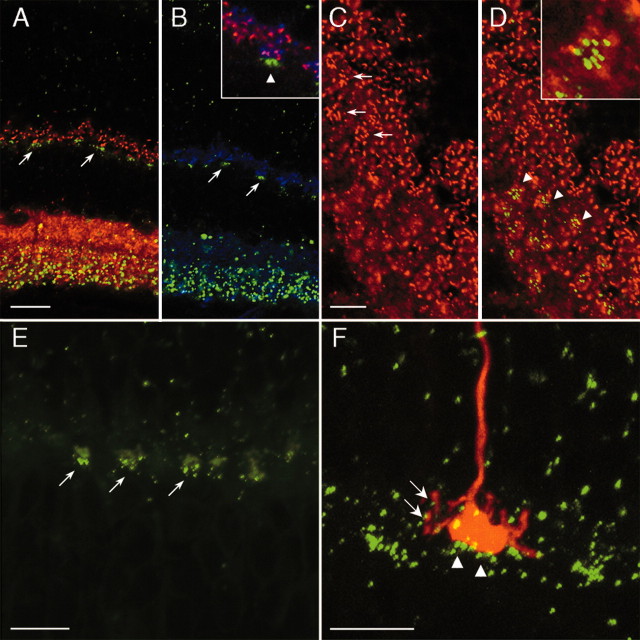
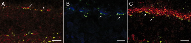
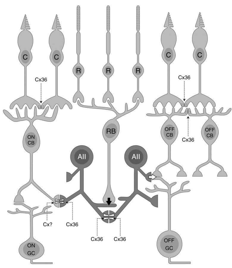
Transgenic technology, immunocytochemistry, electrophysiology, intracellular injection techniques, and reverse transcription PCR were combined to study the expression of neuronal connexin36 (Cx36) in the outer plexiform layer of the mouse retina. Transgenic animals expressed either a fusion protein of full-length Cx36 with enhanced green fluorescent protein (EGFP) attached at the C terminus or exon 2 of Cx36 was replaced bybeta-galactosidase (beta-gal). In the outer nuclear layer,beta-gal-positive cell bodies, which were confined to the most distal region close to the outer limiting membrane, displayed immunoreactivity against S-cone opsin. Cx36-EGFP puncta colocalized with cone pedicles, which were visualized by intracellular injection. In reverse transcriptase PCR experiments, Cx36 mRNA was never detected in samples of rods harvested from the outer nuclear layer. These results strongly suggest expression of Cx36 in cones but not in rods. In vertical sections, Cx36 expression in the vitreal part of the outer plexiform layer was characterized by a patchy distribution. Immunocytochemistry with antibodies against the neurokinin-3 receptor and the potassium channel HCN4 (hyperpolarization-activated cyclic nucleotide-gated potassium channel) displayed clusters of the Cx36 label on the dendrites of OFF-cone bipolar cells. In horizontal sections, these clusters of Cx36 appeared as round or oval-shaped groups of individual puncta, and they were always aligned with the base of cone pedicles. Double-labeling experiments and single-cell reverse transcriptase PCR ruled out expression of Cx36 in horizontal cells and rod bipolar cells. At light microscopic resolution, we found close association of Cx36-EGFP with the AMPA-type glutamate receptor subunit GluR1 but not with GluR2-GluR4, the kainate receptor subunit GluR5, or the metabotropic glutamate receptor mGluR6.


Figures

Figure 1.
Figure 1.
Expression of Cx36 in wild-type and transgenic mouse retina. A, Immunolabeling of vertical sections of wild-type mouse retina with polyclonal antibodies to Cx36. B, Expression pattern of EGFP in Cx36Cx36–EGFP transgenic mice. C, High-power confocal micrograph showing the OPL of Cx36Cx36–EGFP mice counterstained with Cx36 antibodies. Top, EGFP staining pattern; middle, Cx36 immunoreactivity; bottom, superimposition of EGFP and Cx36 immunolabeling. Yellow indicates overlap of EGFP and Cx36 immunofluorescence. D, Expression pattern of β-galactosidase in Cx36LacZ/LacZ transgenic mice. Scale bars: A, B, D, 20 μm; C,10 μm. Nomarski image in A indicates layering of the mouse retina. GCL, Ganglion cell layer.
Figure 2.
Figure 2.
Scotopic ERG recordings. A, Example recordings of the dark-adapted ERG from a Cx36/ EGFP+ transgenic mouse in response to 20 m sec white light flashes ranging from 0.05 to 3800 lux corneal illuminance. B, Intensity–response curves did not differ statistically for both the a-waves (squares, triangles) and b-waves (circles, diamonds) of Cx36/ EGFP+ (filled symbols) and Cx36+/+ EGFP mice (mean ± SEM; n = 3). C, Implicit time versus intensity of the a- and b-waves were also identical.
Figure 3.
Figure 3.
Expression of Cx36 in photoreceptors of Cx36Cx36–EGFP mice. A1, Stack of confocal images displaying the typical morphology of a rod photoreceptor injected with the fluorescent dye Alexa 594. A2, A 200 nm optical section of the rod shown in A1 is superimposed with the EGFP staining pattern. EGFP is not localized in the rod spherule (arrow). B1, Stack of confocal images showing the typical morphology of a cone photoreceptor injected with the fluorescent dye Alexa 594. B2, A 200 nm optical section of the cone shown in B1 is superimposed with the EGFP staining pattern. Cx36–EGFP appears to be expressed in the cone pedicle (arrow). C, Enlarged confocal micrograph of another rod spherule contacted by a rod bipolar cell. D, High-power confocal micrograph of the cone pedicle shown in B clearly reveals localization of EGFP-positive puncta at the cone pedicle (arrows). E, A vertical section of a Cx36LacZ/LacZ mouse retina counterstained with polyclonal antibodies directed against S-cone opsin. Both cell bodies (arrows) and inner segments of S-cones contain β-galactosidase-positive puncta. Very few S-cone somata do not express β-galactosidase (arrowhead). F, Localization of Cx36–EGFP on dye-injected cone pedicles results in a distinct peak in the center of the three-dimensional plot. z-axes numbers indicate brightness of the respective pixels. G, When a mirror image of the EGFP staining pattern is superimposed on the injected cones, colocalization of EGFP on cone pedicles is at chance levels, as indicated by the absence of a central peak. Scale bars: A, C, D, 5 μm; B, 10 μm; E, 20 μm.
Figure 4.
Figure 4.
Agarose gel electrophoresis of reverse transcriptase PCR products for Cx36 mRNA. A, Micrograph of the ONL of wild-type mouse retina. The dotted line separates the inner, rod-containing half of the ONL (IH) and the outer half (OH), which is composed of both rods and cones. B, Cx36 mRNA was detected in samples taken from the outer half of the ONL. The molecular weight of the amplicon corresponds to the expected value (447 bp). C, Samples harvested from the inner half of the ONL were negative for Cx36 mRNA. Ret, Positive control performed with transcripts from whole mouse retina. Scale bar: A, 10 μm.
Figure 5.
Figure 5.
Expression of Cx36–EGFP patches in the OPL. A, B, Double labeling of Cx36–EGFP vertical section with antibodies against the presynaptic proteins bassoon (A) and synaptophysin (B). EGFP puncta appear as clusters that do not colocalize with presynaptic immunocytochemical markers (arrows). B, Inset, An enlarged confocal micrograph of a triple labeling, indicating postsynaptic location of Cx36–EGFP (arrowhead). C, Labeling of Cx36–EGFP horizontal sections with antibodies against bassoon. The section cuts the OPL in a slightly oblique way, with the INL in the bottom left and the ONL in the top right corner. Cone pedicles are easily identified according to the rosette-like arrangement of synaptic ribbons immunolabeled by bassoon (arrows). D, In the same section, EGFP clusters appear as round or oval accumulations of individual puncta, which do not colocalize with the bassoon-labeled cone pedicles, but are confined to a different focal plane (arrowheads). D, Inset, An enlarged confocal micrograph of EGFP-positive puncta on the diffuse background of an out-of-focus cone pedicle. E, Labeling of cone pedicles with PNA–FITC in a vertical section of Cx36Cx36–EGFP mouse retina. EGFP puncta appear clustered at the base of fluorescein-labeled cone pedicles (arrows). F, Dye-injected cone pedicle displays punctate (arrows) and clustered (arrowheads) Cx36–EGFP label on the presynaptic and postsynaptic side, respectively. Scale bars: A, B, 20 μm; C–F, 10 μm.
Figure 6.
Figure 6.
Cx36 is not expressed in horizontal cells and rod bipolar cells. A, Vertical section of a Cx36LacZ/LacZ mouse retina processed for β-galactosidase and calbindin-like immunocytochemistry. Cell bodies of horizontal cells do not contain β-gal reaction product (arrows). B, Confocal micrograph of PKC-like immunoreactivity in a vertical section of Cx36Cx36–EGFP mouse retina. Staining patterns of EGFP and PKC do not overlap (arrows), indicating that Cx36 is not expressed in rod bipolar cells. Scale bars, 20 μm.
Figure 7.
Figure 7.
Expression of Cx36 in different types of OFF-cone bipolar cells. A, Vertical section of a Cx36Cx36–EGFP mouse retina immunolabeled with polyclonal antibodies to the neurokinin-3 receptor NK3R. Patches of EGFP colocalize with NK3R-positive dendrites of OFF-cone bipolar cells (arrows). B, Localization of β-galactosidase in a dye-injected OFF-cone bipolar cell (arrow). C, A 200 nm optical section obtained from a different type of OFF-cone bipolar cell demonstrates dendritic localization of EGFP puncta (arrows). D, Vertical section of a Cx36Cx36–EGFP mouse retina immunolabeled against the potassium channel HCN4. EGFP-positive puncta are localized to type 3 OFF-cone bipolar cell dendrites (arrows). E, Vertical section of a Cx36LacZ/LacZ mouse retina immunolabeled against HCN4 and superimposed on a transmission image. Most cell bodies of HCN4-positive OFF-bipolar cells express β-galactosidase (arrows), although few somata are devoid of the β-gal reaction product (arrowhead). Dotted lines indicate the INL–IPL border. Scale bars, 10 μm.
Figure 8.
Figure 8.
Spatial association of Cx36 with glutamate receptor subunits in the OPL. A, Confocal micrograph of a vertical section through Cx36Cx36–EGFP mouse retina labeled with polyclonal antibodies against GluR1. Arrows indicate close spatial relationship of Cx36–EGFP label and GluR1 immunoreactivity. B, Vertical section of a Cx36Cx36–EGFP mouse retina labeled with polyclonal antibodies against GluR5. Cx36–EGFP label appears associated with GluR5 but confined to a region below the GluR5 immunoreactivity (arrows). C, Vertical section of a Cx36Cx36–EGFP mouse retina labeled with polyclonal antibodies against the metabotropic glutamate receptor subtype mGluR6. mGluR6 is expressed on the dendrites of rod bipolar cells and ON-cone bipolar cells and is not associated with Cx36–EGFP (arrows). Scale bars, 10 μm.
Figure 9.
Figure 9.
Schematic drawing indicating the distribution of Cx36 in the outer retina. Cone pedicles make homotypic gap junctions containing Cx36 with neighboring cone pedicles in the scleral part of the OPL. In addition, OFF-cone bipolar cells make homotypic, Cx36-containing gap junctions with other OFF-cone bipolar cells in the vitreal part of the OPL. These gap junctions appear restricted to an area outlined by the cone pedicle. Cx36-containing gap junctions in the rod pathway are also indicated. C, Cones; R, rods; CB, cone bipolar cells; RB, rod bipolar cells; AII, aII amacrine cells; GC, ganglion cells.

Similar articles

Cited by

References

    1. Benardo LS (1997) Recruitment of GABAergic inhibition and synchronization of inhibitory interneurons in rat neocortex. J Neurophysiol 77: 3134–3144. - PubMed
    1. Blanks JC, Johnson LV (1983) Selective lectin binding of the developing mouse retina. J Comp Neurol 221: 31–41. - PubMed
    1. Brandstätter JH, Löhrke S, Morgans CW, Wässle H (1996) Distributions of two homologous synaptic vesicle proteins, synaptoporin and synaptophysin, in the mammalian retina. J Comp Neurol 370: 1–10. - PubMed
    1. Brandstätter JH, Koulen P, Wässle H (1997) Selective synaptic distribution of kainate receptor subunits in the two plexiform layers of the rat retina. J Neurosci 17: 9298–9307. - PMC - PubMed
    1. Brandstätter JH, Fletcher EL, Garner CC, Gundelfinger ED, Wässle H (1999) Differential expression of the presynaptic cytomatrix protein bassoon among ribbon synapses in the mammalian retina. Eur J Neurosci 11: 3683–3693. - PubMed

Publication types

MeSH terms

LinkOut - more resources